
Content
- General characteristics of the disease
- Reasons for the appearance
- Pathogens and transmission routes
- Symptoms and Signs
- Infected or not
- Classification of tuberculous pleurisy of the lungs
- Allergic
- Perifocal
- Pleural tuberculosis
- Fibrous
- Exudative (effusion)
- Purulent
- The clinical picture of the course of the disease
- Diagnostic methods
- X-ray
- Basic principles of treatment
- Traditional methods
- Recovery Predictions
- Rehabilitation and features of the body's recovery
- Possible consequences and complications
- Prevention methods
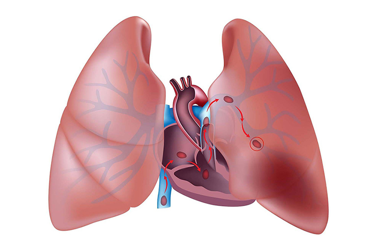
In tuberculosis, the pleura is often affected and tuberculous pleurisy develops. It can be a complication or an independent clinical form of this disease. Most often (70-75% of cases) develops in children and adolescents.

General characteristics of the disease
Pleurisy of tuberculous genesis is a specific reaction of the pleura to infection with mycobacterium tuberculosis. Over the past decade, the incidence rate has increased. In children with tuberculosis, the level of pleurisy is 70-75%.
Contrary to earlier studies, which showed that pathology is most often diagnosed in the young age group (20-40 years), the disease is "aging". Today more than 1/3 of patients are over 60 years old. According to the results of the last 7 years, the peak of the disease falls on the age group of 21-50 years.
There is also a gender dependence in the development of pathology. Men get sick 2.5 times more often than women.
Reasons for the appearance
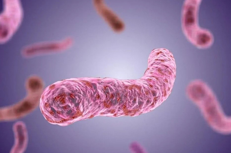
Inflammation of the pleura is caused by Koch's bacillus. The pathological process can be triggered as the direct penetration of an infectious agent from the lungs, and with tuberculosis of internal organs, after a chest injury leading to pneumothorax or after collapse therapy.
In the pathogenesis of the disease, 2 main factors play a role:
- allergic reaction;
- direct invasion of the infectious agent into the pleural layers.
A prerequisite for the pathogenic effect of Koch's bacillus is a decrease in local immunity and the whole organism.
Pathogens and transmission routes
A person can become infected by contact with a patient with an open form of tuberculosis. When coughing and sneezing, the pathogen is released into the air with sputum droplets and settles on dust particles. When inhaled, they become infected. An infectious agent enters the pleural cavity from the lungs during tissue decay in the lesion.
In addition to the contact route of infection, mycobacteria can enter the pleura with the blood flow (hematogenous route) or with lymph (lymphogenous).
Symptoms and Signs
The clinical picture depends on the stage and type of the disease. In some patients, there are no manifestations of the disease, especially at the initial stage, and it is detected by chance (during the passage of a medical examination). With the progression of the pathology, there are signs of damage to the respiratory system:
- shortness of breath, chest pains of varying intensity associated with breathing, movement;
- cough;
- general weakness;
- excessive sweating, especially at night;
- a feeling of heaviness and discomfort in the chest on the side of the lesion;
- an increase in body temperature from subfebrile to febrile digits.

When the costal pleura is involved in the pathological process, the patient complains of pain from the sides of the chest on the side of the lesion.
With damage to the interlobar pleura, pain is felt at the level of the III-IV rib (paravertebral pain point) or in front of the sternum, respectively, IV-VI rib (anterior-lower pain point). With deep breathing or pressure on the intercostal spaces, it increases.
When the pleura is damaged in the diaphragm, the pain syndrome is shingles in nature and radiates to the trapezius muscle and the shoulder joint. If the exudate accumulates on the right side, patients complain of pain in the upper abdominal cavity, similar to the symptoms of an acute abdomen. They are aggravated by loud conversation, sneezing, coughing, after eating.
Important information: Basic principles of treatment of tuberculosis of the spine and bones in adults
With the accumulation of fluid in the upper part of the pleural cavity, symptoms of brachial plexitis and periplexitis are noted. In the phase of acute pleurisy, the patient tries to breathe shallowly, faster, in order to reduce the severity of pain. In the event of pleural adhesions, shortness of breath becomes constant, lying down the person takes a forced position - on the unaffected side of the body.
Infected or not
Contagiousness depends on the form of pleurisy. If the pathological lesion is limited to the pleural cavity and the exudate is not released to the outside, there is no risk of infection.
When the cause of the disease is cavernous cavities in the lungs or the discharge of effusion through the respiratory tract during the formation of fistulas in the pleura, the risk of infection increases. In the effusion, a greater number of mycobacteria of tuberculosis and other nonspecific representatives of microflora are found than in uncomplicated forms of the disease.

To determine whether a patient is contagious or not, only a doctor can after examining the sputum secreted by him.
Classification of tuberculous pleurisy of the lungs
Pathology is classified according to several criteria:
- presence or absence of effusion (dry, exudative);
- the nature of the exudate (serous, serous-fibrinous, purulent, hemorrhagic, eosinophilic, cholesterol, chyle);
- the presence or absence of restriction of inflammatory effusion (diffuse, encapsulated);
- localization of the focus.
Allergic
The allergic form develops against the background of the pleural hypersensitization reaction to the effect of pathogen toxins. Under their influence, an inflammatory-allergic process occurs with the release of effusion. There are no characteristic changes in the structure of pleural tissue with an allergic form.
Such pleurisy may be the only manifestation of infection of the body with tuberculous mycobacteria. But it can be combined with various forms of the disease and other pathologies caused by the body's nonspecific response to the pathogen.
This form has features:
- acute onset;
- exudate is often serous or hemorrhagic;
- Koch's sticks are not found;
- the pleura is edematous, hyperemic;
- fibrinous deposits are absent;
- cure occurs quickly with the use of anti-inflammatory and antihistamines, incl. hormonal.
After recovery, structural changes do not remain on the sheets.
Perifocal
This form is an infection of the pleura in the area adjacent to the cavities of the lungs and can complicate the course of different types of tuberculosis. The perifocal type has its own characteristics:
- long course, prone to recurrence;
- serous exudate;
- starts as dry pleurisy, and then turns into exudative;
- one of the forms of pulmonary tuberculosis is necessarily present;
- mycobacteria in the exudate are absent (the integrity of the pleura is not violated);
- hyperemia and compaction (edema) of the pleural sheets with fibrin deposition.
The reverse dynamics of the process is slow, pleurisy resolves, leaving a large number of pleural adhesions.
Pleural tuberculosis
This is the infection of the pleura with mycobacteria and the development of the tuberculous process in the tissues. The form may be the only manifestation of tuberculosis or be combined with its other types. Features of the disease:
- slow flow;
- gradual accumulation of serous effusion;
- mandatory presence of mycobacteria;
- numerous small or large foci of whitish color on the pleura;
- slow recovery with adhesion formation and tissue fibrosis.
With a diffuse process and the decay of large tuberculous foci, a violation of the process of resorption of effusion, purulent pleurisy may develop.
Fibrous
The dry (fibrinous) type is characterized by a small amount of exudate. The effusion contains a large amount of fibrin. After resorption of fluid, it is deposited on the pleura, forming whitish layers. They are subsequently the basis for widespread or local foci of adhesion of the pleural sheets and limitation of its mobility (adhesive pleurisy).
Important information: Features of the development and course of miliary pulmonary tuberculosis
The features of the clinical picture of dry pleurisy are:
- acute onset of the disease;
- signs of intoxication of the body, accompanied by febrile (less than + 38 ° C) temperature;
- dry hacking cough;
- pain in the chest in the projection of the pleural lesion.
Dry pleurisy is most often the initial phase of the exudative form. In rare cases, it can act as an independent variant of the pathological process.
Exudative (effusion)
Effusion pleurisy, regardless of the localization of the focus, has common symptoms:
- Oiled start: slight malaise, increased sweating, decreased appetite, normal temperature.
- After a few days (sometimes 2-3 weeks), signs of an acute illness appear: the temperature rises to febrile (+ 39... + 39.5 ° С), chills, fever, heaviness and pain in the projection of the affected area, cough causing pain, dyspnea.
- With a slow onset and progression of the pathological process, symptoms increase gradually.
- The patient tries to take a position in which shortness of breath and pain is less, tries to breathe shallowly.
- On visual examination of the chest, its asymmetry is noted - the intercostal spaces on the side of the lesion are smoothed or convex.
- In the process of breathing, the affected side lags behind the healthy side in movement.
- At the initial stage of the disease, the sound of rubbing pleura is heard, which eventually decreases or disappears due to an increase in the amount of effusion.

After resorption of the effusion, symptoms of dry pleurisy appear.
Purulent
This form (pleural empyema) has its own specific features:
- there are pronounced symptoms of intoxication of the body;
- the exudate contains pus, mycobacteria and the corresponding pyogenic microflora;
- thoracoscopy reveals large areas of caseous necrosis, fusion sites, tuberculous tubercles.
The content of the tuberculous pleural empyema does not dissolve against the background of anti-tuberculosis therapy and therefore always requires surgical intervention.
The clinical picture of the course of the disease
Acute onset of the disease occurs in 80% of cases and is more common in children over 12 years of age and adults. Gradual, with mild symptoms - in 12% of cases, and asymptomatic is typical for children under 7 years of age (in 1/3 of cases).

The severity of the clinical picture and the rate of regression depend on the nature, stage of the disease and the volume of effusion in the pleural cavity.
Diagnostic methods
The formation of effusion in the pleural cavity is characterized by a number of diseases that are not associated with tuberculous etiology. To establish an accurate diagnosis, it is necessary to study the medical history and differential diagnosis.
The diagnosis is established according to the results of complex studies:
- physical: examination, percussion and auscultation;
- radiation: X-ray, CT, MRI, ultrasound;
- Mantoux samples;
- thoracoscopy;
- perfusion lung scintigraphy;
- immunological tests;
- fine needle biopsy of the pleura.

With ultrasound, there is a high probability of detecting even a small (less than 10 ml) effusion in the pleural cavity. Ultrasound is used to diagnose encapsulated pleurisy, since it allows you to determine whether the seal is fluid or infiltrate. The method also helps to determine the exact localization of the lesion and the place where the needle is inserted during puncture.
CT has limited diagnostic value because it can differentiate between pleural adhesions and peripheral lung tumors. Despite the high information content of radiation and laboratory studies, it is not always possible to establish an accurate diagnosis. Trial therapy can help in the diagnosis.
Important information: Clinical picture and nuances of the development of infiltrative pulmonary tuberculosis
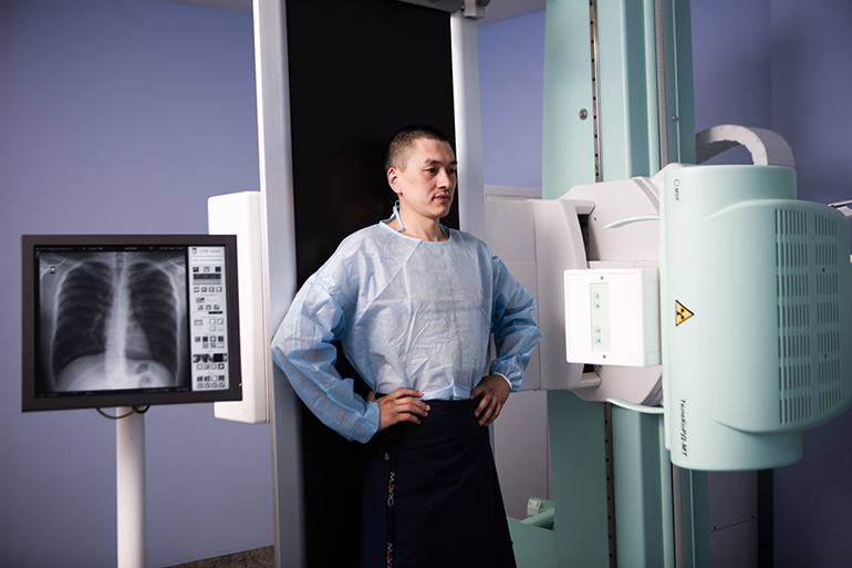
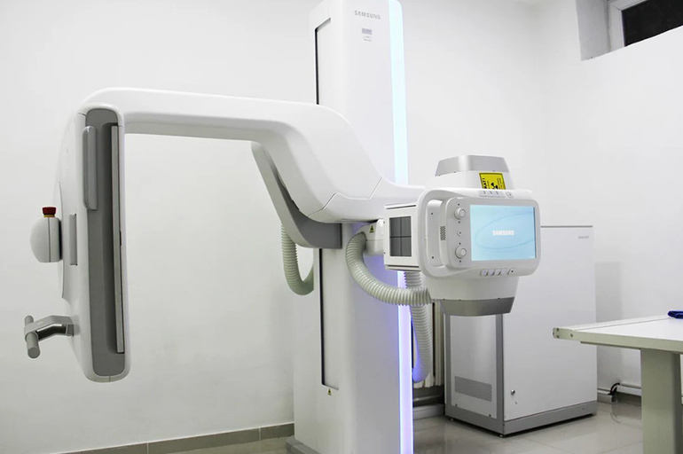
X-ray
X-ray is not only a highly informative method for examining the chest organs, but also a cheap, affordable diagnostic method. Radiography is performed in 3 projections: vertical, straight, lateral. The method makes it possible to detect pleural effusion (at least 250 ml), study the state of lung tissue, determine the localization of exudate and the position of the mediastinal organs.
With the help of condition monitoring (radiography within 1.5-3 months), it is possible to establish:
- regressive course of pleurisy;
- the formation of residual pleural overlays;
- one-sidedness or two-sidedness of the lesion.
The accuracy of the diagnosis in the assessment of clinical and radiological studies is 90.4%.
Basic principles of treatment
The treatment program includes:
- bed rest (in the acute phase);
- diet food low in salt, liquid and high in protein;
- vitamin therapy;
- the use of anti-tuberculosis drugs: Isoniazid, Rifampicin, Streptomycin, Isoniazid, Pyrazinamide, Ethambutol (3-4 drugs are prescribed at the same time);
- administration of corticosteroids with profuse effusion;
- application of methods of evacuation of effusion - drainage and puncture of the pleural cavity;
- NSAIDs, physiotherapy, breathing exercises (during the period of effusion resorption).

With a complicated course with the formation of adhesions, fistulas, as well as with a purulent form, surgical treatment is necessary.
Traditional methods
In folk medicine, there are a large number of recipes for diseases of tuberculous etiology. This is the use of medicinal plants in the form of vapors, infusions, inhalations. It is recommended to take natural biogenic stimulants: aloe juice, bee products, wax moth infusion.
Numerous recipes have different efficacy and safety, require prior consultation with a specialist and cannot replace official treatment.
Recovery Predictions
With the timely detection of pathology and adequate treatment, the prognosis is favorable. The most dangerous form of the disease is chronic pleural empyema. With her, the mortality rate is 1-2%.
Rehabilitation and features of the body's recovery
A significant violation of the respiratory function and vital activity occurs with the formation of adhesions and pleural fibrosis.

To restore respiratory function, it is recommended to undergo a physical rehabilitation course, consisting of:
- therapeutic massage;
- Exercise therapy;
- breathing exercises;
- physiotherapy.
To strengthen the immune system and the whole body, it is necessary to introduce vitamin and mineral supplements.
Possible consequences and complications
With the wrong treatment strategy or non-compliance with the doctor's recommendations, pathology can lead to various complications:
- pulmonary tuberculosis;
- the formation of adhesions, abscess, fistulas, cavities;
- tear or rupture of the pleura during formed breathing under conditions of high physical activity;
- the occurrence of spontaneous pneumothorax;
- the development of skeletal pathologies: scoliosis, retraction of one part of the chest, narrowing of the intercostal spaces;
- displacement of the mediastinal organs and impairment of their function.
With purulent pleurisy, there is a high probability of blood poisoning and death.
Prevention methods
Preventive measures are the same as for tuberculosis:
- timely vaccination;
- tuberculin diagnostics in children, adolescents;
- regular X-ray and fluorography of the chest cavity;
- avoidance of contact with patients with tuberculosis.
It is necessary to closely monitor the state of health and seek medical help in a timely manner, i.e. because most cases of the disease are detected during compulsory medical examinations (1/3) and treatment (1/3) to to the doctor.