Content
- Characteristic
- Functions and properties
- Anatomy and structure
- Possible diseases and pathologies
- The gap
- Classification
- Symptoms and Signs
- Causes
- Treatment methods
- Stretching
- Classification
- Symptoms and Signs
- Causes
- Treatment methods
- Ligament diseases
- Inflammation
- Autoimmune diseases
- Infection
- Video about the structure of the ankle
Understanding ankle anatomy, consisting of bones, muscles, ligaments, is important for the correct diagnosis and choice of treatment.
Characteristic
The ankle is a complex joint made up of three bones, cartilage, tendons, and the lining of the joint. Functionally, it is a hinge joint that provides dorsal and plantar flexion of the foot. Although it has some side-to-side movement, most of this movement is performed at the joint below, which is called the subtalar joint.
The ankle is full of muscles and ligaments that give it strength, flexibility, and range of motion.
Ligaments are tough connective tissue made up of collagen fibers arranged in parallel bundles that increase the strength of individual fibers. Collagen bundles are attached to the outer covering that surrounds the bones. Ligamentous tissue has low vascularity, or poor blood supply, which slows down its healing compared to other types of soft tissue.
Ankle ligaments, the anatomy of which lies in the fact that they are the connecting link between the articular bones, very strong, flexible and resistant to damage when stretched or squeezing.
Functions and properties
The ankle joint and the ligaments involved with it can be visualized as a ring in the coronary (vertical) plane:
- The upper part of the ring is formed from the articular surfaces of the fibula and tibia.
- The lower part of the ring is formed from the subtalar joint (at the junction of the calcaneus and talus).
- On the sides of the ring, the medial and lateral ligaments are involved.
Collagen fibers connect bone structures to each other. All bone structures of the leg can be represented as a chain. The thighbone is one link in the chain.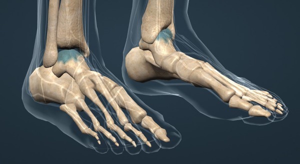
The lower leg bone, lower leg, is another link. The ligaments of the knee joint form another link connecting the femur and tibia. As in a chain, individual links can move freely, but they must remain consistent and cannot be disconnected, which is what the ligaments are responsible for. This allows the joint to move, preventing it from moving.
The ankle joint is the most important part of the body, as it supports the hinge joint between the leg and foot formed by the ankle, subtalar and tibial joints. Ligaments help them in this, and all structures together constitute a large working block.
Between the tibia bones are the tibiofibular ligaments, on the inside there is a large deltoid ligament, on the outside - the talofibular and calcaneofibular. They are composed of collagen and elastic fibers, thanks to which they stabilize the joint and at the same time allow it to perform complex movements.
Thus, the ankle ligaments perform 2 functions:
- Mechanical: allow the ankle to move only in a certain plane, limiting excessive or abnormal movement of the joint.
- Proprioceptive function: transmission of signals to the nervous system regarding the position of the joint. Signals from the ligaments reach the nervous system, which, in turn, signals the corresponding muscles to contract to return the leg to a level position.
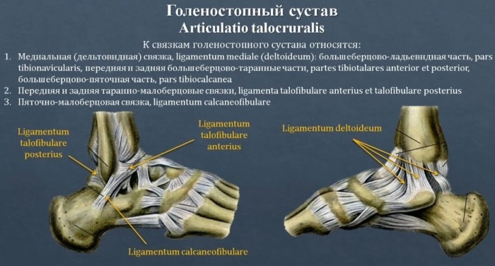
Anatomy and structure
The tibia, talus and fibula, articulating, represent the structure of the ankle. They slide and rotate relative to each other. They are surrounded by a thick lubricating coating - the joint capsule. It retains synovial fluid, which, together with the smooth cartilage lining the ends of each bone, allows the ankle to move with very low friction.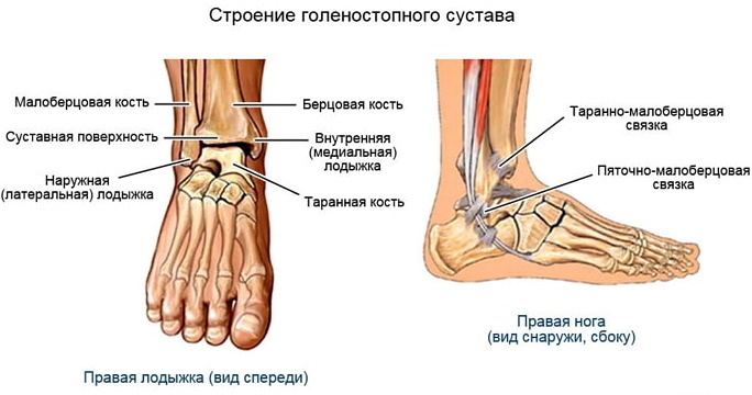
At the top and bottom of the ankle, there is a large group of ligaments that connect the bones at the ankle. They are subdivided into medial and lateral. On the outside of the ankle, there are 3 ligaments.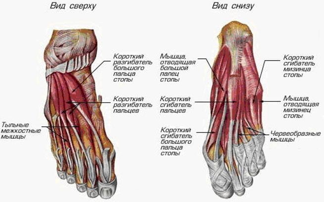
There are five of them on the inside of the ankle. There are also 4 ligaments that connect the two bones of the leg together, the tibial and the fibula. This compound is called syndesmosis. Syndesmosis is technically considered a joint, but is actually formed by the ligaments that support the stability of the ankle joint.
The main ligaments involved in the ankle joints are:
| Outer ankle: 3 ligaments making up the lateral ligament complex | Anterior talus, which connects the anterior part of the talus to the long bone of the lower leg called the fibula. |
| The calcaneofibular ligament, which connects the corresponding bones. | |
| Posterior talofibular, connecting the talus to the fibula. | |
| The deltoid, thick ligament that supports the entire medial or inner side of the ankle. | |
| The anterior inferior tibial ligament, which connects the peroneal and tibial bones. | |
| Two posterior fibular ligaments that intersect the posterior part of the tibia and fibula | Posterior inferior tibial ligament. |
| The ligament located under the tibiofibular is transverse. | |
| The interosseous ligament, which runs the entire length of the tibia and holds them together. |
Ankle ligament injuries occur whenever any of them goes beyond elasticity.
The anatomy of the ankle ligaments is important for proper diagnosis. Joint injury is the most common cause of acute ankle pain. Chronic pain is caused by weakness in one of the ligaments in this area.
Possible diseases and pathologies
External ligament injuries occur when the foot turns downward and inward and the ankle twists outward. These injuries are among the most common types of injuries. If the ligament is over-rotated, it can partially stretch or rupture completely.
When the primary ligament (the anterior talofibular) ruptures, the secondary external ligament, called the calcaneofibular ligament, also stretches partially or completely. In some severe cases, a rupture of the posterior talofibular ligament occurs.
The gap
Violation of the integrity of the ligament in the ankle joint occurs due to twisting of the ankle in accidents or during sports. First of all, the external peroneal ligaments are affected. The second most common tear occurs in the area of the calcaneal ligament, which connects the fibula to the outside of the heel.
Classification
Ankle ligaments, with complex connections and interactions in the anatomy, can rupture as a result of injury. In places where the integrity of the fibers is broken, isolated closed ruptures are more common.
In medicine, there are 3 degrees of disruption of the connective tissue that holds parts of the skeleton:
- 1 degree. Small sprain, ligament rupture occurs at a microscopic level. After a few days, the victim will be able to move their leg normally, which heals within 2 weeks.
-
2nd degree. Multiple fiber breaks. It can take 6 to 8 weeks to heal.
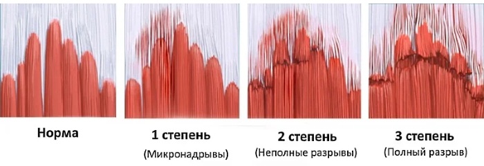
- Grade 3. The fibers are completely torn apart. It may take 3 to 6 months or more to regain strength and mobility.
Symptoms and Signs
Soreness and immobility are signs of rupture, indicating a serious problem. A popping sound is often heard when the fibers break. In cases of severe damage, the pain is sudden and severe enough, the leg becomes motionless. If hematomas, swelling appear, the victim should immediately seek medical help.
Causes
Tears occur when the ankle moves outward and the foot rolls inward or, less commonly, when the joint moves inward and the foot outward.
Treatment methods
Therapy depends on the degree of damage to the connective tissue.
As a rule, conservative treatment with 4-6 weeks of immobilization in an orthosis is sufficient, which includes:
- novocaine blockade at the site of the lesion (10-15 ml of a 1% solution);
- the imposition of a circular plaster cast on the surface of the leg from the tips of the toes to the upper third of the ankle;
- immobilization for a period of 1.5 to 2 months.
In the event of a complete rupture of the ligaments, when treatment and rehabilitation are ineffective, surgery may be required. If ligament replacement is planned, ankle arthroscopy is performed in advance. The doctor checks the cartilage and removes the stuck ligament residues.
Ankle ligaments (fiber anatomy is of great clinical importance) can become chronic inflammation with frequent ruptures. In this case, an operation is performed on the ligamentous apparatus. Recovery occurs after 2 months, and postoperative treatment is carried out by methods of conservative therapy.
The recovery period includes carrying out physiotherapeutic procedures aimed at regenerating ligaments, restoring mobility.
There is always the possibility that the joint will not fully recover. In such cases, the victim will need a bandage or other support during physical activity.
Stretching
Stretches occur when the foot is suddenly twisted and the fibers of the connective tissue are overstretched. This most often occurs in the outer part of the foot. 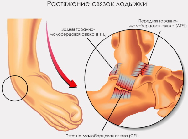 Stretching of the internal ligaments is less common.
Classification
Injuries are classified according to the position of the ankle at the time of the injury, the direction of the fall of the body, the type of footwear, the activity at which the injury occurred and the force with which it occurred. There are cases when different types of injuries are combined.
There are 3 main categories of ligament injuries:
- Stretching of the lateral ligaments.
- Damage to the middle ligaments (deltoid muscle).
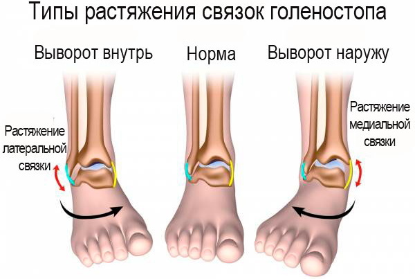
- Stretching the syndesmosis.
Symptoms and Signs
Stretching is characterized by the following symptoms:
- Localized pain at the site of injury. For example, if the deltoid ligament has been stretched, pain will appear inside the ankle. The pain is often described as sudden and sharp, and is aggravated by movement or exertion of the ankle.
- Edema. If the deltoid ligament is damaged, the inside of the ankle swells up noticeably. Swelling can occur on the outside of the ankle if one or more ligaments in the lateral ligamentous complex are stretched.
- Hematoma. The development of blue, red, or purple skin over the site of the sprain indicates a rush of blood to the area.
- Limited motor ability of the ankle. In more severe sprains, patients cannot turn or bend the leg or foot.
Causes
Risk factors for the development of sprains and deformities of the ankle joint:
- Poor athletic performance. Anyone who tries to engage in intense sports without prior preparation, such as regularly performing stretching and strengthening the ankles and calves, is at increased risk of sprains or strains of the ligamentous muscles apparatus.
- Fatigue. When muscles and ligaments fatigue towards the end of vigorous activity, people are at a higher risk of injury if they “overcome” fatigue in pursuit of more activity instead of rest.
- Ignoring warm-ups before physical activity. Athletes who immediately engage in strenuous exercise without first warming up their muscles are at high risk of sprains. Without a warm-up period, muscles and ligaments will remain tense, and less flexibility means a greater risk of injury.
- Being overweight increases the shock load on the joints when walking, running and jumping, which increases the chance of strains or fiber breaks.
- Several studies have shown that women over the age of 30 are exposed to significantly higher more likely to develop sprains than men in this age group, regardless of their mass index body.
- Inappropriate footwear, especially high heels in women when worn continuously, increase the risk of sprains.
- People who have had frequent sprains in the past have a higher risk of recurring the same injury than people who have never had ankle deformities.
Treatment methods
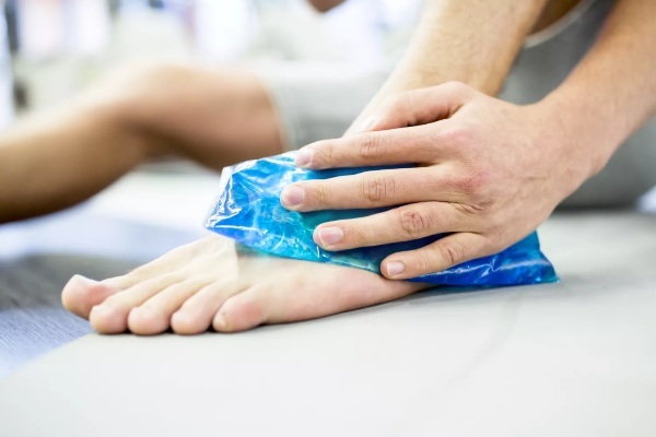
For grade 1 and 2 sprains, most doctors recommend the RICE protocol (Rest, Ice, Compression, Elevation):
- Rest. Patients are advised not to walk or stand on a stretched ankle until complete healing. The rest period can be from several days to several weeks.
-
Ice. It is recommended that you begin applying cloth-wrapped ice packs to the injury site as soon as possible. Ice reduces swelling and pain. The bags are applied for 20-30 minutes from 1 to 4 times a day. It is not recommended to apply ice directly to the skin.
- Compression. Wrapping the ankle with elastic bandages, compression bandages immobilizes and maintains the site of injury, which prevents exacerbation of the process. Patients with mild sprains can wrap their ankles themselves, while more serious injuries need to be treated by a healthcare professional.
- Height. Raising the ankle in a supine position above the level of the heart during the first 48 hours after injury will reduce swelling, eliminate discomfort, and accelerate the healing process.
Patients with grade 1 or 2 sprains sometimes need to take NSAIDs such as Ibuprofen, Acetaminophen, which reduce inflammation and relieve pain at the same time.
Ankle ligaments (the anatomy of which differs from the structure of the tendons) during stretching are successfully treated with ointments containing menthol, petroleum jelly: Diclofenac, Voltaren, Emulgel. Grade 3 sprains and strains are usually unstable and require longer healing time.
The following treatment protocol is used:
- In addition to resting and elevating the joint, the first phase of healing involves fixation of the foot, ankle, and lower leg. A traditional cast or air bandage can be used. Many patients will need crutches.
- Rehabilitation. Patients will require a number of restorative procedures such as electrical stimulation and ultrasound.
- Isometric strengthening exercises that promote proprioception (awareness of body position) are especially important for grade 3 sprains because they reduce the likelihood of re-injury. Therapeutic exercises can be water-based. They are useful for those who are seriously injured.
For severe or chronic sprains that do not respond to first-line treatment or rehabilitation, one or more types of surgery may be required:
- Arthroscopic surgery, in which the surgeon examines the ankle joint from the inside using a special camera. The doctor checks to see if part of the damaged ligament, muscle, or tendon is stuck inside the joint capsule.
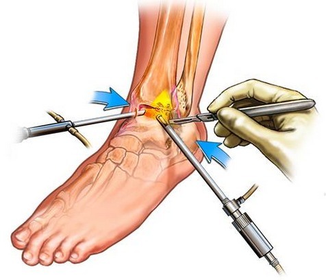
- Surgery can be reconstructive, in which the surgeon repairs torn ligaments, muscles, or tendons with stitches. In this case, the surgeon can transplant tissue from other parts of the patient's foot and ankle.
After the operation, additional time will be required for recovery and physical therapy, the classes of which stretch out for several months. Some patients with severe or chronic sprains are unable to return to their previous level of activity.
Ankle ligaments (the anatomy and functions of which have an inhibitory and directing effect) sometimes do not recover well after surgery associated with the plastic of damaged fibers. Patients with extremely painful grade 3 sprains or those recovering from surgery may also receive strong or weak pain relievers.
Ligament diseases
Since the ligaments are intercellular connective tissue with a very small number of cells, nerves and blood vessels, their diseases are mainly associated with inflammation, autoimmune diseases and infections.
Inflammation
In an inflammatory process, the body repairs damaged tissue and fights infection. When the body undergoes an inflammatory response, blood begins to actively enter the damaged area, bringing in nutrients and immune cells.
But the nature of the ligamentous tissues, dense, with limited blood supply, leads to the fact that this process causes damage due to swelling and prolonged degradation of the tissue. All this further damages the ligaments. They become susceptible to injury and stretch easily under normal physiological conditions.
Autoimmune diseases
Some diseases associated with a malfunction of the immune system affect the connective tissues and the ligaments are affected. Rheumatoid arthritis, mixed connective tissue disease, polychondritis, systemic lupus erythematosus and scleroderma, all of these autoimmune diseases lead to damage to the ligamentous tissue.
Infection
Infection of the fibrous tissues of the ligaments occurs directly through wounds or internal infections. Direct infections of mating are rare. Bacteria, viruses that cause necrotizing fasciitis, or bone infections can affect the ligaments and, although rarely, pose a serious threat to these structures. Most often, an infection of a joint or adjacent tissue causes an inflammatory response in the ligaments and tissue damage.
Some diseases indirectly affect the damage to the ligamentous apparatus. With tendinitis, fibromyalgia, osteoarthritis, connective tissues, joints or muscles are affected.
Proper ankle mechanics requires not only muscles, but also the ligaments that hold the bone structures together, as well as the tendons that attach the muscles to the bones. Knowledge of anatomy, recognizing musculoskeletal problems before they develop into serious injuries, is the key to an active, pain-free life.
Video about the structure of the ankle
Anatomy of the foot. Joints and ligaments: