
Content
- What is the eyeball, functions and anatomy
- Eye structure
- Does the eye grow throughout a person's life?
- The growth and development of the organ of vision from birth
- A period of active growth
- Normal sizes of the eyeball in children and adults
- How old does the eyeball grow?
- Development pathologies and their causes
- Cataract
- Keratitis
- Myopia
- Retinopathy
- Loss of the eyeball
- Retinoblastoma
- Video about the structure of the eyeball
Human eyeball - This is a mobile organ of vision, which begins to actively grow even during the period of intrauterine development, and also continues its formation until the end of the phase of puberty of the body. The study of the functions, features of the anatomical structure and eye diseases is the science of ophthalmology.
What is the eyeball, functions and anatomy
The eyeball is a paired organ of the human body, through which information is collected and transmitted about environmental conditions. The image of the outside world is transmitted to the supersensitive receptors of the optic nerve, passes through the structure of the chiasm, everything membranes of the eye, and then enters the occipital parts of the central nervous system, where the final picture is already processed and formed happening.
The human eyeball is an important part of the general visual system, which provides a continuous overview of objects in the environment. The most active growth of the eyes occurs in childhood, when the rapid development of the whole organism takes place.
The visual system is designed in such a way that for it to work properly, a person must have 2 healthy eyeballs. In this case, high-quality stereoscopic vision is provided, capable of quickly processing information about the state and conditions of the environment.
The right segment of the retina of each eye carries out operational transmission of data about the image of the external world from the right side in relation to the organ of vision. The information signal goes directly to the optic nerve receptors. The processing of the data collected by the right retina takes place in the occipital centers of the brain, located in the right hemisphere.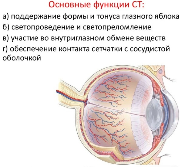
The left retina of the eyeball works in exactly the same way. During the processing of information collected from the right and left retina of the eyeball, synchronization of individual fragments occurs, which are combined into a single picture.
Due to these functional features of the eyes and centers of the brain, a person gets the opportunity to see the world around him in a three-dimensional spectrum. Each eyeball sees its environment differently. Impaired mobility of one of the eyes or their incorrect location in relation to each other leads to the appearance of binocular vision disorders.
The following main functions of the human eyeball are distinguished:
- formation of a unified optical system of the visual analyzer;
- projection of the image of the environment;
- interaction with the centers of the brain responsible for the implementation of visual function;
- providing the structural elements of the eyes with useful substances and blood supply;
- protection of the organ of vision from the effects of negative environmental factors by active lacrimation (ingress of small particles of dust, dirt, insects, allergens into the eye).
The eyeball grows in sync with the formation of the facial bones, which hold the organ of vision in its anatomically correct position. The preservation of the functions of the eyes allows for the full vital activity of the whole organism.
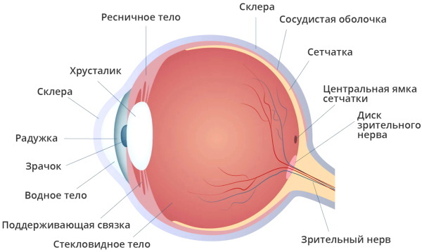
Eye structure
The human eyeball is a very complex optical device of biological origin. The main task of this paired organ is to quickly and correctly transfer the initial image of the environment to the optic nerve receptors. Then the process of complex processing of the received information is started, in which all the structural elements of the eyes are involved. The table below details the structure of the eyeball.
| The structure of the organ of vision | The main functions of the structural units of the eyeball |
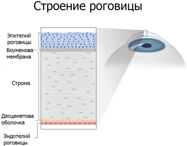
| Cornea | The cornea is a transparent and very thin outer membrane that covers the front of the eye. In this part of the organ of vision, even the smallest blood vessels are completely absent. The cornea has a high refractive power of light and forms a single optical system of the human eyeball. This part of the eye is in contact with the outer absolutely opaque shell of the organ of vision - the sclera. The structure of the cornea of the eye includes the following components:
The last structural element of the cornea is responsible for the trophism of the eyeball. The endothelium regulates the saturation of the tissues of the eyeball with a sufficient volume of fluid. In conditions of excess moisture in the eyes, the cells of the endothelial layer remove excess moisture, preventing edema of the organ of vision. |
| Anterior chamber of the eye | The anterior chamber of the eye is a kind of space located between the iris of the eye and its cornea. This part of the organ of vision is completely filled with thick intraocular fluid. |
| Iris | The iris is a collection of muscle tissues of the eyeball, during the natural spasm and relaxation of which there is a change in the physiological size of the pupil. In its shape, this part of the eye resembles a circle with an inner hole (the pupil is located inside). The iris forms the base of the shell of the eyeball, which is made up of many blood vessels. This part of the organ of vision is responsible for the color shade of the eyeball. If a person has blue eyes, this means that a small number of pigment cells are concentrated in this membrane. In people with brown eyes, the concentration of pigment cells in the iris is very high. According to its functional purpose, the iris of the eyes plays the same role as the diaphragm in any camera, it regulates the light flux.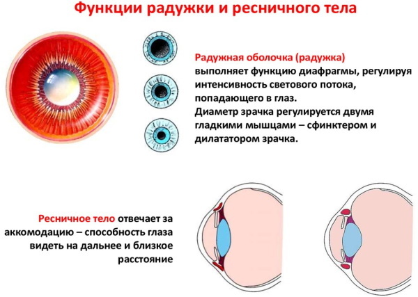
|
| Pupil | The pupil is an opening inside the iris, the physiological size of which depends on the degree of illumination of the environment. The more saturated the space with light, the smaller the pupil becomes. |
| Lens | The lens is the biological lens of the eyeball. This part of the organ of vision is ideally transparent, elastic, with the ability to quickly change shape and focus on environmental objects. The presence of a healthy lens allows a person to see well at close range, as well as to identify objects that are at a great distance. The biological lens of the eyeball is located inside the capsule, and its attachment is carried out by the ciliary band. The lens body is part of a single structure of the optical system of the eyeball.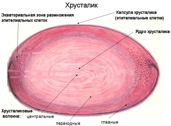
|
| Vitreous | The vitreous humor is a completely clear and gelatinous fluid located in the posterior part of the organ of vision. This structural element of the eyeball is responsible for maintaining its anatomically correct shape, takes an active part in the intraocular metabolism of nutrients. The vitreous humor is also part of the optical system. |
| Retina | The retina is a collection of complex photoreceptors and peripheral cells that are highly sensitive to light rays. These receptors are located inside the retina, and are also divided into 2 classification types. These are eye sticks and cones. In cells of this type, a unique photochemical process occurs for the synthesis of the enzyme rhodopsin, with the help of which the energy of the light rays is converted into electrical impulses of the nervous tissue. Retinal rods are characterized by increased sensitivity to light, which allows a person to see in low light conditions. Cones are responsible for the implementation of the functions of central vision, they need a larger volume of light flux, but it is their presence that allows a person to identify small details and objects.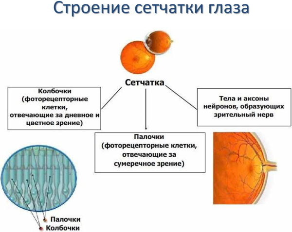
|
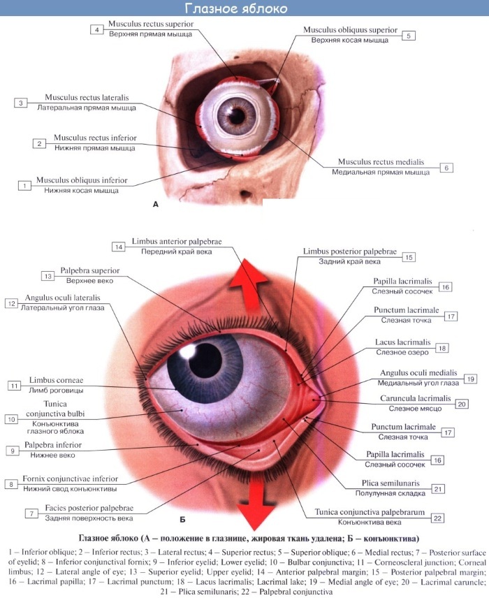
| Sclera | The sclera is the outer shell of the organ of vision, which has an opaque structure. 6 muscles of the oculomotor type are attached to the surface of this area of the eyeball. A small percentage of blood vessels and peripheral nerves are concentrated within the sclera. |
| Choroid | The choroid is interconnected with the surface of the retina. This structural part of the eye lines the entire posterior portion of the sclera. The main function of the choroid is to provide blood supply to the intraocular tissues. Pathological damage to the retina often leads to the spread of the disease to the circulatory system of the eye. Nerve endings are completely absent in the structure of the choroid. Under the conditions of the development of the disease, a person does not experience any pain, which often leads to severe complications and dysfunctional visual impairments. |
| Optic nerve | The optic nerve performs the function of transmitting electrical impulses from peripheral nerve endings to towards the centers of the brain, in the structure of which further image processing takes place the surrounding world. |
The eyeball grows simultaneously with the development of other senses. All structural units of the eyes are interconnected, and their well-coordinated work allows a person to see well near and at long distances.
Does the eye grow throughout a person's life?
The active growth of the eyeball begins from early childhood, and continues until the age of 18-20 years. From this point on, the volume of the organ of vision remains stable. If, after 20 years, a person has a further increase in the volume of the eyes, then this indicates the presence of ophthalmic pathology. In this case, the development of progressive myopia is possible, as well as the appearance of concomitant diseases of the organ of vision.
The growth and development of the organ of vision from birth
The process of growth and development of the organ of vision starts in the first days of the independent life of a newborn child. Already at 2-3 weeks, the baby shows the first signs of a reflex complication of the work of the visual system. Object, spatial and color type of vision becomes more perfect.
There is a constant development of the tissues of the eyeball. For 2-3 months. life, the child has a central vision function. As the cerebral cortex develops, the infant acquires new functions of the eyeball to recognize new objects and geometric shapes. In the same time period, the baby begins to recognize the mother's breast.
For 4-6 months independent life, the newborn's visual system begins to identify the faces of the people around him. By 10 months. the development of the eyeball, the child acquires the skill of recognizing complex geometric shapes in the form of a pyramid, cube or cone.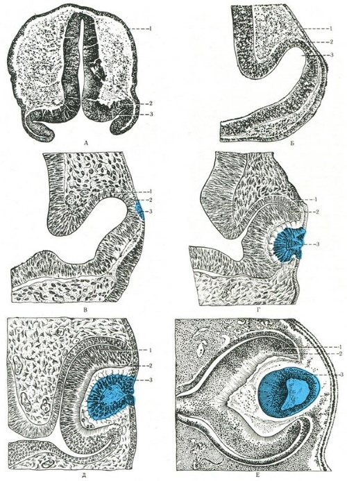
At 2-3 years of age, the optical system of children allows them to perceive objects that are drawn on a board or sheet of paper. A full-fledged visual acuity in a child of the younger age group is formed by the age of 5-6 years. This is the period of time when children enter primary school, acquire new skills that contribute to a more active development of the organ of vision.
According to scientific studies, visual acuity in a newborn child is at a very low level. On average, this indicator ranges from 0.005 to 0.015 units. The first 3 months of life, the quality of visual function rises to 0.01-0.03 units. By the age of 2, it already reaches the level of 0.2-0.3 units.
In children of the age group from 7 to 11 years, the indicator of visual acuity is in the range of 0.8-1.0 units. The improvement of the functions of the eyeball and the further development of the optical system continues until the child comes of age. During this period, visual function may improve or deteriorate.
A period of active growth
The eyeball grows most actively in children aged 2 to 7 years. This is the first stage in the rapid formation of the child's visual system. The next surge in the physiological activity of the growth of the organ of vision falls on the phase of puberty. In the period from 13 to 16 years, all structural elements of the eyeball are characterized by active growth, which is due to a sharp increase in the level of female and male sex hormones.
Normal sizes of the eyeball in children and adults
The eyeball of an adult who does not suffer from ophthalmic pathologies has the following parameters:
- diameter 24 mm;
- length in the sagittal axis 24 mm;
- horizontal length 23.6 mm;
- vertical length 23.3 mm.
The volume of the eyeball in an adult corresponds to 7.448 cm3, and its mass is 7-8 g. These sizes are the same for all people. Differences in the above parameters do not exceed a fraction of a millimeter.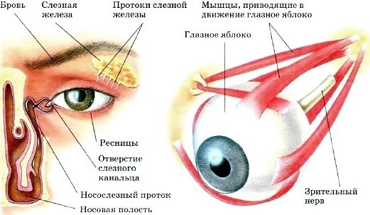
In infants, the average eye diameter is 16.2 mm. By the 1st year of independent life, this indicator increases to 19.2 mm. By the age of 3, the child's eyeball has a diameter of 20.5 mm. By the age of 7, the organ of vision increases to a level of 21.1 mm.
Upon reaching the age of 10 years, the diameter of the eyeball is 22 mm. By the age of 15, the adolescent's organ of vision acquires a size of 23 mm. By the age of 20, a healthy young man or girl has an already formed visual system with an eyeball diameter of 24 mm.
How old does the eyeball grow?
The eyeball grows only until a person reaches the age of 18-20 years. From this time on, reverse processes are triggered, associated with the gradual aging of the optical system of the eyes. The growth phase stops completely.
Development pathologies and their causes
There are a large number of ophthalmic diseases that lead to visual impairment. Some pathologies take their development directly within the structure of the eye, while others are a complication of concomitant diseases of internal organs or life support systems of the body. For example, secondary damage to the human visual system causes hypertension, diabetes mellitus, infectious diseases that have spread to the tissues of the eyes.
Cataract
Cataract is a serious ophthalmic disease, the development of which is characterized by the clouding of the structure of the lens of the eye. The emergence of this pathology is associated with protein denaturation.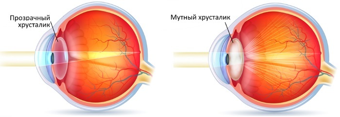
Cataracts can lead to complete loss of vision, and the reasons for its appearance are caused by the negative effects of the following factors:
- elderly age;
- previous eye injuries;
- hereditary predisposition;
- harmful working conditions;
- living in a region with an unfavorable ecological situation.
Prerequisites for the development of cataracts have every 6th inhabitant of the globe, whose age is over 40 years. Treatment of this pathology involves a surgical operation to replace the affected lens with an artificial lens.
Keratitis
Keratitis is an acute or chronic inflammation of the cornea of the eyeball, which is characterized by opacity and ulceration of its surface. This disease is manifested by attacks of severe pain, active lacrimation and photophobia. The main reasons for the development of keratitis are associated with damage to the cornea as a result of an eye injury, or its infection with infectious microorganisms.
The most common causative agents of inflammatory processes in this part of the organ of vision are the influenza virus and tuberculosis bacillus. Keratitis treatment should be completed in a timely manner. Otherwise, a thorn will form on the surface of the human eyeball, or it will completely lose its visual functions.
Myopia
Myopia is an eye condition that is also called myopia. This visual defect differs in that a person sees perfectly near, and objects in the distance are blurred, their image is blurred.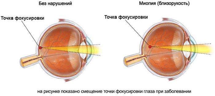
The reason for the development of myopia is associated with the fact that the refraction of the image of the environment occurs not on the surface of the retina, but in front of this part of the eyeball. The solution to this problem lies in the use of glasses or contact lenses. The development of myopia is facilitated by intense loads on the organ of vision, prolonged reading of literature, working at a computer and other electronic devices.
Retinopathy
Retinopathy is a severe ophthalmic disease that is manifested by damage to the retina of the organ of vision. The development of this pathology is based on vascular disorders that cause negative changes in the blood supply of the retina.
In most cases, retinopathy occurs as a complication of the following diseases:
- hypertension;
- diabetes;
- infection with infectious microorganisms;
- an inflammatory process that has arisen against the background of an injury or after a surgical operation.
The progression of this disease leads to a significant decrease in visual acuity. Treatment of retinopathy includes the use of anti-inflammatory drugs, as well as surgery.
Loss of the eyeball
Loss of the eyeball is a pathological process that manifests itself as the eyes go beyond the boundaries of the bony orbit. The cause of this ophthalmic anomaly is trauma to the organ of vision and displacement of the bones of the skull as a result of their damage. Treatment of this pathology requires an emergency surgical operation to restore the anatomically correct position of the eyeball.
Retinoblastoma
Retinoblastoma is a malignant neoplasm that affects the retina of the eye. In most cases, this pathology develops in children of the younger age group. Scientific research shows that this type of tumor of the eyeball begins its development even at the stage of the embryonic formation of an infant.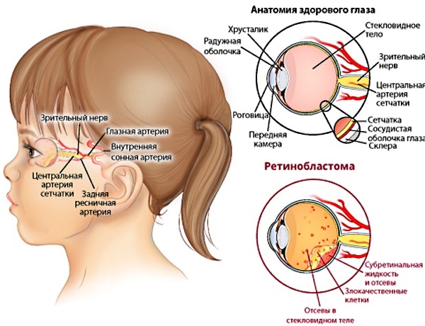
Most often, this disease is diagnosed in children aged 2 years. Almost all cases of retinoblastoma are found in patients under 5 years of age. The method of treating pathology depends on the stage of the tumor process. Chemistry preparations and surgical removal of the affected tissue are used.
The eyeball is a complex human optical organ, the functions of which are to perceive information about the world around us with its further transmission to the centers of the brain. The process of active growth of eye tissues starts in the first months of a newborn baby's life and continues until his adulthood. The diameter of the adult eyeball is 24 mm.
Too large parameters of this organ of vision indicate its pathological state, the development of ophthalmic diseases. Treatment of eye diseases is carried out using anti-inflammatory, vitaminizing drugs, as well as using the method of surgical intervention.
Video about the structure of the eyeball
Anatomy of the eye: