Content
- Anatomy
- Structure
- The main purpose
- What doctors treat pathologies?
- Possible diseases and symptoms
- Diagnostic methods
- Treatment
- Surgery
- Drug treatment
- Iliac Vein Video
Common iliac vein is a rather large paired vessel located in the pelvic area at a great distance from the skin surface. Damage and diseases of this vein are characterized by a low frequency of occurrence, but most often they are dangerous in nature. Due to its deep location, diagnosis and treatment are usually difficult, which increases the time until qualified medical care is provided and can have fatal consequences for a person.
Anatomy
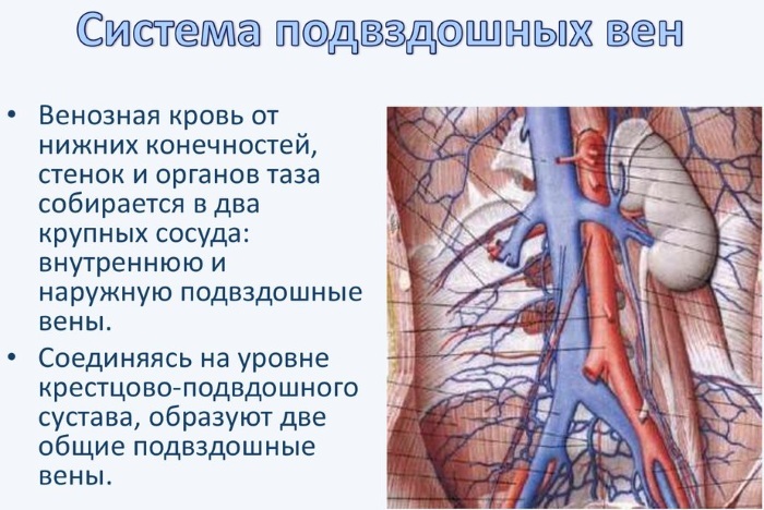
The common iliac veins (PV) on the right and left are combined with each other at the body of the IV lumbar vertebra, where form the inferior vena cava, which runs slightly to the right of the midline, so the left PV is longer right. At the sacroiliac junction, they are both formed from two veins: the internal and external iliac veins.
The iliac vein is located along its entire length in close proximity to the artery of the same name (this applies to both right and left), which is also quite large in size, which has a huge clinical meaning.
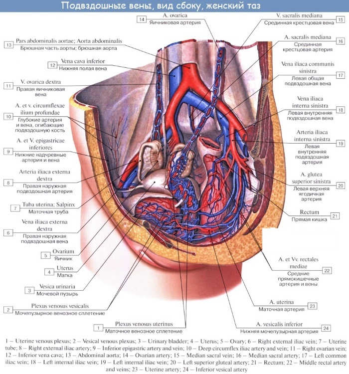
The internal PV is composed of several venous plexuses that communicate with each other, and the external PV, in turn is a continuation of the femoral vein and at the same time, before merging with the internal one, receives 2 more inflows from the abdominal organs cavity.
| Vessel | Inflows in men | Inflows in women |
| Common iliac vein | Internal and external iliac veins. | |
| Internal iliac vein | Sacral, rectal and urinary venous plexuses. | |
| Prostate venous plexus. | Uterine and vaginal venous plexus. | |
| External iliac vein | The inferior epigastric vein and the deep vein surrounding the ilium. |
Structure
Unlike arteries, the blood flow in the veins is not pulsating, but constant, wave-like, moreover, the veins of the lower extremities need to overcome the gravity of the blood and "lift" it from the bottom up, which determines the features of their structure:
- consist of three layers (inner - intima, middle - media and outer - adventitious);
- while small and medium-sized veins have valves to prevent backflow of blood, PVs are large and there are no valves in them, and the movement of blood occurs due to muscle contractions;
- the iliac veins contain pronounced muscle elements (myocytes) in all three layers, but despite this a significant effect on blood flow is still due to the contraction of the muscles of the legs, abdomen and pelvis.
The main purpose
The iliac vein is located deep in the pelvic region and carries venous blood from the lower limb, the pelvic organs from their side (except for the ovaries in women and the testes in men: their veins on the right flow directly into the inferior vena cava, and on the left - into the left renal vein), some of the lower intestine, located next to the PT, and from the abdominal muscles, as well as the nearby pelvic muscles.
Venous blood characteristics:
- depleted in oxygen;
- saturated with carbon dioxide;
- darker than arterial;
- has a reduced amount of glucose;
- lower pH;
- contains end products of tissue metabolism (urea and others).
What doctors treat pathologies?
An angiosurgeon is engaged in identifying the causes of pathologies of the entire human cardiovascular system and their treatment, performs surgical interventions, including endoscopic ones.
Of the numerous areas of his work, several of the most common and significant ones can be distinguished: stenting (installation of special devices (stents) into the lumen vessel for its expansion and improvement of blood flow at the place of fixation, it is more often used for atherosclerosis and coronary heart disease), endoprosthetics (with an artificial prosthesis the damaged part of the vessel is replaced, usually performed with aneurysms) and dilatation (temporary mechanical impact from the inside on the walls of the vessel for its expansion and restoration cross-country ability).
Phlebology is one of the divisions of vascular surgery that investigates venous diseases.
The following diseases are responsible for the competence of a phlebologist:
- phlebitis;
- thrombophlebitis;
- varicose veins;
- thrombosis;
- trophic ulcers.
Accordingly, the difference between a vascular surgeon and a phlebologist is quite large.
Possible diseases and symptoms
The iliac vein is located far from the surface of the body, therefore, its direct damage is possible only in case of injuries to the pelvic region (gunshot and non-gunshot, penetrating), or mediated damage by fragments of broken bones of the pelvis or the head of the femur and nearby movable structures (arteries, organs, pathological formations).
Like many other veins, the iliac veins can be affected by diseases such as acute and chronic thrombosis (with the development of later thrombophlebitis and post-thrombotic disease), phlebitis from the effects of various external factors (for example, after injections), varicose veins or chronic venous failure. Their symptoms are mostly similar: pain, swelling, increased venous pattern, sometimes there is a change in skin color over the affected area.
The iliac veins, due to their location, are prone to pathologies inherent only to them:
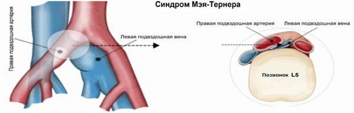
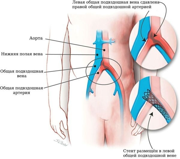
- in May-Turner syndrome, the iliac artery and the body of the V lumbar vertebra clamp together the left common PT. This syndrome is difficult to diagnose, most often it manifests itself as chronic pain of various localization: in the pelvic region, in the scrotum (can be both unilateral and spilled) and in the lower left limbs. As a result of this condition in men, the formation of an unstable erection is possible, which is often the cause of infertility against the background of a tense recurrent varicocele (usually just a left-sided one);
- constant exposure to the pulsation of the common iliac artery on the vein wall will certainly cause inflammation. In response to this, elastin and collagen will begin to be deposited in the PV lumen, on its inner shell. as protection, which will subsequently cause fibrosis of the intima with the formation of venous spurs in it and membranes. At the next stage in the development of inflammation, a narrowing of the lumen of the vessel occurs, followed by stagnation of blood below the level of narrowing, and then, as result, persistent unilateral leg edema, which can cause venous thrombosis and subsequently pulmonary embolism (PE, life-threatening condition). The situation is complicated by the fact that the symptoms are usually erased due to the depth of the vessels, which delays the moment of correct diagnosis of the disease and, accordingly, the beginning of treatment.
Diagnostic methods
- Interviewing the patient, collecting anamnesis of life and disease. The most common complaints: pain of various localization (in the leg, groin and pelvic regions, perineum, lower abdomen), edema of the lower limb (usually unilateral) and changes in the state of the skin over the site of the lesion of the vein, there may also be infertility, erectile dysfunction and an increase in size testes in men.
- Examination and manual (with the help of a doctor's hands) examination of the patient. Visually, it is possible to note the swelling below the site of the lesion of the vein, its symmetry, the color of the skin over the affected area, the vascular pattern. Palpation (with the help of palpation) determines the tension of the skin, its temperature and local soreness.
- The use of a tonometer and a stethophonendoscope has little information content.
Special examination methods:
- Ultrasound examination (including duplex, in vascular mode) makes it possible to assess the state of blood flow and vascular walls, to determine the degree of their narrowing, the presence of thrombi, their location, size and nature of attachment (a floating thrombus is the most dangerous, with one end it is not attached to the vein wall, but freely fluctuates in the lumen, in in the future, such a blood clot has a greater chance of breaking off, migrating through the heart into the pulmonary artery and leading to the development of PE, which in a large percentage of cases ends death of the patient).
- Angiography, which, in addition to its diagnostic value, is also a method of treatment. When filling the vessel with a contrast agent and determining the narrowing area, it is possible to immediately conduct a balloon dilatation in a hospital of a vascular surgical center using an endovascular interference.
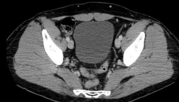
- Magnetic resonance imaging (MRI) makes it possible to accurately visualize the vessels and determine where the vein is narrowed and whether surgical attention is required. MRI is not the method of choice for the diagnosis of venous disease, due to the fact that it takes too long, expensive, has a number of limitations to its use and has a slightly less informative value in comparison with angiography.
- Among laboratory methods that examine blood, the greatest diagnostic benefit is the determination of the level of D-dimer.
As a rule, this is enough to confirm the diagnosis and start treating the disease.
Treatment
The iliac vein is located at a considerable distance from the surface of the body, so external influences are usually not very effective. Most often, patients seek medical help in the late stages of the disease, when it is no longer possible to do without surgery.
The main tasks in this case are to eliminate the root cause of the pathology that has arisen, to prevent the occurrence of various kinds of complications (for example, pulmonary thromboembolism arteries), stop the development of limb edema, which is able to squeeze nerve fibers so much that it will cause neurological disorders, and carry out prevention relapse.
Surgery
The most effective method of treatment today will be an operation.
Help can be obtained both in a planned manner and in an emergency, but only in a hospital setting during hospitalization in the surgical department.
- The optimal therapeutic tactic is minimally invasive endoscopic, including endovascular, surgical intervention, when after the operation the patient has several small unobtrusive scars. Such operations are as gentle as possible and have the best cosmetic result after the healing of skin incisions.
- Patients with May-Turner syndrome, for example, are recommended intravascular stenting of the left iliac vein.
- When the pulsation of the artery affects the wall of the PV, it is necessary to disconnect the nearby vessels, by leaving a so-called spacer between them for isolation of a vein, as well as elimination of pathology in its lumen: removal of a spur or membrane, removal of a thrombus and restoration of patency of the narrowed vessels. Speaking of the pad: it stays in the body forever, like any structure like a stent, with the help of which the diameter of the vessel has returned to its previous size.
Drug treatment
Along with surgical methods, the use of drugs, physiotherapeutic treatment (including physiotherapy exercises) is no less necessary, especially in the postoperative period.
It is necessary to start any therapeutic measures with a change in lifestyle (to receive adequate, and most importantly, regular physical activity, eat balanced and varied, optimize work and rest, and reduce obesity body weight).
Drug therapy is carried out mainly with the use of anticoagulants. The first 10-30 days, low molecular weight heparins are applied subcutaneously, under the control of a coagulogram for several later, oral forms are added to them: warfarin or one of the inhibitors of blood clotting factor Ha.
- Low molecular weight heparins:
- enoxaparin - 1.5 mg / kg body weight once a day (maximum single dose - 180 mg);
- dalteparin - 200 IU / kg body weight once a day (single maximum dose of 18,000 IU);
- nadroparin - 170 IU / kg body weight once a day.
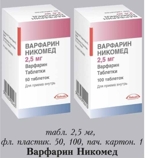
- The first 2 days warfarin at a dose of 10 mg, then the INR is determined and depending on the value the dose is adjusted for long-term use of the drug, while the INR must be maintained at in the range of 2.0 - 3.0.
- Selective coagulation factor Xa inhibitors:
- rivaroxaban (Xarelto) - 15 mg 2 times a day for the first 3 weeks of taking, after 20 mg 1 time a day;
- apixaban (Eliquis) - 10 mg 2 times a day in the first week, then 5 mg 2 times a day, or 2.5 mg twice a day if prolonged use is expected for more than 6 months;
- dabigatran (Pradaxa) - 150 mg 2 times a day, it can be replaced with heparin after 5 days of use.
Warfarin has both a number of advantages and disadvantages in comparison with Xa inhibitors: it is much cheaper (about 150 rubles. for a month's course of treatment versus 3000 thousand rubles), but at the same time constant monitoring of INR is necessary (2 times a month), and since treatment with these drugs it is supposed to be long, at least 3 months, then the patient needs to make a choice taking into account his financial opportunities.
The use of infusion therapy is usually unjustified, since excess fluid introduced from the outside will give additional stress on the veins of the lower extremities, which can provoke a second case diseases.
Wearing compression hosiery in the postoperative period is advisable after certain types of surgical treatment, however, you must strictly follow the prescribed schedule for its use and not exceed the time indicated by the doctor.
Physiotherapy is also useful in rehabilitation, it is aimed at reducing pain, swelling and inflammation. As an analgesic measure in the acute phase of the disease and to increase vascular tone, it is used ultraviolet irradiation, daily for 10 days, 4-5 biodoses, with a wavelength of 280-320 nm, and local cold.
Anti-inflammatory effect is observed when using a low-intensity UHF electric field, heparin electrophoresis (10,000 U, duration 20-30 minutes, course of 10 procedures) or Dimexide, sodium thiosulfate solution locally over the affected plot.
Over time, other treatments may be used to relieve pain and inflammation, such as: radiation red laser, Sollux lamp, paraffin and ozokerite applications, radon baths, darsonvalization and diadynamic therapy. In terms of its effect on deep veins, the alternating magnetic field of low frequencies has proven itself well, which significantly reduces the risk of recurrent thrombus formation.
For the treatment of trophic ulcers, ultrasonic waves are widely used through the aquatic environment, diadynamic therapy, oxygen barotherapy, ultraviolet irradiation, an infrared laser and even autotransfusion of blood irradiated with ultraviolet light (blood is taken from the patient's vein, irradiated and poured back) while improving nutrition of tissues in the area around the ulcer, the oxygen content in the blood and its antimicrobial properties increase, the wound is better cleaned, a denser scar is formed and pain decreases.
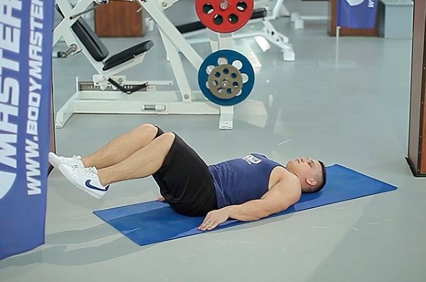
Physiotherapy exercises in different variations are shown in the early (breathing exercises and minimal activity on the first day after surgery) and in the late postoperative period. The main emphasis is on the development of the muscles of the lower extremities, to a greater extent it concerns the lower legs, to enhance their auxiliary "pumping" function in order to facilitate venous and lymphatic outflow. Below are 2 sets of exercises (in a lying and standing position) for daily execution.
Complex number 1:
- Lie on your back, give your legs an elevated position with the help of pillows (tilt angle 15-20 degrees) and stay in this position for 10-15 minutes.
- Remove the pillows and slowly breathe “belly” (when inhaling, the belly rises, while exhaling, it falls) 3-5 times.
- Alternately bend and unbend the legs at the ankle, knee and hip joints, as well as the toes, repeat 3-4 times in each zone.
- Raise your arms through the sides upwards while inhaling and slowly lower them downwards while exhaling, 3-4 times. Then raise your arms forward and up, inhale, lower your arms, exhale, also 3-4 times.
- Lower and raise the legs bent at the knees alternately to the left and to the right 6-8 times.
- Alternately take to the side and bring the arm and leg of the same name, 3-4 times on each side.
- Bend and unbend the feet of one leg, then the other, 8-12 times.
- Fold your arms behind your head and simulate cycling, make 3 sets of 6-10 movements.
- Pull one leg to the chest, straighten it vertically upward and slowly lower it, without bending at the same time at the knee joint. For each leg, 3-6 reps.
- Raise the leg up and perform 2-3 circular movements clockwise and counterclockwise, the same on the other side, 3 sets.
- Take each hand to the opposite side with a turn of the body, 3-6 times.
Complex number 2:
- Stand facing a wall or the back of a chair at arm's length and hold on tight.
- Rise on your toes, then smoothly roll onto your heels and back onto your toes. So repeat 5-7 times.
- Transfer body weight to one leg, standing on toes (the other leg is on the floor to maintain balance), then shift the load to the other leg. Carry out 3 sets of 5-8 times.
- Stand on your toes and sit down 3-5 times, while making sure that your knees do not come down inward.
- Stand with your right side to the stop, hold on tightly with your right hand, and swing back and forth with your left leg. Do the same with your right foot, while turning your left side. The movements are not too fast, without jerking, 8-10 swings with each leg.
- In the same position, sideways, make circular movements inward and outward with each leg 3-5 times.
For a variety of workouts, you should alternate them with walking on toes, heels, the inner and outer sides of the foot, with a high hip lift and a wide stride. The duration of such a walk should not be less than 3 minutes.
Thus, the iliac vein, which is located, it would seem, in an extremely advantageous position (deep from body surface, protected from all sides by the pelvic bones and other sufficiently strong structures) can still be vulnerable. And despite the low prevalence of the diseases described above, you should not think about them at the last turn, wasting precious time to provide care, while the patient's condition is steadily and rapidly worsens.
Iliac Vein Video
Internal and external iliac veins in 2 minutes: