
Content
- Indications
- Contraindications
- What documents are needed
- Do I need a compulsory medical insurance policy
- Do I need a referral from a doctor
- Where do they make for free
- Training
- Procedure step by step
- Decoding the results
- How often to do
- Video about fluorography
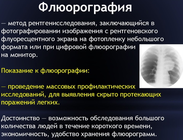
Fluorography is one of the methods a preventive or diagnostic method of x-ray examination of the chest and lungs for the presence of pathologies or foreign bodies. It is recommended that X-rays are taken for each person, no more than 1 time per year. Fluorograms are pictures with dimensions of 70x70 or 110x110, which are less informative, than standard radiographs, but only the examined patient receives less radiation exposure.
Today, digital examination technology is being actively introduced, which is accompanied by a minimum dose load, but giving the highest quality image, the ability to archive images and their subsequent storage. You can take a picture in a specialized clinic for a fee or free of charge by visiting the X-ray room in the clinic, presenting a medical policy.
Indications
It is recommended to make fluorography for free according to the policy if it is necessary to examine the heart, lungs, and mammary glands. In rare cases, this technique may be recommended for examining bones.
During the examination, it is possible to identify in the early stages:
- neoplasms in the chest or lungs;
- tuberculosis;
- pneumonia;
- pleurisy;
- areas of the inflammatory process;
- sclerosis;
- bronchial obstruction;
- hernia of the dome of the diaphragm;
- lung abscess;
- fibrosis;
- pleural layers;
- foreign body.
Pneumonia in humans is most often accompanied by a sharp increase in body temperature and a severe cough; with such symptoms, fluorography is prescribed. But oncology and tuberculosis most often do not manifest themselves in the initial stages, in this case, only an examination will reveal the disease as early as possible and begin effective therapy. It is for this reason that everyone, without exception, is recommended to undergo an examination once a year.
Contraindications
Teenagers from 15 years old and adults can do fluorography free of charge under the policy, but there are also contraindications to its use:
- pregnancy;
- severe shortness of breath;
- the inability of a person to be in an upright position;
- fear of confined spaces is claustrophobic.

You can find out more about contraindications for the procedure from a family doctor who prescribes a referral or from a specialist in an office with an X-ray machine.
What documents are needed
You can make fluorography free of charge under the policy, but you must first:
- Make an appointment with a therapist, visit him and take a referral for the examination. In the direction, the place of the examination, date and time must be indicated. Also, the doctor can write a presumptive diagnosis in question if there are already assumptions after taking an anamnesis.
- At the time indicated on the direction, come to the clinic for the examination, having with you an identity document - a passport and a compulsory medical insurance policy.
You can pass the examination without a referral, but in this case you will have to find out on your own where it is go through, what are the conditions of passage, what documents you should have with you and the requirements for it implementation.
Do I need a compulsory medical insurance policy
You can do fluorography without presenting a policy, but this is only if a person has applied for this service to a paid clinic. But in this case, no one will conduct an examination for him free of charge. It can be offered free of charge only if VHI is available.
If there is no insurance policy, then the service will only be paid. In a state polyclinic, if you do not present a policy, no one will do fluorography.
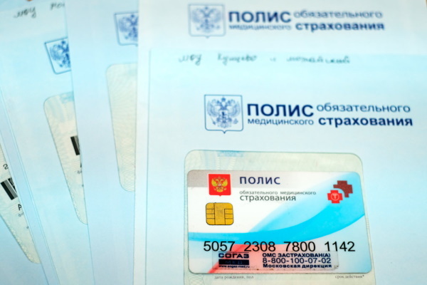
If you have a policy, a person can be examined for free in any city in Russia. If he was refused, then this amounts to a violation of human rights, and he can file a complaint with the control authorities.
Do I need a referral from a doctor
You can make fluorography free of charge according to the policy in any clinic and without a referral. This examination is considered a preventive procedure that everyone should undergo 1-2 times a year. Medical indications are not required for its passage, therefore, it is not necessary to take a referral. And without a referral, the doctor will conduct him, the main thing is that there is a passport and medical insurance.
It is especially important to regularly undergo fluorography for people at particular risk:
- having pathologies of the respiratory system;
- diabetics;
- with HIV-positive status;
- taking corticosteroids or receiving radiation therapy;
- food industry workers, doctors, educators, teachers;
- adolescents aged 15-18.
But it so happens that a person goes to the clinic not at the place of his registration, in this case, an employee of the fluorography office may require a referral from the attending physician. If a person does not have this with him, then he may be denied to undergo an examination.
In order not to stand in long queues for a referral for examination, you can visit the State Services portal and make an appointment with a therapist at the place of registration in your personal account. If this is not the case, then it will need to be created, which will take a few minutes.
Where do they make for free
It will not be possible to do a fluorography in any polyclinic in the city, because not every one of them has the equipment that allows you to undergo this examination. It is also worth remembering that almost all private clinics offer everyone to go through examination on the most modern equipment, but only for this service you will have to pay. The policy is not valid in such institutions.
You can make a free fluorography using an insurance policy in your city:
- in the clinic at the place of registration of the person;
- at the TB dispensary, but only if the X-ray equipment does not work in the clinic for some reason.
In the clinic at the place of registration of a person, he can be examined without a referral from a therapist, the main thing is that you must have an identity document and a compulsory medical insurance policy with you.
People who have come from another city, but who have an insurance policy with them, must apply to a tuberculosis dispensary. People from rural areas can be examined at the district clinic.
In large cities, there are specialized mobile points for fluorography. Many large enterprises of the city order mobile points for their employees for routine examination. They can also undergo fluorography free of charge with an insurance policy.
If there is no policy, then in any private clinic you can do fluorography at any convenient time for a fee. The average cost of an examination varies between 500-1000 rubles.
Training
If you have not had to deal with fluorography before, then you need to initially take a referral from a doctor, and then visit the X-ray room at the appointed time. No preparation is required for this study. 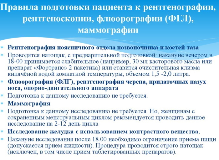 The patient simply enters the office at the appointed time, undresses to the waist, removes all metal objects and jewelry that may interfere with obtaining a high-quality and clear picture. In most cases, if the patient has a chain, then it is recommended to simply take it in the mouth during the procedure so as not to remove it.
Procedure step by step
After all the preparatory measures have been completed, the patient is offered to go to a special apparatus and strictly follow all the recommendations of the specialist:
- enter the device;
- take the necessary position, which involves pressing the chest against the equipment screen, the chin rests on a special support;
- at the request of the radiologist, the patient will need to hold his breath for a few seconds;
- all research passed.
 The patient exits the apparatus and gets dressed. The results are issued the next day. If during the study no pathologies were identified, then the person receives a certificate with a seal. If, during the examination, serious changes were revealed, then the patient is recommended to undergo a complex examination to make it easier to find out exactly what is the cause of pathological changes in the organs of the chest cells.
Decoding the results
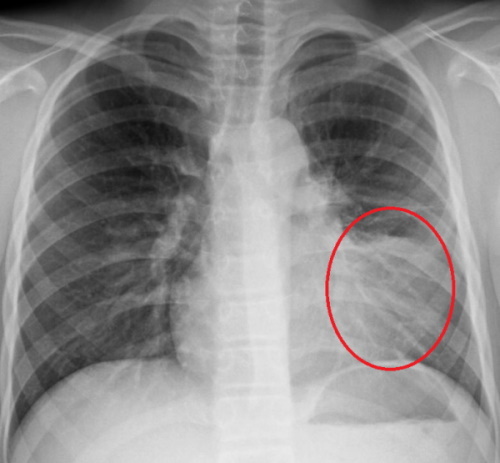
The picture in the image obtained with the help of special equipment can change if the density of the tissues being examined is changed. Most often, on fluorography, connective fibers are observed in the lungs themselves.
Their appearance can vary depending on which part of the organ they are localized in. They can have a different appearance and be localized in different parts of the organ. Depending on their location in medicine, they can be called: strands, fibrous blotches, scars, adhesions, sclerosis.
If connective fibers are observed in the bronchi, then they allow them to maintain their shape, if the patient is diagnosed with asthma, and the vessels to prevent stretching in hypertension. All of this can be seen in a photograph that was obtained using any type of X-ray equipment.
The image shows more clearly the densest tissues, which may indicate the presence of calcifications, malignant neoplasms, cysts, abscesses, emphysematous phenomena.
Not at every stage of the disease can it be detected using fluorography, for example, pneumonia is visible in the picture only in a developed form.
It takes time to develop the X-ray film. That is why the result after the examination is handed out only in a day. If everything is normal, then a paper form with a seal and an inscription is healthy is issued. And if there are changes, then the patient is offered to undergo a comprehensive examination.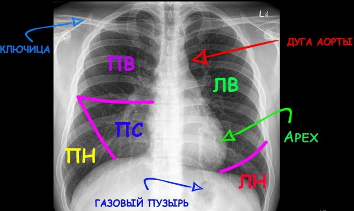
The table below provides a list of the phenomena that may be visible in the picture. Correct reading of them will allow you to accurately diagnose.
| Snapshot changes | Decoding |
| Dense roots | The roots in medicine are called organs that are localized at the entrance to the lungs. These include: bronchial arteries, vein of the lungs, main bronchus, lymph vessels, lymph nodes. More often, if compaction is observed, then expansion is detected. If only compaction is observed, then this indicates a chronic process in the body. Consolidation can be observed with swelling of large vessels, enlarged lymph nodes, which is characteristic of the development of inflammation of the bronchi and lungs. These symptoms are often seen in heavy smokers who may feel quite healthy. |
| Tie in the root area | Heaviness is a common pattern for people suffering from chronic inflammation of the bronchi, in heavy smokers. If a similar symptom is accompanied by others, then this may indicate that the person has an occupational lung disease, COPD. |
| Vascular pattern is pronounced | Any picture should show a pattern of blood vessels. If it is pronounced, then this indicates that blood flow is increased in this area. Such a picture may indicate acute inflammation, which may not cause serious harm or be the first signal for the development of cancer. Sometimes an additional examination may be ordered to clarify the diagnosis. A pronounced pattern is observed in people with heart disease, mitral stenosis and heart failure. But these pathologies are accompanied by other pronounced symptoms, and if they are not there, then an increase in the pattern can be observed after a cold or flu. |
| Fibrous tissue | They are more often seen on images of patients with pneumonia or tuberculosis. They can also be observed after surgery or injury. Fibrous tissue is not dangerous. |
| Hearths |
 Dark spots appear in the picture. They are not uncommon for various pathologies. If they are localized in the lower lobe of the lung, then inflammation can be suspected. If you pay attention to other symptoms, then there will be no difficulties in making an accurate diagnosis. The lesions in the upper lobes indicate tuberculosis. Dark spots appear in the picture. They are not uncommon for various pathologies. If they are localized in the lower lobe of the lung, then inflammation can be suspected. If you pay attention to other symptoms, then there will be no difficulties in making an accurate diagnosis. The lesions in the upper lobes indicate tuberculosis. |
| Calcifications | These are round areas in the image that resemble bone tissue. Sometimes they are confused with calluses on the rib. More often, calcifications are found in places where tissues have become inflamed due to the presence of the causative agent of tuberculosis. The body turns on a defense reaction, limiting the area where microbes can reproduce. Sometimes a part of the lung may look like this if there is a foreign body in it. If there are a lot of calcifications, then this may indicate that the subject had close contact with a person infected with tuberculosis, but his body was able to overcome the disease. |
| Adhesions | They appear on the pleura of the lung after an inflammatory process. They do not cause fear and do not worsen a person's well-being. |
| Layering | This occurs with thickening of the pleura, and is observed more often in the upper lobes of the lung. More often this may indicate that the inflammatory process has passed and does not cause concern. |
| Sinus condition | Sinuses are cavities in the pleura that are formed due to the presence of folds in the pleura. Usually this indicator is taken into account during the survey. In a healthy patient, the sinuses should be free. If liquid is found in them, then additional research is required. If the sinus is sealed, then this indicates a previous pleurisy, trauma or other pathology. If the general well-being of the patient is normal, then this is not dangerous. |
| The shape of the mediastinal shadow is changed | The shadow of the mediastinum is the most important of the indicators in the fluorography picture. The mediastinum is the place between the lungs. It is in it that the lung, esophagus, aorta, trachea, vessels, lymph nodes and thymus gland are located. If the shadow of the mediastinum is enlarged, then this indicates an increase in the volume of the heart. |
How often to do
If you follow the legislation of the Russian Federation, then it is recommended to undergo fluorography once a year. In this case, the procedure will not cause any harm to the body. But there are a number of cases when it is allowed to undergo such an examination more often, for example, if the picture turned out poor quality and because of this, the doctor cannot give the patient an accurate diagnosis and choose an adequate treatment. People with chronic pathologies are recommended to undergo an examination 2 times a year.
Fluorography can be done completely free of charge according to the medical policy in the clinic at the place of residence. The main thing is to have a policy and a passport with you. If a person who has a medical policy ends up in another city, then he can also undergo a study there, but by contacting a specialized clinic rather than a polyclinic. If you go to a private clinic, then you will have to pay for the study, the policy does not work in them.
Video about fluorography
Differences between fluorography and X-ray: