
Content
- Characteristic
- Mechanism of occurrence
- Functions and properties
- Anatomy and structure
- Possible violations
- Chiasm (chiasm) pathology
- Hemianopsia
- Physiological anisocoria (uneven pupils)
- Damage to the III cranial nerve
- Pupillary reflex video
The transmission of impulses from the retinal photoreceptors to the nerve endings of the brain and the pupillary light reflex passing along the reflex arc are two main pathways, with which the eye perceives changes in the environment and reacts to them. Multiple information processing signals are efficiently transmitted from the cornea to the brain, and any damage to the pupillary reflex pathway can lead to visual pathology.
Characteristic
There are certain actions in the body that are spontaneous and do not require brain processing. Such actions or reactions are called reflex actions.
Reflex actions are involuntary actions that occur without conscious thought processes. For example, when foreign particles enter the eye, tears immediately wash them away (glandular secretion).
Reflexes are of two types:
- Naturals are reflexes that do not require previous experience or training.
- Conditioned reflexes are reflexes that develop throughout life through experience or learning.
Difference between natural and conditioned reflex:
| Natural (simple) reflex | Conditioned (acquired) reflex |
| Congenital, requires no previous experience. | Developed from experience or learning. |
| Directly related to the stimulus | Caused by a condition completely different from the direct initial stimulus. |
| Simple reflexes are similar in all people. | Varies from person to person depending on training and experience. |
The path along which nerve impulses (irritation) pass to the executive organ is called a reflex arc. The pupillary reflex, the reflex arc of which follows a fairly simple path, is represented by a number of neural links.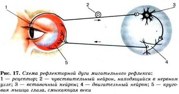
Reflex arc components:
- A receptor is a protein molecule, usually embedded in the surface of the plasma membrane of a cell, that receives chemical signals from the outside.
- Sensory nerve - carries a message from a sensory neuron to the spinal cord.
- The relay center is an intermediate neuron in the spinal cord that transmits an impulse from a sensory neuron to a motor neuron.
- Motor nerve - transfers a message from the spinal cord to an executive organ, muscle or gland.
- Effective (executive) organ - receives a signal from the motor nerve and acts in accordance with it.
The reflex arc can be represented as follows:
Stimulus -> receptor in the sense organ -> afferent nerve fiber -> central nervous system -> efferent nerve fiber -> muscle -> gland.
With reflex actions, the arc is formed by impulses from the receptor that reach the spinal cord, and the corresponding reflex impulse, which is then sent by the spinal cord to the muscles. The impulse is not sent to the brain in order to shorten the response time.
Mechanism of occurrence
Pupillary reflex (the reflex arc allows the pupil to narrow 0.5 s after a directed bright light) is a reaction that controls the diameter of the pupil when it is exposed to light of different intensity. This allows the eyes to adapt to bright or dim light.
If you go into a room and turn on the light, then all objects are clearly visible. After turning off the light, looking at the darkness, a person can still identify various objects in space. While he may not be able to see them as well as before, it will be enough to not stumble in the dark.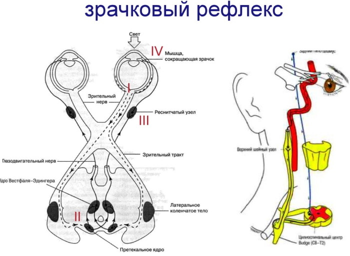
In this way, the pupil adapts to the lighting in the environment, allowing you to see in both bright and dark rooms. Pupillary response is a reflex that adjusts the diameter of the pupil when it is exposed to different intensities of light.
Functions and properties
The iris contains two sets of smooth muscles - the circular and radial muscles.
The circular ones are arranged in concentric rings around the pupil, and the ray ones run radially. These muscles are antagonistic.
The pupillary light reflex is the reflex contraction and relaxation of the antagonistic muscles of the iris in response to a change in light intensity, which causes a change in pupil size.
The pupil dilates at low light intensities and contracts at high light intensities. This allows enough light to enter the eye for dim light vision while filtering out excess light at high light intensity to prevent damage retina.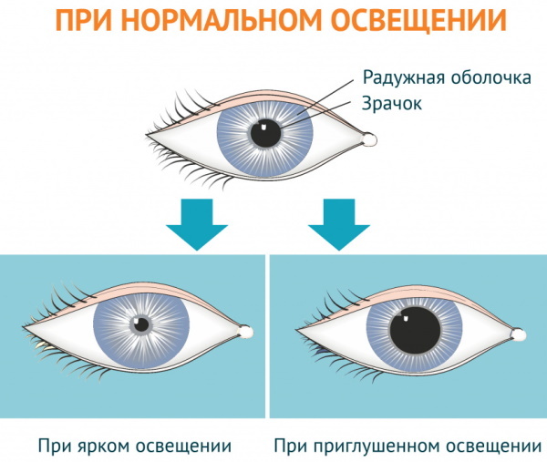
The pupil shape of a normal human eye remains circular when narrowed or dilated. Changes in the shape, size and speed of reactions of the pupil are of diagnostic value in eye diseases.
Anatomy and structure
Vision is a complex process that involves coordinated and simultaneous brain and eye activity. Light entering the eye is converted into an electrical response called a nerve impulse, which travels to the brain along the optic nerve to create the final image. The optic nerve is the second pair of cranial nerves that carry visual information from the eye to the brain.
To better understand how light travels from the retina to the brain, one must understand the anatomy of the eye.
The most important parts of the eye include:
- iris;
- cornea;
- lens;
- retina.
Pupillary reflex reflex arch
The eye or eyeball is in the orbit, but only the front of the eye is visible:
- The white part of the eye is the sclera, which is the visible part of the outer layer of the eyeball.
- The colored portion is the iris, which has a small disc-shaped shape with an opening called the pupil.
- The pupil is black, through which light passes to the lens, which subsequently focuses it on the retina. It either shrinks or expands, depending on the amount of light falling on it.
- A transparent layer called the cornea covers the iris and pupil. The cornea is like a dome around the iris, and behind the cornea is a fluid called watery moisture. which helps to clear the eyes and provides essential nutrients substances. The cornea also protects the eye from debris and injury.
- The eyelids and eyelashes are protective.
Since the eye is an optical device, it has complex structures:
- Lens - natural lens - attaches to muscles with strong fibers. Contraction of these muscles changes the shape of the lens. The light rays passing through the pupil reach the lens located behind it. The role of the lens is to focus light on the retina. The path of light entering the eye changes (refracts) to varying degrees depending on the shape of the object. Refraction occurs when light hits different environments. In the eye, light travels from the air into the fluid of the cornea, which causes a change in its path. The process of changing the path of light is called accommodation, and it allows the eye to focus on objects that are either close or far away.
- Retina Is the innermost light-sensitive tissue of the visual system. The retina contains millions of sensory cells, including light-sensitive cells called photoreceptor cells, referred to as rods and cones. The former work in dim light and provide black-and-white contrast, while the cones function in a well-lit environment and are able to perceive different colors.
The pupillary reflex, the reflex arc of which starts from the retinal photoreceptors, is the body's normal response to irritation. Refraction caused by the lens creates a sharp image on the retina. The sensory cells in the retina receive these light signals and transmit them to the brain via the optic nerve.
The main role of the optic nerve is to transmit visual information from the retina to the visual centers of the brain using electrical impulses. The optic nerve is made up of nerve cells and is an important part of the central nervous system.
Simply put, nerve signals from rods and cones are sent to the brain via the optic nerve. Inside the brain, these signals are converted into images that a person sees. In this way, light enters the eye and travels along the optic nerve to the brain to create the final image.
The reaction of the eye to different intensities of light is not the same. When bright light is directed directly into the eye, the pupil constricts almost instantaneously, thereby protecting the retina from dangerously bright light.
On the other hand, in a dimly lit place, the pupil dilates, allowing more light to enter the eyes.
The main players in the vision are rods and cones. Rods are extremely effective in dim light, as even a small amount of light can cause them to fire. They are able to accurately detect light, contrast, and movement, but cannot detect color.
The eye regulates the light entering it, like the diaphragm of a camera.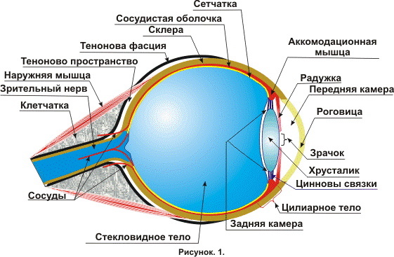
The pupillary reflex is a very unique and interesting way in which the eye captures images and controls light exposure. Much like a camera, it regulates the amount of incoming light, which leads to the formation of an image by the brain.
The anatomical structure of the visual pathway is represented by a number of neural links.
The reflex arc, the afferent (ascending) part of which starts from the cones and rods, carries information along the ascending pathway to the brain.
The first steps of the pupillary light reflex along the way:
- Light travels through the cornea, anterior chamber, pupil, lens, and posterior chamber, eventually reaching the retina.
- Photoreceptor cells (rods and cones) in the outer layers of the retina convert light stimuli into neural impulses.
- These signals are then transmitted to bipolar cells (neurons with 1 dendrite and 1 axon), which interact with ganglion cells (transmitting nerve impulses). The latter, in turn, merge to form the optic nerve head. The disc sends impulses to the brain for further processing and image recognition.
Next steps:
- The optic nerve forms the optic chiasm, which diverges into the left and right optic pathways.
- Retinal fibers located next to the temporal zone continue to move along their side, and the nasal (located on the side of the nose) retinal fibers intersect with the opposite side of the visual path.
- The intersection of the optic nerves is the nerve structure in which the fibers of the optic nerve intersect. The fibers are arranged in such a way that the nasal fibers on both sides intersect and move to the opposite side of the brain. The fibers of the temporal half of the retina remain on the same side, while the fibers of the nasal half are crossed. As a result, the left optic tract contains the nasal fibers of the right eye and the temporal fibers of the left eye. Right - nasal fibers of the left eye and temporal fibers of the right, respectively.
- Information from the visual tracts is connected together on an optical plate, and then the signals pass into the border area between the midbrain and diencephalon, where the nuclei involved in the analysis of moving objects.
- Each pretectal (borderline) region sends bilateral signals to the parasympathetic (where ganglia or nerve nodes are located) nuclei of the midbrain.
Efferent (descending, transmitting impulses from the cerebral cortex) fibers move along the oculomotor nerve, which sends axons (nerve fibers that connect different cells) to directly innervate the sphincter muscles iris. The contraction of the iris sphincter muscle leads to constriction of the pupil (miosis).
In dim light, the muscle fibers of the pupil contract and dilate the pupil. The muscles of the pupil dilator are innervated by the sympathetic fibers of the ciliary nerve.
Possible violations
The pupillary reflex, the reflex arc of which is the anatomical substrate of the pupil's response to light, indicates, first of all, the health of the eyes. Pupil examinations are an important part of routine ophthalmic, neurological, and general medical examinations.
Due to the proximity of the pupillary pathways to various anatomical structures, pupillary dysfunction can be caused by a variety of diseases. Because of the differences in the passage of the pupillary and sensory fibers, eye tests can help localize the lesion in the optic pathway. First of all, the ophthalmologist identifies pupil disorders and determines further research.
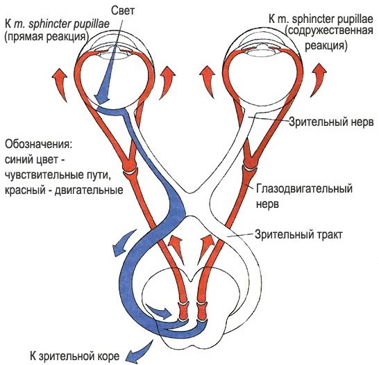
The physiology of normal pupil constriction is the balance between the sympathetic and parasympathetic nervous systems. The visual pathway of the light reflex is a complexly coordinated scheme, in which many components participate, performing their actions with high precision.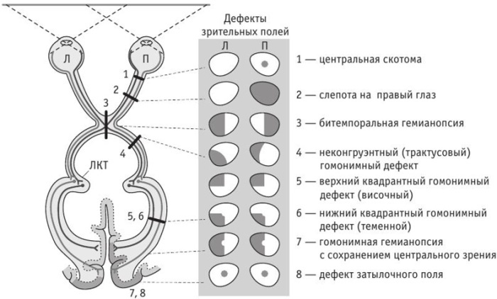
The first innervation leads to pupil dilation, which is controlled by a muscle group in the peripheral 2/3 of the iris. Sympathetic innervation begins in the cerebral cortex.
Parasympathetic innervation leads to constriction of the pupils. A circular muscle called the pupillary sphincter performs this task.
Any violation on this difficult path leads to visual impairment. The location of the pathology is associated with the type and degree of the disorder.
There are several ways to study the pupil's response to light. Some methods are based on the asymmetry of the afferent visual pathway, while others are based on the study of the visual field by measuring the light response of the pupil to focal light stimuli.
Chiasm (chiasm) pathology
Chiasm is formed as a result of the intersection of the right and left optic nerves. The axons of the optic nerves are redirected to the chiasm to form the right and left optic tracts. The intracranial optic nerves and chiasmus rise at a 45 ° angle from the base of the skull. Chiasma is shaped like the Greek letter χ, hence its name. The chiasm is approximately 4 mm thick, 12 mm wide, and 8 mm long.
Chiasm lesions are manifested by primary optic nerve atrophy, which leads to visible pallor of the optic nerve head and loss of a layer of nerve fibers over time. The reason may be compression of the optic nerve cross section under the influence of a brain tumor, multiple sclerosis, or a skull injury.
Hemianopsia
This is bilateral blindness in half of the field of view of one or both eyes.
Under normal conditions, the left side of the brain processes visual information from both eyes about the right side of the visible world. The right hemisphere of the brain processes visual information from both eyes about the left side of what a person sees. Damage to any part of the optic chiasm can cause partial or complete blindness in the visual field.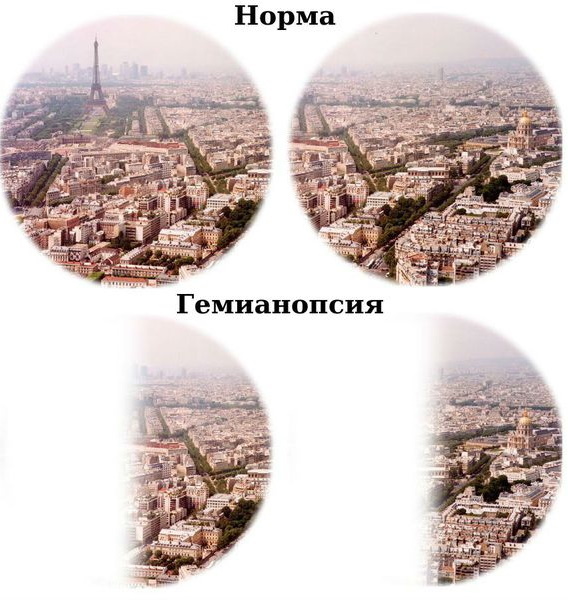
Hemianopsia can occur if there is damage to:
- optic nerves;
- intersection of the optic nerves;
- brain imaging areas
Less commonly, damage is caused by:
- aneurysm;
- an infectious disease;
- exposure to toxins;
- neurodegenerative disorders;
- transient events such as seizures or migraines.
The condition of hemianopsia is caused by problems in the brain, and not by a violation of the eyes themselves.
Physiological anisocoria (uneven pupils)
Anisocoria is a term for pupils of different sizes. Many people have the same pupil size, and both pupils will get smaller or larger to let light through at the same time. The presence of anisocoria may be normal (physiological) or a symptom of an underlying medical condition. During a full eye examination by an ophthalmologist, pupil size and response to bright and dim light are checked. Based on the assessment, the doctor may conduct additional tests to make a diagnosis.
Anisocoria usually does not need to be treated as it does not affect vision or eye health. Treatment is prescribed if there is an underlying disease.
Damage to the III cranial nerve
Pathology can lead to damage to the parasympathetic fibers going to the muscle of the pupillary sphincter, disrupting, such way, the efferent arch of the pupillary light reflex, which leads to insufficient constriction of the pupil on the affected side.
Pupillary light reflex abnormalities range from isolated benign manifestations to precursors of serious, even life-threatening conditions. Complete understanding of the reflex arc pathway, neuroanatomy underlying the innervation of antagonistic pupillary sphincters and dilator muscles, it is necessary to detect and determine the importance of a particular anomaly pupils.
Abnormalities can be found with damage to the optic nerve, oculomotor nerve, brain stem lesions such as tumors, and medication such as barbiturates.
Pupillary reflex video
Pupillary reflex. Anatomy, reflex assessment:


