Content
- Characteristic
- Anatomy and structure
- Internal
- Outdoor
- Front
- Functions and properties
- Possible diseases and pathologies
- Phlebectasia
- Aneurysm
- Thrombosis
- Phlebitis and thrombophlebitis
- Jugular Vein Videos
The circulation of blood in the human body provides branched venous system, streams of blood from the head through the neck carry away the channels called the jugular. These vascular trunks are paired, refer to the structure of the superior vena cava. Important internal highways that supply blood to the face and heart are located in the thickness of the cervical zone. Information about the structure and properties of veins will help to avoid problems with their functioning.
Characteristic
The set of jugular vessels is responsible for the quality of the outflow of blood from the organs located in the head, the flow moves through the cervical region to the venous bed located under the collarbone. The main task of the jugular vein is to prevent stagnation of blood in the brain in order to protect a person from serious pathological changes.
The vascular structure consists of 3 paired veins - anterior, external and internal, with different widths and specific functions. The main volume of the venous flow flows from the head through the neck, entering the cavity of the right and left internal jugular veins. The outer vessel (jugular) is enriched with the blood of 3 venous channels - the occipital vein, suprascapularis, posterior ear. The anterior pair of canals that drain blood from the outside of the head through the neck can be seen when the larynx is straining when coughing or singing.
Anatomy and structure
Blood volumes from all human organs are transported to the right chamber of the heart through two large venous trunks (upper and lower). In addition to the superior and inferior vena cava, blood enters the right atrial cavity through the heart's own arteries. Together with many vessels, the superior and inferior vena cava form a complex venous system.
| Name | short information |
| Vienna upper | Blood collected from the organs of the upper body is concentrated in its cavity. Branches of all three jugular veins are also involved in the process, into each of which the channels of the organs of the head and neck area flow into. |
| Vienna lower | A large vessel that opens into the cavity of the right atrium collects blood from organs located in the retroperitoneal space and below. Therefore, the jugular bundle does not participate in the blood circulation process. |
For doctors of different professions (dentists, surgeons, neuropathologists) knowledge of the anatomical structure of the venous structures of the head and cervical spine are the clinical basis for the correct diagnosis and operations. Information about the peculiarities of the location and functioning of the jugular vein system will help patients notice deviations in time in order to seek help from a specialist.
Internal
The jugular vein, located deep within the neck, is the widest venous channel responsible for regular blood flow from the head to the heart. With a maximum width of 20 mm, the wall of the inner vessel (v. jugularis interna) are rather thin, which allows easy contraction. If you look at the jugular vein from the inside, you can find several valves that facilitate the free flow of blood.
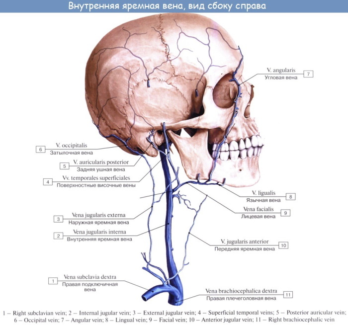
The largest of the jugular streams is an extension of the sigmoid venous collector (sinus), and leaves the jugular fossa, located in the bony base of the skull. Further, the blood bed expands, forming a bulbous connection (upper bulb), as moving down to the location of the sternoclavicular joint, which covers with its muscles vein.
Throughout the cervical v. jugularis interna fits inside a wide connective tissue container, where the carotid artery and the vagus nerve are located. In this case, the location of the jugular (internal) vein in a powerful vascular bundle is more superficial and lateral in relation to the artery (lower neck).
Behind the sternoclavicular junction, the diameter of the internal bed increases again, as evidenced by the presence of a second large expansion - the lower bulb. Here the jugular bed is combined with the subclavian, from where the brachiocephalic vein originates, expanding in the lower part. At the confluence with the subclavian canal, there are valves, usually only 2-3 of them. The branches of the tributaries of the jugular bed are divided into two types - intracranial and extracranial.
Intracranial vascular streams pump blood from the following organs:
- venous collectors (sinuses) of the dura mater;
- diploic (branched) veins of the bones of the skull, devoid of valves;
- cerebral trunks that distribute blood from the brain to the sinuses;
- meningeal arteries of the meninges;
- orbital, auditory and pharyngeal veins, upper and middle thyroid.
The group of extracranial highways includes the vessels that lie in the front and outside of the skull.
The venous flows of the following structures are connected to the internal jugular vein along its course:
- pharyngeal plexus and lingual trunk, coming from the root of the tongue;
- the upper or middle part of the thyroid blood line;
- the facial zone, receiving blood from the soft structures of the mucous membrane;
- pterygoid plexus emerging from the infratemporal recess.
The jugular vein is in the path of the powerful blood flow. The vascular trunks (internal, external and anterior) start at the base of the cranium, carry out venous blood from the head through the neck to the superior valveless vein, which is short and hollow.
The intracranial and extracranial arteries are interconnected by the so-called graduates, the processes of which penetrate the cranial bones. External tributaries provide the movement of blood from the outer surfaces of the head, skin, face, and their combination with deep channels becomes a guarantee of good venous outflow. At the same time, the extensive network of the circulatory system can become a source of infection.
Outdoor
The site of localization of the external artery (vena jugularis externa) from the jugular vein family is the cervical tissue. The task of a small vein in diameter is the outflow of blood from the face area, the outer surfaces of the head and cervical integuments. The venous line starts behind the plane of the auricle, closer to the line of the mandibular angle, is formed due to fusion two external streams:
| A type | How is formed |
| Front | The bed is connected (anastomosis) with the lower jaw bed, which wedges into the internal jugular vessel. |
| Rear | The ear canal merges together with the occipital stream, which flows into the cavity of the vertebral vein. |
The further path of the outer trunk runs down (obliquely) along the surface of the muscle structure that connects the sternum with the clavicle under the cover of the subcutaneous muscle of the neck. After crossing the thin layer of the pretracheal plate, it turns out to be at the confluence of two ducts - the subclavian and internal jugular, flowing into the cavity of the subclavian vein.
On the way, the external jugular vein receives blood flows from several vascular channels:
- posterior ear, extending from the outer section of the carotid artery and passing behind the auricle;
- anterior jugular, consisting of the right and left channels, which sometimes merge into the middle vessel;
- suprascapular, collecting blood from the muscles of the periosteal and subosseous type;
- transverse veins of the neck, flowing into the external jugular or subclavian artery.
In the cavity of the outer trunk, there are two valves - one paired valve closes the initial section of the venous vessel, the second is located approximately at the level of the middle part of the neck. The vena jugularis externa is filled with occipital, ear and suprascapular blood flows. It is in the space of the trunk of the external vein that a catheter is installed if a long-term intravenous supply of medicinal solutions is required.
Front
The jugular vein, represented by a bundle of paired vessels, is located in the thickness of the neck, wedges into the system of the superior vena cava. The anterior section of the jugular vein (v. jugularis anterior) is a fairly large inflow of the external channel. It is formed by small, fused skin vessels that lie in the thickness of the chin area.
Uniting with the facial veins, the bed of the anterior blood bundle is directed downward, without departing from the midline of the neck. The venous flow first moves along the external muscle bundle, which ensures the work of the maxillofacial apparatus. Passing the maxillary-hyoid muscle node, it wedges into the surface of the cervical fascia plate (connective tissue) above the sternal jugular notch.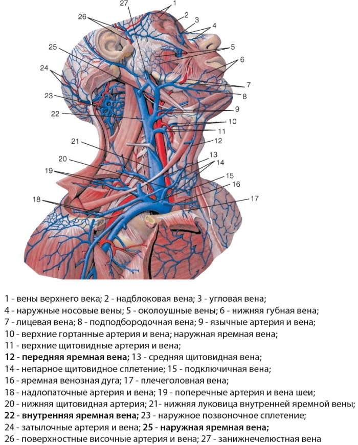
In the lower region of the neck, the jugular veins of the anterior type, going to the right and to the left, firmly anastomose (connect), forming a rounding in the outer layer (arcus venosus juguli) - a venous arch. Then there is a deviation of the jugular line to the outside, then it moves behind the muscles of the sternum and clavicle, wedging into the external jugular bed. The connection of the anterior flow with the subclavian and brachiocephalic veins is rare.
In rare cases, the anterior canal is reticulate, but both jugular veins are poorly developed. In this case, a branched network of thin vessels is observed, abundantly anastomosing with the venous ducts. Sometimes right-sided and left-sided blood bundles can merge into a single bed to form the midline vein of the neck.
Functions and properties
The circulatory system of the paired jugular veins is part of the system of the main vessels that provide a full outflow of venous blood from the brain and organs located in the cranium.
Despite the different locations and structural features, all types of branches of this type differ in a standard structure:
- the inner layer of veins is formed by endothelial tissues and receptors responsible for the stability of regulatory mechanisms;
- the thickness of the middle layer is represented by elastic collagen-type fibers, as well as smooth muscle cells that maintain a constant blood flow;
- outside, the venous bundles are covered with connective tissue permeated with elastic collagen threads, which stabilize the strength of the vessel walls.
Features of the topography and details of the anatomical structure of the multichannel jugular line depend on the location of the vein. The general task of the bloodstream of this type is to drain blood from the head and neck section into the trunks of the brachiocephalic veins, which along the way they flow into the space of the superior hollow artery.
The jugular vein is located in front of the cervical zone, and provides the outflow of the main part of the blood (venous) from the brain.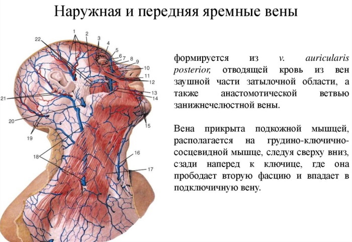
Each of its three paired veins, which are part of the upper hollow canal, are assigned especially important functions:
- ensuring the correct circulation of cerebral blood flow, supplying the brain with blood at regular intervals;
- creation of favorable conditions for the implementation of the reverse outflow of blood saturated with oxygen and nutrients;
- evacuation of toxins accumulating in the intercellular fluid from the organs and tissues of the head, as well as the cervical spine.
The collection of blood by the jugular veins is carried out mainly through the network of carotid arteries, as well as the vertebral trunks. For the life support system, the most important are two paired vessels - external and internal, their pronounced muscle layer withstands the powerful pressure of blood well. However, the development of pathological conditions affecting the veins of the multichannel jugular complex leads to the threat of irreversible changes that not only worsen the state of health, but also pose a threat to life the patient.
Possible diseases and pathologies
The venous system often suffers from excessive stretching, which is manifested not only by inflammation, but also by the formation of blood clots and lesions. Diseases are susceptible to both adults and children, but more often older patients suffer from damage to the jugular vessels. In young patients, traces of congenital abnormalities usually appear. The most common venous problems include phlebectasia, aneurysm, thrombosis, phlebitis, and thrombophlebitis.
Phlebectasia
The pathology of the jugular vein is accompanied by an inflammatory process, as a result of which dilatation (expansion) of the external vessel occurs. The disease is not due to either age or gender, it can be congenital or acquired.
Probable causes of phlebectasia include the following problems:
- impaired blood circulation after an injury to the skull or neck, spine, leading to the development of inflammation;
- the presence of cardiovascular pathologies and compressive neoplasms that reduce the elasticity of blood vessels;
- consequences of a concussion of the brain, prolonged psychological stress, improper installation of a catheter.
 The jugular vein is located between the base of the skull and the sternoclavicular zone, localized along the lateral parts of the neck in the form of a right and left stream. Malformations of their development, in particular ectasia, are usually associated with improper formation of the walls of the vein or its valves.
Symptoms are not very bright, they can manifest themselves as discomfort when the neck is strained, in case of neglect, the operation of the valves is disrupted. Usually, the disease does not threaten with serious consequences, it is treated with methods of conservative therapy (antihistamines and anti-inflammatory drugs, antibiotics). In the case of a severe course with a risk of complications, surgery is indicated.
Aneurysm
The variant of a true aneurysm (protrusion of the vessel wall) is quite rare, sometimes young children aged 2-7 years suffer from pathology. The reasons for the uncommon abnormal condition are presumably associated with the malformation of the fascia of the base of the venous bundle during intrauterine development of the fetus. As a result, the choroid swells, not withstanding blood pressure, which is manifested by a swelling in a vein on the side of the neck.
A rare pathology does not signal any special manifestations; symptoms of anxiety and headache, accompanied by sleep disturbances, can be observed. However, the danger of aneurysm is explained by the high risk of rupture of the choroid. Therefore, the treatment is surgical - aneurysm resection, anastomosis, branch prosthetics.
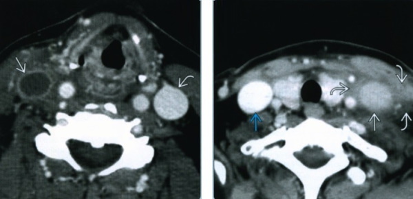
Thrombosis
The causes of the pathological condition are due to the presence of chronic diseases (autoimmune or hereditary), trauma to the walls of blood vessels (surgery, pregnancy), congestion (sedentary lifestyle, cardiac failure). As a result of these and other vascular problems, a blood clot, a thrombus, is formed in the lumen of the affected venous bed. Thrombus formation can affect any jugular vein, but more often the external bed suffers.
The greatest severity of symptoms is distinguished by the acute period of the lesion of the jugular vein located on the neck:
- the skin itches, acquires a bluish tint;
- subcutaneous vessels swell, causing facial edema;
- on the reddened neck and cheeks, the venous pattern is enhanced.
 The intensity of the manifestations of thrombosis depends on how much the lumen of the vessel is blocked by a clot. The choice of treatment method depends on the severity of the disease. With non-threatening symptoms, drug therapy is prescribed with anticoagulants, phlebotonics, antibiotics, antispasmodics. Severe thrombosis with persistent blood clots is treated with surgery.
Phlebitis and thrombophlebitis
Both pathologies are characterized by inflammation of the venous wall, but most often the inflammatory process affects the bulb of the jugular vein. The most likely cause of the disease is the development of purulent inflammation in the middle ear and the tissues that form the mastoid process. It is also possible that infection from other sources can be ingested. Symptoms depend on the type of vein affected by the inflammation, but in general, patients complain of pain in swallowing, high temperature, and swelling of the neck.
The course of the disease leads to infection of the internal tissues of the vessel (phlebitis) or the resulting thrombus (thrombophlebitis), and purulent exudate spreads with the bloodstream to other internal organs. As a result, the septic process becomes generalized, the treatment of which is surgical - the inflamed vein wall is removed along with thrombotic formations.
For any problems with veins, you should immediately contact a physician. If the patient's complaints concern the jugular vein, the family doctor will refer you to a phlebologist and cardiologist for an accurate diagnosis. After all, damage to the blood vessels in the neck area leads to poor circulation, which increases the risk of life-threatening complications.
Jugular Vein Videos
Internal, external and anterior jugular veins: