Content
- Symptoms and Signs
- Causes
- Diagnostics
- X-ray
- Ultrasound
- Treatment methods
- Conservative
- Surgical
- Soft tissue surgery or open reduction
- Surgical removal of the tendon or tenotomy
- Varus osteotomy
- Endoprosthetics
- Possible consequences and complications
- Video about Coxa valga in children
Hallux valgus or Coxa Valga develops in children if hip dysplasia has not been treated.
During a routine examination of the baby, the orthopedist sometimes detects hip dysplasia, although it is not very common. The cause is an abnormality of the epiphyseal cartilage under the femoral head.
The femur is a very strong and stable bone connected to the pelvis through the hip joint. It is divided into a head, a shaft, a femoral neck and a lower end. A large number of clinical signs in the hip joint are congenital or develop at a young age. The displacement of the head and glenoid cavity relative to each other leads to coxa vara or coxa valga. Mixing from the femoral neck can be congenital or acquired.
Coxa Valga in children is sometimes asymptomatic and does not require treatment. In moderate to severe cases, physical therapy, a walker, canes, or crutches are used to make walking easier. The operation is performed if conservative treatment is not enough to relieve pain.
Symptoms and Signs
Hip joint abnormalities do not cause pain in infants and are therefore difficult to identify.
Parents may notice:
- The child's thigh makes a popping or popping sound that can be heard.
- In infants, you may notice that one leg is longer than the other.
- One hip or leg does not move in the same way.
- The skin folds under the buttocks or on the thighs are not aligned.
- The child limps when he starts to walk.
Coxa Valga in children
Any of these signs should be addressed to your doctor. In rare cases, the pathology causes pain or discomfort. The disease often goes unnoticed, and in most cases, Coxa Valga does not cause any symptoms with bilateral involvement. But with unilateral deformity, a waddling gait develops, a pulling pain occurs in the hips, and the gait is disturbed.
Causes
The hip is a hinge joint. The rounded head of the femur forms a ball that fits into the socket of the acetabulum. The thigh forms the primary connection between the bones of the lower extremities and the axial skeleton of the trunk and pelvis.
The three pelvic bones - the ilium, ischium, and pubic - together form the acetabulum. The joint itself may not completely ossify or harden until the person is 25 years of age, which is the reason why early detection is important when treating hip disease.
The bone structure of a child in the uterus must develop so that the head of the femur is exactly in the center of the acetabulum. The acetabulum should cover the head of the femur as if it were a ball inside the cup. In congenital hip dysplasia, the development of the acetabulum allows the head of the femur to extend upward from the socket, especially when the child begins to walk. The result is a shallow rosette, which results in improper displacement of the femur because the acetabulum does not sufficiently cover the head of the femur.
If this is not corrected, the hip joint will be unstable. In some cases, dislocation may occur. Over the years, when the femoral head and acetabulum move without proper alignment, the cartilage in the joint is worn out prematurely and unevenly, which further leads to osteoarthritis.
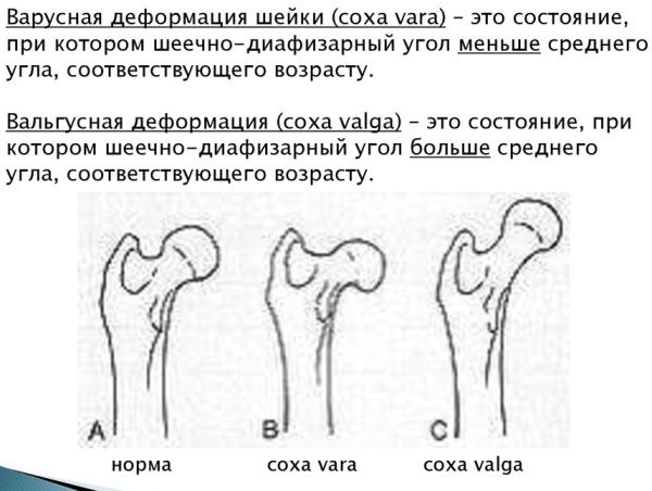
The position of the femoral neck in relation to the femoral shaft is important for correct mechanical loading of the hip joint. This position is measured as the intersection of a straight line through the neck and thigh axis. The angle between them changes throughout life: while in a newborn it is about 150 °, by adulthood it smoothes out to about 120 °. In old age, the angle becomes even flatter due to a decrease in bone substance.
Thus, the normal femoral neck angle is 120–140 °, depending on age. At steeper angles (> 140 °), they speak of valgization or coxa valga, at more obtuse angles (<120 °), they speak of varization (coxa vara). Only the extreme options are pathological; they usually occur during childhood.
Changing the angle leads to improper stress on the hip joint and femoral neck. In addition, large deviations often disrupt the balance of the thigh muscles, which in turn can increase inappropriate stress on the bones. Sometimes this creates a vicious circle that can only be broken by surgery.
The exact anatomical features of the hip joint are different for each person. With pathology, both the hip joint and the femur can be affected. In any case, the length of the femur is affected - it is shortened with coxa vara or lengthened with coxa valga. This creates additional imbalance in the hip joint and leads to difficulty walking, pain and stiffness in the joints.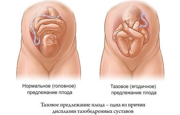
- The most common cause of hip displacement is untreated hip dysplasia.
- In older children, the main causes are diseases such as Perthes disease or slipping of the femoral head.
- Diseases that reduce bone quality, such as rickets or osteomalacia, also cause the femoral neck to sag.
- Coxa valga in children can develop as a result of cerebral palsy. An imbalance in the muscles leads to abnormal growth of the femoral neck.
Hallux valgus can be "symptomatic" and occur due to:
- Little's disease (bilateral congenital spastic diplegia, view from cerebral palsy);
- after polio;
- in the process of progressive muscle dystrophy;
- with the development of tumors affecting the growth of epiphyseal cartilage;
- vitamin D deficiency (rickets);
- impaired metabolism in the mother during pregnancy.
Oxygen deficiency in the embryo also affects the development of pathology. Lack of oxygen inhibits the synthesis of nucleic acids, proteins, which structurally changes the development of tissue in an unborn baby.
Less often, after the completion of growth and in old age, flattening (variation) of the femoral neck is observed. Here, the most common causes are osteoporosis, femoral neck fractures and necrosis of the femoral head.
Diagnostics
The physician receives clinical indications for coxa valga and any associated muscle imbalance by assessing gait patterns and checking the range of motion of the hip joint. At the same time, it is clear that patients cannot well spread the leg or turn it inward. Evaluation includes looking for a family history of similar deformity, trauma, or infection.
X-ray
In children, Coxa Valga disease is confirmed by x-rays. The procedure makes it possible to determine the angle of the femoral neck. Since the femoral neck usually points slightly forward (anteversion angle), the angle measured on the X-ray image is greater than the actual angle. Using special images and a conversion table, the doctor can determine the actual angle of the femoral neck.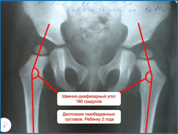
Radiographs will show whether the deformity is unilateral or bilateral. After the X-ray is taken, an instrumental assessment is carried out. With the help of a protractor and a ruler, special measurements are made in the picture. And based on the results, a diagnosis is made.
Clinical diagnosis is not as easy as it seems, therefore, diagnostic procedures must be carried out by a specialist. X-rays are taken in children over 3 months of age and are the definitive method for detecting dysplasia. At an earlier age, the skeletal system is not yet fully developed, it consists of cartilages, which are difficult to fix on an x-ray.
Ultrasound
An orthopedic traumatologist examines a child at 3 months of age and upon reaching 1 year of age. If the likelihood of dislocation is noticed, a certain "click" when abducting the hip, different lengths of the legs, dysplasia, an ultrasound examination is prescribed. The procedure is harmless for young children, it allows you to see early signs and reveal the pathology of the hip joint.
Ultrasound helps the doctor see the joint from different angles, rather than the only two-dimensional image that an X-ray provides. An ultrasound scan can accurately show the position of the ball in the socket, and the doctor can load the hip during the exam to determine the stability of the joint. Ultrasound can also show cartilage in babies who still have little bone.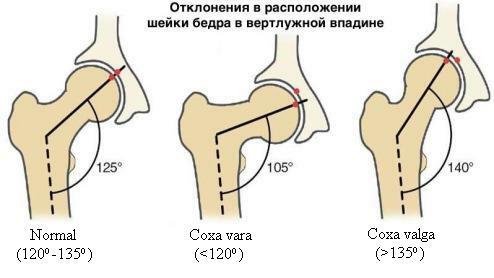
It makes no sense to carry out an ultrasound scan up to 4 weeks, since some pathological features of the bone structures of the hip joint can disappear on their own, during the first month of life child.
The cost of diagnostic procedures:
| X-ray | From 1 thousand 900 to 2 thous. RUB 300 |
| Joint ultrasound | From 1 thousand 500 to 3 thous. rub. |
| MRI of the hip | From 6 thousand rub. |
| Selection of corrective agents for dysplasia | From 1 thousand RUB 300 |
Treatment methods
Coxa valga in children is not necessarily a medical condition requiring treatment if the acetabulum is adequately developed for the age and constitution of the patient. Femoral neck valgus with good development of the acetabulum is common and does not lead to secondary osteoarthritis later in life. Timely examination of the child and diagnostics will make it possible to establish violations in time.
The purpose of treatment at Coxa valga:
- fix the head of the femur in the acetabulum;
- to strengthen the articular ligaments and muscles to exclude too much bone mobility.
If a hip joint disorder is diagnosed in early infancy, the problem is often solved with a padded brace. Surgery may be required for older children and young adults to move the bones into the correct position for smooth movement of the joints.
Conservative
There are many methods of hip joint correction in children. The child's age and the severity of symptoms will determine the best course of treatment.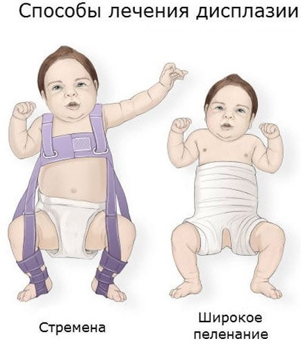
| Physiotherapy | Ozokerite applications, mud therapy, electrophoresis with calcium chloride. |
| Exercise therapy | A set of special exercises depending on the age of the child, swimming, especially on the stomach. |
| Massage | Strengthens the muscular region of the buttocks and thighs. |
| Pavlik's tourniquet | Used for children under 6 months of age. This is a chest strap, 2 shoulder straps and 2 stirrups made of canvas, Velcro and buckles. The design is offered to the child to be worn 24 hours a day for 6-12 weeks. For babies with dislocated hips, the harness can be worn for a shorter period. The success rate of using the Pavlik harness ranges from 85 to 95% for infants under 6 months of age. |
| Closed repositioning of dislocation | Rigid fixation of the hip joint with a coxitis bandage after reduction of the dislocation. It is possible to apply a plaster cast. |
Wide swaddling is often used as an additional method of prevention. Doctors recommend limiting physical activity to such children.
Surgical
Coxa Valga in children is not always treated with surgery. However, the more serious the condition becomes, and the problem is not paid attention to, the faster it can lead to immobility and shortening of the leg.
Surgical options:
Soft tissue surgery or open reduction
Surgical option for children over 2 years of age at the time of the first diagnosis, or for those who have failed to correct the joint by closed reduction. In open reduction, the surgeon cuts the hip capsule and changes the position of the femoral head. After suturing the thigh, the bandage is applied for 4 months or longer to stabilize the thigh. Open reduction and other soft tissue surgeries such as tendon lengthening are usually only possible in infants and very young children.
Surgical removal of the tendon or tenotomy
The intervention ensures correct insertion of the femoral head into the acetabulum. After the operation, a plaster cast is applied for 1.5 months. After 2 months, an X-ray is taken, and in case of successful healing, the plaster cast is removed. If the joint is still unstable, the bandage is reapplied.
Varus osteotomy
Treatment for extreme cases is varus osteotomy, also called femoral osteotomy, in which an orthopedic surgeon cuts the bone and moves it. In this procedure, a wedge-shaped piece of bone is cut out on the inner side of the lower leg of the thigh. This smoothes the angle of the femoral neck and, accordingly, normalizes it.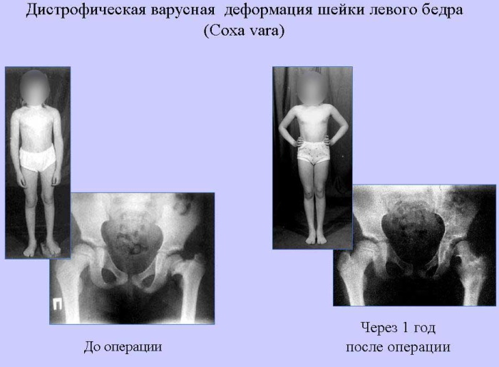
As a result of the operation, the legs are shortened by 1-2 centimeters. Therefore, the victim must wear suitable orthopedic insoles after surgery to compensate for this imbalance. When planning a surgical correction, it should also be borne in mind that the angle of the femoral neck still tends to flatten as it grows.
Endoprosthetics
During the operation, the affected joint is completely replaced with an artificial prosthesis. Osteotomy carries risks such as adverse reactions to anesthesia and infection, and is highly invasive. As a result, surgery is only recommended when the patient clearly requires surgery. Sometimes operations cause complications that can cause severe pain or are fatal.
The goal of treatment for hip dysplasia in infancy and childhood is to prevent subsequent disorders and pathological conditions as the child grows into adolescence and adolescence age.
Possible consequences and complications
If hip dysplasia is not treated or diagnosed properly early on, there is the possibility of some long-term problems.
- In the long term, there is a risk of premature joint wear.
- Adolescents can experience hip pain and discomfort, and many young people develop osteoarthritis of the hip early. Untreated hip dysplasia is the most common cause of early-onset hip arthritis in young women.
- In addition, in case of displacement of the femoral neck, deformity in the knees or crooked legs develops compensatory.
- In most cases, the pathology leads to deformity of the legs. The legs can also be shortened, which can lead to significant limitation of movement and serious complications in daily life.
- This disease significantly reduces the quality of life. The patient suffers from severe pain in the thigh area. They can take the form of rest pain or pressure pain. The muscles in the affected areas weaken and physical activity becomes impossible.
- Sports activities are strictly limited. In the future, excessive stress can lead to a fracture of the femur or sprain. In most cases, treatment with Coxa valga leads to a positive course of the disease, and the symptoms are relieved relatively well.
The deviation is often discovered by accident. In most cases, this has practically no pathological significance. However, an excessively large angle of the femoral neck leads to a change in the load in the joint area. Increased pressure is exerted on the sensitive articular cartilage. As a result, the articular cartilage cannot be supplied with nutrients at this stage. In old age, you are more likely to develop premature osteoarthritis. Coxa valga is one of the diseases called "pre-arthritic deformity".
Coxa valga in children is not a disease per se, but a condition that often goes unnoticed. The intervention of the doctor is mandatory and the appointment cannot be postponed.
Video about Coxa valga in children
Komarovsky about hip dysplasia: