Content
- Etiology of oral leukoplakia and pathogenesis
- Reasons for development
- Smoking
- Spicy, cold, hot food
- Alcoholic drinks
- Mechanical injury
- Galvanic currents
- Adverse effects of occupational and environmental factors
- Taking medications
- Other provocative aspects
- Types and clinical manifestations, symptoms
- Flat leukoplakia
- Verrucous
- Erosive and ulcerative
- Soft
- Simple form
- Smokers' leukoplakia
- Diagnostics
- Treatment of oral leukoplakia
- Drug therapy
- Surgical intervention
- Traditional methods of treatment
- Disease prognosis
- Video about oral leukoplakia
The mucous membrane of the oral cavity can suffer from the effects of irritating factors of an internal and external nature. Most leukoplakia is considered a dangerous pathological condition. The disease is manifested by keratinization of the oral mucosa, which should normally be soft and non-inflamed. External signs are clearly visible visually, in the photo they look like white spots without clear edges or grayish-yellow plaques of a scaly structure. The disease refers to precancerous conditions, requires urgent treatment based on the results of accurate diagnostic data.
Etiology of oral leukoplakia and pathogenesis
Pathology is a chronic disease of the mucous membranes, and changes in the epithelium can spread not only to the oral cavity, sometimes the mucous membrane of the intestine, sinuses, urinary bubble. Most often, doctors diagnose leukoplakia of the oral cavity, the epithelial layer of which undergoes keratinization (hyperkeratosis). Moreover, signs of hyperkeratosis of the tongue are much less common.
The disease is recognized by characteristic plaques without clear boundaries, they are clearly visible on the surface of the tongue and cheeks, affect the palate, corners of the mouth, affect the border of the lips. The formation of keratinized areas can last a week or months, however, the exact mechanism of the development of leukoplakia has not yet been established. Leukoplakia of the oral mucosa mainly affects men who smoke after 40 years, in women, pathology is extremely rare.
The formation of changes on the mucous membranes of the oral cavity starts as a result of constant exposure to a source of irritation, which contributes to the development of chronic inflammation. Under the influence of a provoking factor, complex mechanisms are triggered that change metabolism, and the progressive inflammation of the epithelial layer leads to a thickening of the oral mucosa (keratosis).
The primary symptoms of leukoplakia, which are not particularly pronounced, include:
- areas of incipient keratinization, which have a gray tint;
- the appearance on plaques of an easily removable whitish plaque;
- sensations of foci of compaction on different parts of the mucous membrane.
The development of the disease is rarely accompanied by painful symptoms, so its onset often goes unnoticed, only dryness in the places of keratinization worries. The result of the progression of pathology is a change in the color of the spots - for the most part they turn white and acquire clear outlines.
At the next stage, the formation of erosions and cracks occurs, signaling pain, burning, tingling, fluid secretion. The further course of the disease is accompanied by increased sensitivity during eating, swallowing, the patient's temperature rises, the person becomes irritable, suffers from an unpleasant smell from mouth.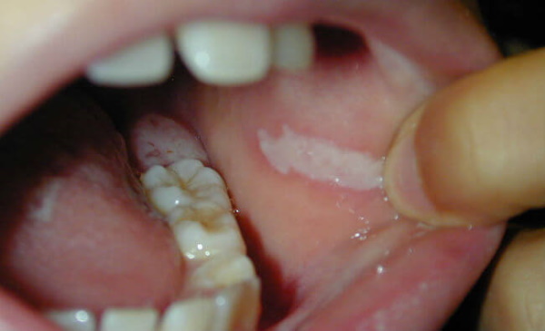
Leukoplakia of the oral cavity (the photo clearly demonstrates its most important symptom - white spots) a month after the initial phase is manifested by plaques, the diameter of which does not exceed 4 cm. They look like bumps without compaction, but over time they lose their shine and become rough. Further transformation of plaques depends on the developing type of pathology. With the onset of severe keratinization, ulcers appear, and the infection process captures other parts of the oral cavity.
Reasons for development
In the medical environment, there is no generally accepted point of view regarding the causal factors that cause the first symptoms of leukoplakia. However, most experts are sure that the most likely cause is the continuous contact of the oral mucosa with constant exposure to an external stimulus. The influence of internal pathologies of the gastrointestinal tract is also not excluded, which contribute to a decrease in the stability of the mucous membranes with a decrease in their protective reaction.
Smoking
The smoking process is accompanied by regular thermal irritation of the mucous membrane. The irritating effect of chemical compounds (resin, phenol, ammonia, nicotine) rapidly destroys the epithelial layer, causing the appearance of nicotinic leukoplakia - Tuppainer's syndrome. Chewing types of tobacco and other plants are no less dangerous.
Spicy, cold, hot food
Food preferences are also provocative aspects. Eating food that is too hot and cold will thermally irritate the mouth. Uncontrolled intake of especially hot spices, salty and sour dishes eventually turns into symptoms of a dangerous disease.
Alcoholic drinks
The use of strong types of alcohol not only burns the mucous membrane, but also contributes to its complete destruction. Therefore, drinkers are also at risk of suffering from their addiction.
Mechanical injury
The epithelial layer can injure the wearing of dental structures (braces or dentures) if they are incorrectly selected. 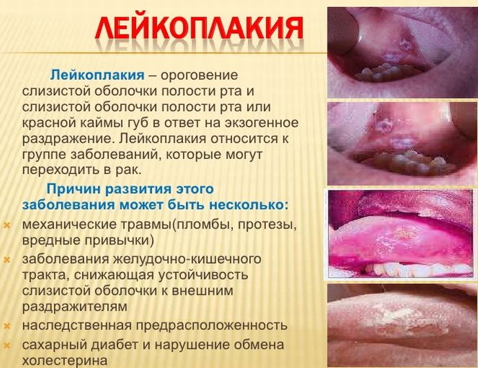 No less dangerous is an incorrect bite, defects in the dentition, when, due to the sharp edges of the teeth, areas of the mucous membrane are damaged. This leads to the appearance of non-healing wounds, the development of allergies.
Galvanic currents
If crowns made of different types of metal are installed in the oral cavity, the occurrence of weak electric currents is not excluded. In this case, saliva becomes an analogue of a galvanic medium, and constant irritation of the mucous membrane turns into its gradual degeneration.
Adverse effects of occupational and environmental factors
Leukoplakia of the oral cavity (photos of characteristic manifestations are given in the article) in some cases becomes the result of living in environmentally unfavorable conditions, when people have to breathe contaminated air. Working with harmful chemicals is accompanied by the inhalation of vapors of aniline dyes, resins, varnishes, gasoline, and other chemical compounds that destroy the inner epithelium.
Taking medications
The appearance of keratinized plaques on the surface of the cheeks, tongue, gums may be associated with long-term therapy with potent drugs. The situation is most common among elderly patients who are forced to take a large number of medications that have many side effects.
Other provocative aspects
In addition to external stimuli, endogenous causes of leukoplakia include metabolic failure, a constant lack of vitamin A, and chronic inflammatory processes. According to the research results, the causal role of hereditary predisposition has been established - in the case of hereditary keratosis, the likelihood of developing leukoplakia is quite high. In addition, the risk of the disease increases in the presence of hypovitaminosis, iron deficiency anemia, diabetes mellitus, hormonal imbalance.
Types and clinical manifestations, symptoms
The initial stage of pathology is rarely signaled by noticeable symptoms, or they can be almost imperceptible. Among the signs that should alert the patient is the appearance of multiple foci of inflammation of different sizes, which are not so easy to notice. Areas of a whitish or grayish hue (plaques) are distinguished by a changed structure - their surface is rough, hard to the touch. However, around the keratinized areas, the mucous membrane looks healthy.
Patients often do not react to the appearance of barely noticeable small plaques, but turn to the dentist when the foci of keratosis increase up to 3-4 cm, pestering especially unpleasant symptoms (pain, taste disturbances, difficulty in chewing, frequent inflammation). The manifestations of the disease can affect any part of the oral cavity, and various types of pathology are signaled by symptoms characteristic of a particular species.
Flat leukoplakia
This form of pathological changes in the epithelial surface is considered the most common option. In most cases, the simple form of the disease proceeds without complaints of discomfort. The dentist detects the disease during a routine examination or during the appointment of therapy for other problems in the mouth.
Keratinization does not differ in particularly striking manifestations, except for the vague outlines of whitish spots. It takes weeks, even months, to form tangible signs.
Mild flattened symptoms have their own characteristics:
- a feeling of dryness and tightness of the surface layer of the mucous membrane;
- signs of slight burning and pressure on the affected areas;
- the appearance of dryness and roughness of spots, clearly marked by contours.
Leukoplakia of this type has a characteristic feature. Disease of the oral cavity is recognized by whitish plaques with unevenly opaque epithelium. The photo shows foci, edged with a fairly clear border. The area of the affected tissue does not yet rise above the surface of the mucous layer.
Verrucous
Leukoplakia of this type can be called the next stage of hyperkeratosis, a continuation of the flat variety in the absence of treatment and elimination of the provoking factor. Symptoms of keratinization become more pronounced, the upper layer of the epithelium becomes more dense, and the lesions are already rising above the line of the surrounding tissues. If you feel the plaques, they are quite hard and slightly mobile, but in the absence of painful sensations.
Verrucous leukoplakia of the oral cavity is classified in two forms:
| Form | Main characteristics |
| Plaque option | Multiple foci of keratosis are in the form of irregular grayish-white plaques with a rough surface. The form of leukoplakia is not very common. |
| Verrucous variant | Dense foci of reborn tissue of white or yellow color look like bumpy growths. This variant of the disease is diagnosed much more often. |
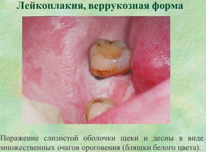 With confirmed pathology of the verrucous type, the results of a histological examination are always required, since the disease is considered the most threatening type of hyperkeratosis. This form is characterized by a high degree of malignancy due to constant trauma to the areas affected by keratosis. The main complaints of patients suffering from the verrucous type of leukoplakia are painful sensations, severe burning sensation accompanying food intake, as well as tightness of the affected areas that have become rough.
Erosive and ulcerative
At the next stage of development, the disease turns into an erosive form, followed by the appearance of ulcerative areas. Ulceration of the mucous membrane forms a severe pain syndrome not only while eating, but also at rest. Traces of erosion and the appearance of cracks, especially bleeding ones, are difficult to treat, and short periods of remission are followed by frequent relapses. The progression of non-healing erosions also ends in malignancy, which is manifested by a sudden compaction (usually one-sided) of the base of the plaque.
Soft
This is a special type of leukoplakia of the oral mucosa, which belongs to benign formations. The leading causes of the disease are considered the influence of neuro-emotional overexcitation and overwork, the habit of biting or licking lips. The disease is characterized by the ability to exfoliate the upper membrane of the affected area, which allows the whitish coating to be easily removed with a spatula. Most often, the course of mild leukoplakia is asymptomatic, not accompanied by inflammation, sometimes patients complain of a change in taste sensations, a feeling of tissue exfoliation.
Simple form
Leukoplakia of the oral cavity (photos of the signs of the disease are given in the article for reference and cannot serve as a self-diagnosis) may accompany lichen planus and lupus erythematosus, then the pathology of the mucous membrane is classified as false leukoplakia. However, the term "simple form" means a flat type of keratosis, which does not bother with unpleasant symptoms, visually manifests itself only as clouding of the epithelium. With leukoplakia of this type, the patient can live to a ripe old age, provided there are no factors that provoke the progress of the disease.
Smokers' leukoplakia
A type of pathology is called Tuppainer's syndrome, its symptoms are diagnosed mainly in men who smoke 10 or more cigarettes per day. Focuses of keratinization of the mucous membrane spread over the soft and hard palate, and can affect the surface of the gums.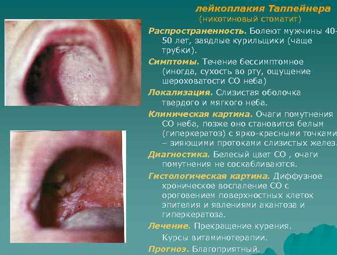
Among the characteristic symptoms are the following manifestations:
- folding of the mucous membranes in the mouth;
- the appearance of a bluish or gray tint;
- blockage of the salivary glands provokes the formation of reddish nodules.
Smoker's leukoplakia is accompanied by signs of an inflammatory process, stagnation due to the accumulation of exudate in the tissues. However, the eradication of a bad habit usually leads to the disappearance of symptoms and the disease itself.
Diagnostics
An in-depth diagnosis of the disease is preceded by a visual examination of the dentist with a clarification of the general state of health, the duration of the appearance of disturbing symptoms, and the identification of risk factors. Then the doctor will need the results of a biopsy, smear for oncocytology.
Due to the specific symptoms, making a general diagnosis is not difficult. Difficulties begin at the stage of precise determination of the form of leukoplakia, its differentiation from other systemic pathologies.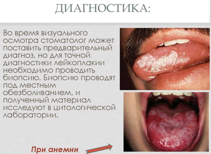
| Type of leukoplakia | Differences from other diseases |
| Flat shape | Chronic lupus erythematosus affects the skin of the face and ears with bright rashes, and the erythema of leukoplakia is characterized by a pale shade. |
| They differentiate from the mild form by the presence of focal zones of a whitish color, looseness of the mucous membrane, traces of abrasions from the habit of biting certain sections of the epithelium. | |
| Hyperkeratosis of the red border of the lips differs from flat leukoplakia by scaly compaction of the surface of the lower lip, limited by a white roller. | |
| Verrucous form | The manifestations of hyperkeratotic lichen planus are not continuous lumpy areas of keratinization of a bizarre shape, as in verrucous leukoplakia. |
| In the hyperplastic variant, the detachment of grayish-white films tightly adhered to the oral mucosa is accompanied by bleeding. Verrucous plaque does not lend itself even to intensive scraping. | |
| Erosive and ulcerative leukoplakia | Pathology should be differentiated from a similar form of lichen planus, manifested by a nodular rash around ulcerated foci. |
| Papules of the secondary form of syphilis are looser than with leukoplakia. Scraping off the plaque of syphilitic papules exposes erosive areas. |
Oral leukoplakia (photos of a progressive form are presented later in the article) is accompanied by a gradual malignancy (malignancy) with traces of abundantly overgrown and hardened foci, the presence of bleeding ulcerated areas. Therefore, when differentiating leukoplakia from Bowen's disease (a variant of intra-epithelial cancer), attention should be paid to the absence of many rashes. A sharply limited focus is always the only one, covered with a grayish-white bloom, and the removal of plaque exposes a velvety red surface.
Treatment of oral leukoplakia
Therapeutic measures to restore the oral mucosa are carried out not only by conservative, but by operational methods. The choice of tactics depends on the specific form of leukoplakia, as well as the stage of the inflammatory process. The size of the areas of hyperkeratosis, the rate of its progression and the characteristic features of the manifestations of the disease are taken into account.
Drug therapy
After quitting smoking, correcting established dental processes, performing restoration of dental units a number of it is necessary to treat the underlying disease, possibly provoking the start of the process of keratinization of the mucous membrane mouth.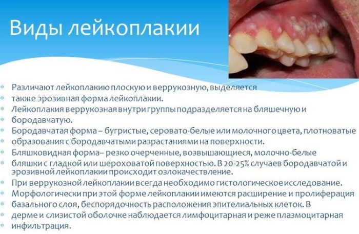
The conservative therapy protocol includes the appointment of the following drugs:
- for the regeneration of the mucous membrane of the affected areas, they are often and abundantly lubricated with oil solutions enriched with vitamin A;
- in order to exclude the spread of the inflammatory process through the tissues of the oral cavity, the affected epithelium is treated with antiseptics;
- relief of painful sensations is performed with systemic anesthetics or with the application of local anesthetics.
As additional methods of treatment, sessions of cryotherapy, laser coagulation, as well as taking vitamin and immunity-enhancing drugs are prescribed. Self-medication of any type of leukoplakia is unacceptable, since the action of some anti-inflammatory medications can provoke malignancy of the plaques.
Surgical intervention
Conservative treatment methods are relevant for combating the manifestations of mild as well as simple forms of leukoplakia. The symptoms of an erosive or verrucous course of pathology are eliminated surgically in a hospital setting. The lesions are excised, followed by sending the removed material for histological control.
Traditional methods of treatment
Home remedies can be used only with the permission of the attending physician, alternative therapy is only assistance to the treatment program prescribed by the dentist.
What is allowed to apply:
- rinsing the mouth with green tea, decoctions of anti-inflammatory herbs that have an antiseptic effect;
- lubricating the affected areas with sea buckthorn or rosehip oil, you can use olive oil;
- foci of the erosive-ulcerative stage can be lubricated with Kalanchoe juice, the plant has bactericidal and healing properties.
In addition to traditional and home methods of dealing with pathology, the diet should be adjusted by enhancing it with vegetables and fruits, fiber-rich foods. Thorough oral hygiene, maintaining the normal functioning of the gastrointestinal tract, and taking medications that strengthen the body are equally important.
Disease prognosis
The primary task of therapy is to eliminate the main irritant factor, the causes of injury to the epithelial layer, then some types of pathology go away on their own. 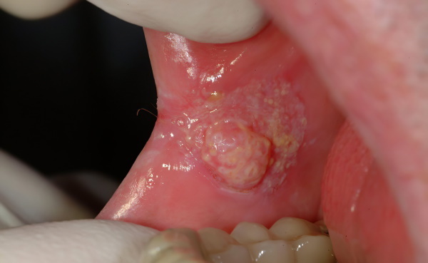 Maintaining a healthy lifestyle, following the recommendations of the attending physician, constant monitoring of the health of the oral cavity will allow you to get rid of the symptoms of leukoplakia forever. At the first symptoms of tissue malignancy, as shown in the photo, it is necessary to urgently remove the lesions in order to stop the spread of the process.
Video about oral leukoplakia
Malysheva about oral leukoplakia: