Content
- What is echocardiography of the heart and the purpose of its conduct
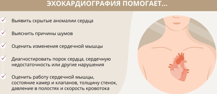
- What abnormalities does echocardiography help to identify?
- Diagnostic value of the study
- Types of echocardiography, study parameters
- Transthoracic ECHO-KG
- Stress echocardiography
- Transesophageal ECHO-KG (PE ECHO)
- ECHO-KG with contrast
- Norms of indicators in adults
- Normal Valve Values
- Cardiac imaging rates
- Decoding the results
- What do deviations from the norm say?
- With heart failure
- In case of heart rhythm disturbances
- Echocardiography to assess the condition of cancer patients
- Comparative characteristics of the information content of echocardiography with other diagnostic methods
- Echocardiography video
Echocardiography is considered the leading method of hardware diagnostics for examining the heart. The procedure is used to assess the structural, tissue, functional parameters of an organ in adults and children. Deciphering the results gives an important treatment and prevention cardiac pathologies information.
What is echocardiography of the heart and the purpose of its conduct
Ultrasound diagnostics reveals functional and morphological changes in the structure of the organ, valve mechanism. The technology is based on the capture and reflection of the signal generated by the equipment by the cardiac tissues.
As a result, an image of the organ structure is formed on the monitor screen. The procedure is performed using high frequency ultrasonic waves and sensitive sensors that perceive the reflected signal. The examination allows you to get a detailed image of blood vessels, cavities, muscle fibers.
Decoding of echocardiography of the heart in adults or children is needed to make a definitive diagnosis.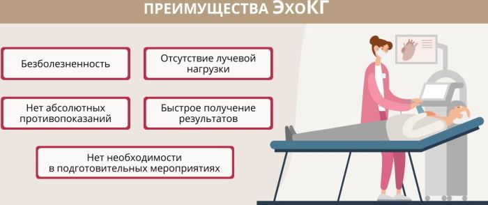
Objectives of the survey:
- quantitative and qualitative assessment of the functional state of the ventricular apparatus;
- determination of regional LV contractility in patients with ischemic syndrome;
- identification of echo signs of atrial hypertrophy;
- calculation of the mass of the myocardium for intrauterine or genetic disorders;
- identification of dilatation (increase in the volume of internal space) of the functional divisions and chambers of the organ;
- elucidation of the degree of morphological changes in the valve mechanism with stenosis, functional insufficiency, prolapse, leaflet vegetation;
- assessment of blood pressure in pulmonary hypertension;
Ultrasound examination is performed to detect pathological changes in the structure of the pericardium. The diagnostic purposes of the technique include the determination of the volume of liquid exudate in the cavities.
What abnormalities does echocardiography help to identify?
The procedure detects intracardiac pathological formations - thrombotic clots, tumor processes, additional abnormal chords.
Abnormalities diagnosed by echocardiographic examination:
- postinfarction cardiosclerosis is a form of ischemia with cicatricial pinching of myocardial muscle fibers;
- dilated or hypertrophic cardiomyopathy, accompanied by uneven lesions of the right or left ventricular section;
- local innervation disorders - bundle branch block, WPW syndrome;
- dysfunction of the interventricular septum;
- systolic disorders;
- insufficiency of the tricuspid valve element;
- dysplasia of arrhythmic genesis of the right ventricular section with replacement of muscle fibers by fibrous-fatty layer;
- fusion of valve cusps.
ECHOKG is used to detect functional and morphological changes in the main or peripheral blood vessels. The wave method is used for hardware auscultation of heart murmurs.
Diagnostic value of the study
ECHOKG is a subjective and operator-dependent procedure. Measurements of cardiac parameters are carried out in various ways that affect the result. The diagnostic value of ultrasound examination depends on the interpretation of the findings by the cardiologist.
Conceived echocardiography is not sufficient to guide the selection of therapeutic strategies. The value of the study is increased by the combination with other instrumental technologies - ECG, X-ray, coronary angiography.
An integrated approach allows you to compile a more complete and reliable clinical picture. Echocardiography is considered the fastest and most affordable way to visualize the heart apparatus.
The technique allows you to obtain important clinical data for:
- Systemic lupus erythematosus. The picture shows a hydropericardium - an accumulation of non-inflammatory transudate in the cavities of the organ. There are destructive changes in the structure of the valve tissues.
- Scleroderma. Echocardiography is prescribed to determine pathological dynamics and identify complications.
- Combined morphological disorders in the connective tissue structures. The diagnostic value of echocardiography is to identify exudative pericardium, valvular dysfunctions.
- Periarteritis nodosa. Dilatation changes in the heart chambers, defects of the valve elements are noticeable.
Decoding of echocardiography of the heart in adults has diagnostic value in pre-infarction, aortic aneurysm, ischemic damage. To increase the reliability of the results, the ultrasound procedure is combined with ECG diagnostics.
Types of echocardiography, study parameters
Apply transthoracic, stress, transesophageal echocardiography. Ultrasound of the cardiac apparatus with intravenous administration of a radiopaque contrast agent is allocated to a separate category.
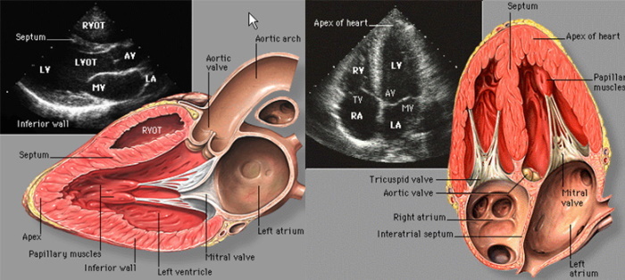
Transthoracic ECHO-KG
This examination uses standard ultrasound transducers attached to the skin of the chest. Transthoracic ECHO-KG is widespread due to the ease of implementation, the availability of equipment.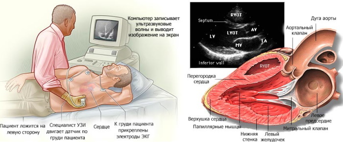
The procedure is difficult if the patient has:
- obstructive pulmonary disease;
- inflammation of the respiratory tract;
- bronchial asthma;
- acute functional heart failure.
The technique provides a relatively high degree of visualization of cardiovascular structures, which makes it possible to recognize dilated or hypertrophic symptoms.
Stress echocardiography
It is used to assess the functional parameters of the heart in conditions of increased activity. Physical activity is created using an exercise bike or simulated with a pharmacological drug.
The first option is preferable because of the physiological naturalness, minimal risk of side effects or allergic manifestations. Non-invasive transthoracic diagnostics through the chest are often performed prior to stress testing.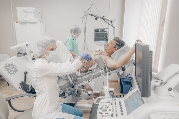
In debilitated patients, physical activity or pharmacological imitation can cause:
- stitching pains in the region of the heart;
- shock state;
- arrhythmic symptoms;
- loss of consciousness;
- vestibular disorders;
- dizziness.
The procedure is carried out with the participation of a resuscitator. The listed negative effects are rare and are quickly eliminated with medication.
Transesophageal ECHO-KG (PE ECHO)
This type of echocardiographic examination involves the use of an endoscope of a special design with an ultrasonic sensor attached to the working end of a flexible tube of small diameter.
Due to its invasive nature, the procedure has a number of absolute contraindications and situational restrictions. PE ECHO is used less often than other types of ultrasound diagnostics of the heart due to technological complexity.
The endoscope is lowered into the esophageal canal through the oral cavity to a predetermined level. The sensitive sensor is pressed tightly against the wall of the cavity. CHEE is prescribed when it is impossible to use another variant of echocardiographic examination.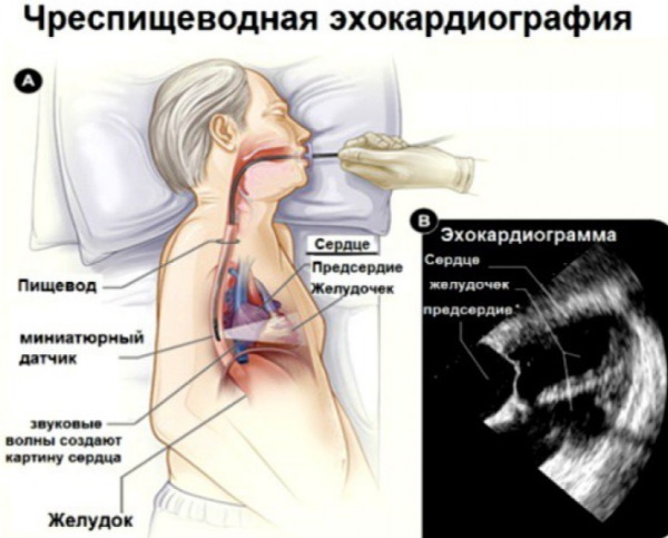
Such diagnostics is effective in the presence of a thick fat layer in the studied area, extinguishing and scattering the ultrasonic signal. The transesophageal procedure more accurately identifies defects in the interventricular septa, atrial sites, and thrombotic disorders not otherwise visualized.
Deciphering of the results of heart examination in adults is carried out by a cardiologist, in children - by a pediatrician. Transesophageal echocardiography is considered a mandatory procedure before using electrical impulse therapy - restoring rhythm using a low-power defibrillator discharge.
Contraindications to ECHO emergency:
- severe or complicated by other pathological processes of the patient's condition;
- inflammation of the esophageal canal;
- malignant or benign formations in the area of introduction of the endoscope;
- stricture (abnormal narrowing) of the cavity;
- diverticula - congenital or acquired deformities of the walls of the esophageal passage;
- varicose veins in the area of diagnostic manipulation;
- esophagitis - inflammation of the epithelium of the esophageal canal.
The procedure does not cause complications. Cases of perforated damage to the walls of the cavity or vocal cords by an inaccurately inserted endoscope are extremely rare. Short-term arrhythmic manifestations are possible.
ECHO-KG with contrast
This examination is performed using the SonoVue preparation, which is a gaseous mixture with microscopic bubbles. Sometimes a neutral saline solution is used as a contrast agent.
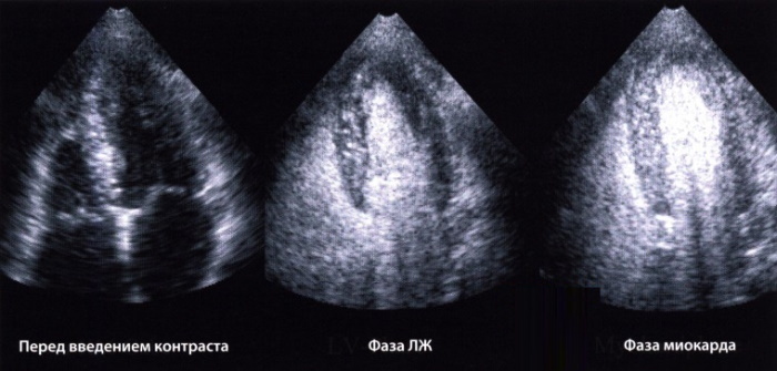
Ultrasonic waves have the ability to be reflected from the boundary of media of different densities. The method provides high-definition visualization. It allows you to determine the size of the hollow parts of the heart apparatus as accurately as possible, to study in detail the defects of the interventricular membranes.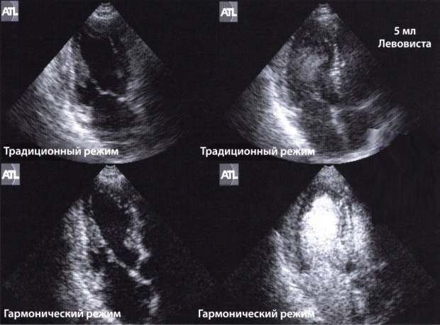
Contrasting is used to study physiological properties, morphological parameters, anatomical structure of the myocardium. This echocardiographic examination is performed with access through the chest or esophagus.
ECHO-KG with the introduction of a radiopaque substance is prescribed as part of the diagnosis of congenital defects cardiac apparatus, to confirm ischemic syndrome with pinched or ruptured tissues myocardium.
Contraindications to the examination:
- acute coronary insufficiency syndrome;
- decompensated angina pectoris;
- recent myocardial infarction;
- severe stage of pulmonary hypertension;
- chronic pathologies of the central nervous system;
- unstable and poorly controlled by medications, increased intravascular pressure;
- connecting the patient to a ventilator;
- period of gestation and breastfeeding;
- a registered anaphylactic response to the constituents of a radiopaque contrast agent.
Side effects or complications of the diagnostic procedure are limited to short-term skin reactions in the puncture area for drug administration. There is slight redness, intradermal hematoma, slight swelling.
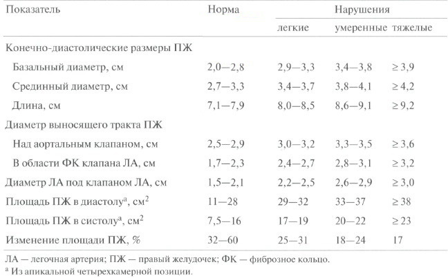
Norms of indicators in adults
The anatomical and physiological parameters recorded by echocardiographic examination differ depending on age, gender, and the clinical state of the cardiac mechanism. Reference values for adults are shown in the table.
| Parameter | Standard for men | Indicator for women |
| End-diastolic value of the ventricle | ≤ 59 mm | ≤ 53 mm |
| Ventricular mass | 135-182 g | 95-140 g |
| Intraventricular cavity volume | ≤ 155 ml | ≤ 104 ml |
| The thickness of the walls of the ventricles during contraction | 10-16 mm | 8-14 mm |
| Ventricular wall thickness during relaxation | 8-11 mm | 6-9 mm |
| Blood ejection fraction | ≥55% | ≥53% |
| Stroke volume of hematological fluid | 45-100% | 45-100% |
| The volume of blood pushed out per stroke | 60-65 ml | 55-60 ml |
| Aortic ring diameter | 20-40 mm | 18-38 mm |
| Atrial wall thickness | 19-40 mm | 17-38 mm |
 Ultrasound diagnostics determines the mass index of the walls of the left ventricular section. The indicator is the ratio of weight to body surface area of the patient. For men, the reference value is 71-95 g / m22, for women 70-90 g / m2.
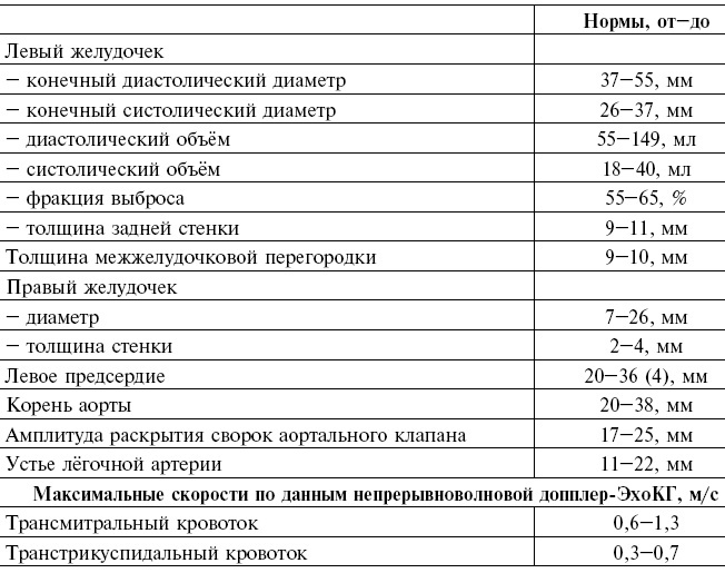
Normal Valve Values
Interpretation of echocardiography of the heart in adults normally demonstrates the absence of valvular insufficiency and complete closure of the leaflets. Their discrepancy by 2-3 mm is considered a pathological deviation.
With such data, the I degree of valvular insufficiency is diagnosed, the 4th corresponds to a value over 9 mm. Stenotic pathologies recorded by ultrasound examination are common, and the fusion of the valves prevents blood flow.
Reference values of the limiting velocity of movement of hematological fluid in the heart valves:
- mitral - 0.4-1.3 m / s;
- tricuspid - 0.3-1.0 m / s;
- pulmonary - 0.5-1.8 m / s;
- ascending and descending aortic - 0.5-1.5 m / s.
When measuring valve parameters, the atrio-gastric cycles of extrasystoles and the following ectopic contractions, which atypically shorten myocardial fibers, are not taken into account.
Cardiac imaging rates
Contrasting means clarify the echocardiographic parameters of valve elements, increase reproducibility examination results with non-optimal visualization, increase the possibility of correlating data with other methods of equipment diagnostics.
Among other results of echocardiography, the parameters of the interventricular septum are of great diagnostic value. The reference indicator in a calm state is set at 0.75-1.1 cm. The parameter is called diastolic thickness.
Another norm for echocardiographic imaging of the heart is an excursion of the interventricular membrane, reflecting its movement during the myocardial contraction / relaxation cycle. The reference indicator is considered to be 0.5-0.95 cm.
The size of the left atrium is 18.5-33 mm, the size index is 1.45-2.9 cm / m2. Echocardiographic diagnostics allows you to analyze the full cycle of heart contractions - diastolic with systolic. With a heartbeat of 70 beats / min, this parameter is normally 0.85 seconds.
Decoding the results
The data obtained during ultrasound examination are interpreted by the cardiologist according to the established reference values, taking into account:
- the general physiological state of the body;
- the presence of concomitant pathologies;
- the age of the patient;
- gender identity;
- medications taken.

The doctor examines the size of the individual elements of the heart apparatus, their mass, the volume of the internal cavities. The cardiologist pays attention to the placement of parts of the organ at rest and under stress.
Morphological analysis of tissue structures, functional state of valves, hemodynamic parameters is carried out. Only after a thorough study of all factors, comparing the results of echocardiography with the data obtained by other methods, is the final diagnosis made.
In severe or specific cases, a medical council is assembled, which collectively deciphers and interprets the echocardiographic information. To study the rate of distribution of blood through the heart chambers, dopplerometry is additionally used.
What do deviations from the norm say?
Parameters going beyond the reference values indicate different pathological processes, hemodynamic disorders, disorders of contractile activity.
Deviations from the reference indicators are observed when:
- ischemic syndrome;
- heart failure;
- congenital defects;
- myocarditis - inflammation of muscle tissue;
- endocarditis - an inflammatory lesion of the connective tissue structures;
- arrhythmic pathologies;
- preinfarction state.
The discrepancy between the results and the established standards occurs for physiological reasons, with systemic diseases. ECHOKG allows timely detection of tumor processes, cicatricial destruction.
Deciphering echocardiography of the heart in adults reveals abnormalities like WPW syndrome. If necessary, the doctor prescribes additional instrumental examinations and laboratory tests. The tactics of therapy depend on the clinical characteristics of the diagnosed disorder.
With heart failure
Ultrasound examination of the heart determines the causes of CHF. In case of functional organ failure, echocardiographic equipment records a violation of the blood ejection fraction in the structure of the left ventricular cavity.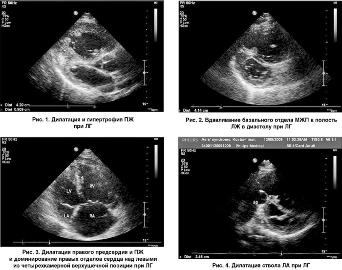
Ultrasound of the heart assesses the parameters of diastolic function, the size of the internal space. In chronic insufficiency, there is a deviation from the norm of the systolic pressure of the pulmonary artery. The reference value is ≥50 mmHg. Art. A liquid exudate is found in the pericardial cavity.
In case of heart rhythm disturbances
Tachycardic dysfunction complicates echocardiographic diagnosis and reduces the reliability of the data obtained. With a rapid heartbeat, transthoracic or transesophageal manipulation is indicated.
The volume of the atria and ventricular cavities, thrombotic formations are of importance. Evaluation of blood ejection fraction in tachycardia is not carried out due to low reliability.
Echocardiography to assess the condition of cancer patients
In malignant neoplasias of any localization, echocardiographic examination records tumor intoxication of the cardiac apparatus. After chemotherapy, traces of the drugs used are found.
Therapeutic radiation leaves signs of cardiotoxic damage, which are established by an ultrasound examination of the heart. The procedure is indicated for cancer patients before starting radiation treatment or chemotherapy, after completing the course.
Comparative characteristics of the information content of echocardiography with other diagnostic methods
For the final diagnosis, lethal clarification of the clinical picture and pathogenesis of the disease, one ultrasound examination of the heart is not enough.
Subjective echocardiographic findings are complemented by:
- Electrocardiogram. The simplest and most widely available option for diagnosing heart disorders. ECG is distinguished by high information content in detecting arrhythmic phenomena, contractile dysfunction.
- X-ray. Used to determine the boundaries and outlines of the organ, diagnose cardiomegaly.
- MRI. The most accurate and informative diagnostic method. Magnetic resonance imaging is used to monitor and verify ECHOKG data, study morphological structure of the organ, diagnostics of myocardial pathologies of non-coronary genesis, cardiomyopathic abnormalities.
- Coronary angiography. Effective for detecting stenosis, thrombosis, and other vascular disorders. Often used in combination with echocardiography.
A rarely used but highly informative method for examining the heart in adults is positron emission tomography. Applied in combination with echocardiography. EPT clearly visualizes areas of insufficient blood supply. Deciphering the results allows you to clarify the clinic of myocardial infarction or ischemia.
Echocardiography video
Echocardiography basics: