
Content
- Characteristics of the circulatory system of the perforating veins
- Functions and properties
- Anatomy and structure
- Possible diseases and pathologies
- Insufficiency of the perforating veins of the lower extremities
- Symptoms and Signs
- Causes
- Treatment methods
- Dilation of perforators
- Symptoms and Signs
- Causes
- Treatment methods
- Venous insufficiency
- Symptoms and Signs
- Causes
- Treatment methods
- Phlebeurysm
- Symptoms and Signs
- Causes
- Treatment methods
- Microbial varicose eczema
- Symptoms and Signs
- Causes
- Treatment methods
- Video about the veins of the lower extremities
Lower limb vein anatomy is divided into 3 systems: deep venous system, superficial and perforating veins.
Characteristics of the circulatory system of the perforating veins
Constant blood flow in all structures is ensured by the system of muscular venous pumps and the work of the bicuspid valves.
Deep veins are located near the center of the legs and are surrounded by muscles and fascia. They are well supported and resistant to dilatation and valvular failure. Therefore, in most patients, the deep veins are normal, which is good, because the deep venous system is responsible for returning 90% of the blood to the lower extremities.
The two main superficial veins, large and small, collect blood from the suprafascial tissues, after which it enters the deep lines. Veins are located under the skin and are not supported by muscles or fasciae, therefore they are prone to dilatation and valvular insufficiency. In most cases, venous insufficiency occurs in the superficial system.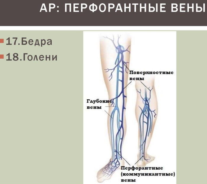
There is also a third structure that forms communication channels between the superficial and deep venous systems. These are perforating (communicating or communicative) veins that connect 2 systems like rungs on a staircase.
The perforating veins of the lower extremities are so called because they perforate the deep fascia of the muscles. On each lower limb of a person, there are many small perforating vessels with different locations, connections, and sizes. There are over 150 of them and they are grouped based on their topography.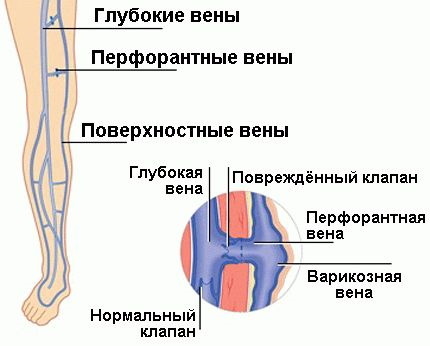
In the area of the lower extremities, a person has perforating veins:
- feet;
- shins;
- knee;
- hips;
- gluteal muscles.
Historically, perforators were classified according to their anatomical location and were named after their founders such as Hunter, Dodd, Boyd, and Cockett. Later studies determined that these veins are anatomically more complex than the original descriptions, and thus were divided into direct and indirect.
Perforated veins contain valves that usually direct blood from superficial veins to deep veins. If these valves fail, high pressure deep venous blood returns to the superficial veins, causing severe superficial venous hypertension.
Functions and properties
Not all of the 150 perforators are clinically important, but some of them can cause serious complications in humans. Clinical significance is observed in 5-10 vessels located mainly on the lower leg. Their role is primary in maintaining proper blood flow through valves that prevent blood from flowing back (reflux) from deep veins to superficial veins.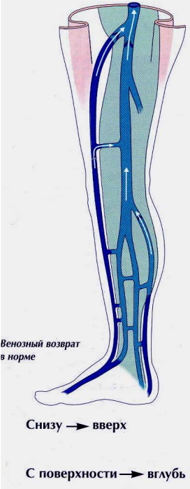
The communicative vessels are located along the entire length of the leg and there are more of them in the calf than in the thigh. They are believed to play a fundamental role in the development of varicose veins.
Anatomy and structure
Perforating veins of the lower extremities straight are the classic vessels connecting the superficial and deep structures, while the indirect ones are more diverse in their location and connection.
Perforators are small in size, rarely exceed 14-15 cm in length, and look like a kind of bridge-steps between the vessels.
Veins are thin-walled expanding tubes that are made up mainly of living cells and their products (including collagen and elastic fibers). The walls are made up of 3 different layers of fabric called tunics.
The outer layer or adventitia passes into a flexible membrane. Below it is the medial (middle) layer and the inner membrane. The inner lining or intima is the last layer.
Adventitia is a skeleton woven from collagen structures and a small number of longitudinally located muscle cells.
Collagen fibers are not involved in maintaining the tone in the internal space of the vein and do not affect motor function. The task of collagen is to maintain the configuration of the veins and preserve them under adverse conditions.
Smooth muscle cells are responsible for vascular turgor, the ability to restore normal shape after deformation. The medina or the intermediate vein sheath is represented by smooth muscle muscles. It is located in a spiral manner along the entire length of the vein. Muscle cells are networked with collagen fibers.
Most perforating veins have one-way valves that regulate the direction of the bloodstream. Thanks to these valves, blood enters the deeper channels and systems. The natural movement of blood flow is centripetal, directed towards the heart.
The valves are formed from two leaflets, which are composed of connective tissue supported by a flexible membrane. When closed, they form folds, since their length exceeds the diametrical size of the vein. Normally, the venous section is 0.1-4 mm. In pathological conditions, the diameter increases to 7-8 mm.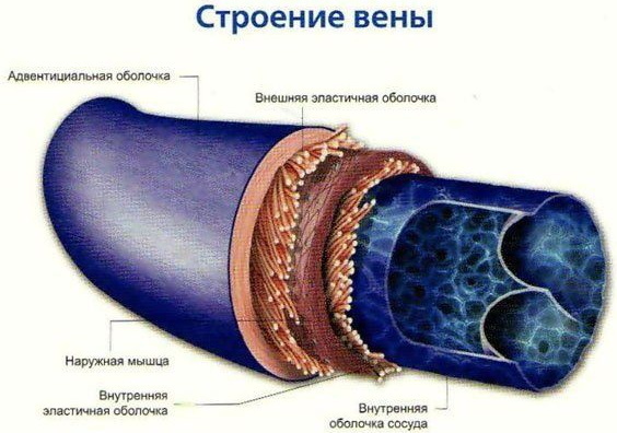
When the valves of the communicative veins become dysfunctional (incompetent), they can cause insufficient blood flow. The resulting reflux causes a rapid deterioration of existing varicose veins and causes the development of venous ulcers.
Perforators located in the foot have no valves.
Possible diseases and pathologies
System dysfunction may result from degeneration of the vein wall, post-thrombotic valve damage, chronic venous obstruction, or muscle pump dysfunction.
The veins of the lower extremities in case of dysfunction of the valves can be excessively filled with blood. Changes in the structure of the valves lead to a volumetric injection of blood into the perforating veins and their stretching. The formation of dilated areas in the absence of valve regulation leads to disruption of the normal flow of blood.
Reverse reflux is formed, which is accompanied by a chain of disturbances in the structure of deep and superficial veins. All this leads to venous congestion, increased venous pressure, valve failure, thrombosis, potential embolization and tissue damage.
The appearance of the first symptoms in the form of heaviness in the legs, burning sensation or increased fatigue is a sign of the disease. A phlebologist is involved in the treatment of the vessels of the lower extremities. Therapy for mild stages is carried out with medication or using compression underwear. Complex cases require surgery.
Insufficiency of the perforating veins of the lower extremities
Usually, the disease develops together with the appearance of varicose veins of the saphenous veins or post-thrombophlebitic syndrome.
Late seeking medical help leads to disability, deterioration of the general condition of the patient and the development of concomitant pathologies.
Symptoms and Signs
The first signals of the disease differ depending on the location and degree of perforator vein failure.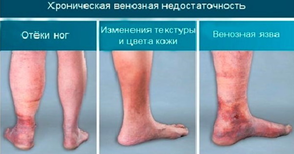
The clinical picture is similar to varicose veins:
- Expansion of the veins. Large, bluish, swollen veins develop around the lower leg and foot. They are painless unless complications arise.
- Leg ulcers. The increased pressure in the superficial veins due to the incompetence of the perforators leads to damage to the skin and tissue, further development of ulcers around the ankles.
- Edema. Backflow of blood into superficial veins can lead to accumulation of blood in the legs and feet. Fluid is forced out of the bloodstream into the surrounding tissues, resulting in edema. Such edema is called "pinpoint", because when pressure is applied to the tumor, traces remain.
- Skin discoloration. It usually occurs around the ankles and is due to venous insufficiency.
- Leg pain. Caused by walking or running, sometimes painful leg cramps can occur for no reason. Both types of pain develop due to poor circulation in the lower extremities.
The final stage is the formation of a necrotic ulcerative defect. At this stage, pronounced itching, a burning sensation, intense pain, heaviness and swelling accompany the patient constantly.
Causes
The perforating veins of the lower extremities sometimes weaken at the point where they join the superficial veins, known as reentry points. This causes the perforating veins to dilate, eventually leading to reflux. The development of the disease is associated with genetic characteristics and lifestyle disorders.
Treatment methods
Instrumental diagnostics includes determination of the lesion site using duplex scanning with color karyotyping. In the absence or insignificant degree of changes, elastic compression is prescribed.
In severe cases, surgery is required. During the operation, the trophic ulcer is closed after ligation of the communicative vein. The endoscopic dressing method can reduce the number of complications and shorten the recovery period.
Dilation of perforators
The development of the disease is associated with valve failure, which leads to disorganization of the blood flow. Dilation is a type of varicose veins.
Symptoms and Signs
The disorder is accompanied by the appearance of a feeling of heaviness, excessive fatigue, burning sensation, distention in the lower extremities. The lesion develops slowly, over time, evening swelling appears.
Causes
The main reason lies in the formation of blood flow reflux. Reflux is an abnormal movement of blood due to improper or defective functioning of the valves.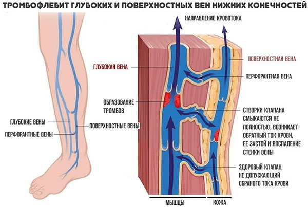
On the cellular structure, a violation occurs at the level of communication between smooth muscle cells and collagen fibers. The increasing blood volume provokes hyperextension of the perforating vessels and the development of pathologically enlarged areas.
Treatment methods
Therapy can be performed both operatively and conservatively.
In milder stages, less invasive treatments are used:
| Compression therapy | It is aimed at increasing vascular tone, decreasing capillary permeability and improving lymphatic drainage. |
| Drug treatment | It leads to a decrease in the diameter of the dilated veins and a decrease in the volume of ballast blood in the extremities. |
| Sclerotherapy | The treatment is carried out with a special apparatus that glues the veins. |
With an average severity of the disease, treatment is carried out surgically. Phlebectomy involves removing the source of reflux and dilated tributaries. Radiofrequency coagulation of veins is performed without incisions and is an effective minimally invasive method.
Venous insufficiency
The disease is a precursor to complete varicose veins.
Symptoms and Signs
Symptoms of venous insufficiency include:
- dull pain, heaviness, or cramps in your legs;
- itching and tingling;
- pain that gets worse when standing;
- edema;
- pain that goes away when the legs are raised;
- thickening and hardening of the skin on the legs and ankles (lipodermatosclerosis);
- slow healing wounds, ulcers on the legs or ankles.
Causes
It develops as a result of primary valve failure or due to constant excess pressure in the saphenous veins. This leads to an increase in blood flow through the perforating vessels and a change in its direction.
The blood flow is directed in the opposite direction, moving from deep veins to superficial ones. A vicious circle is formed: the venous pump of the lower leg is overloaded and loses the rest of its functionality, which leads to an even greater increase in pressure in the superficial veins.
Treatment methods
An operation is performed, during which the intersection and ligation of the retrograde blood flow is carried out. In the initial stages, treatment consists of coagulation, clipping or hardening of damaged areas of perforators. Minimally invasive methods can reduce the patient's recovery time after surgery.
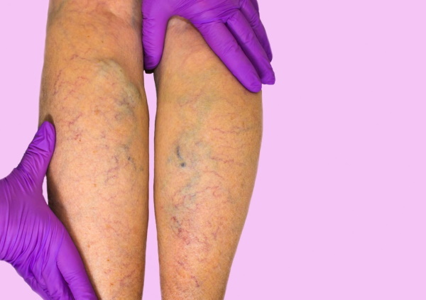
Phlebeurysm
When the valves don't work properly, blood builds up in the veins and increases pressure in the venous system, causing the veins to bloat. A constant increase in venous pressure in the lower extremities can lead to the development of varicose veins. (dilated, elongated and tortuous saphenous veins), especially in the long and short saphenous veins, and their tributaries.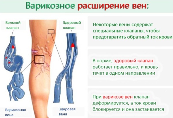
The disease occurs due to the failure of the valves of the superficial, deep or perforating veins. The vessels are in a permanently dilated state, their diameter is> 3 mm, when the person is standing.
Symptoms and Signs
In the initial stage, there is a feeling of heaviness, convulsive sensations and swelling of the lower extremities. Due to the increased fluid content, blood cells begin to penetrate into the subcutaneous tissue, changing the color of the skin.
Areas of compaction and hyperpigmentation (darkening) of the skin are found. As the situation worsens, a disruption in tissue supply leads to stagnation of blood and cell necrosis. As a result, trophic ulcers are formed.
Causes
The trigger mechanism consists in the formation of a return flow of blood due to the failure of the valves. Genetic predisposition or a combination of negative factors (sedentary lifestyle) leads to an imbalance in the proportion of muscle tissue and elastic fibers.
The increasing pressure promotes the formation of lacunae - enlargements - in the perforating veins. As a result, a stable stretching is formed, leading to a violation of the blood supply to the tissues.
Treatment methods
Perforating veins of the lower extremities are treated surgically in case of advanced conditions. Under general anesthesia, the surgeon - phlebologist makes the intersection and removal of the enlarged areas.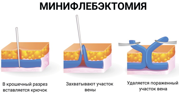
If a disease is detected at an early stage, it is allowed to use minimally invasive methods. Laser coagulation consists in guiding a light guide into the vein and restoring normal blood flow.
Microbial varicose eczema
Varicose eczema is a common skin condition that affects the lower legs. If left untreated, ulcers may form on the skin.
Eczema is commonly seen in middle-aged and older people, but it can also occur in young people if they have a genetic predisposition to varicose veins. This type of eczema is most likely to occur if the person has high blood pressure or varicose veins, or has had deep vein thrombosis, phlebitis, or cellulitis in the past.
Symptoms and Signs
Eczema usually starts on the ankles.
The disease is characterized by:
- swelling that diminishes during sleep;
- itching;
- the appearance of reddish-brown speckled spots on the skin, which become hot and itchy;
- thin and fragile skin around the ankle becomes shiny and flaky.
If left untreated, varicose eczema can lead to open, oozing ulcers and infection.
Causes
Varicose eczema develops when the veins in the legs cannot pump blood back to the heart. Over time, or due to certain factors, including being overweight or pregnant, the valves responsible for sending blood back to the heart weaken, causing blood to collect in the veins. Factors that can contribute to the development of the disease include high blood pressure, kidney failure, heart disease, and a blood clot in the leg.
Both conditions - varicose veins and varicose eczema - fall under the broad category of venous insufficiency. Another side effect of venous insufficiency is not only the bulging of the veins, but also the leakage of blood into the skin.
Treatment methods
The disease recurs if the source is not treated (eg, varicose veins). Therapy is aimed at relieving symptoms. Topical steroids and antihistamines can counteract inflammation and itching. If the eczema has developed into a weeping ulcer, the patient may apply medicated wraps to speed up the healing of the area. In severe cases, a skin graft may be needed to close a large ulcer.
Your doctor may prescribe an antibiotic to prevent a bacterial infection known as cellulitis, which affects the deeper tissues of the skin. People with varicose eczema are also more likely to develop contact dermatitis, in which the skin becomes extremely sensitive to touch.
The best way to prevent eczema is to treat varicose veins. Addressing the underlying cause of venous insufficiency significantly reduces the risk of dry, patchy skin that can lead to ulcers.
Detection of diseases of the lower extremities at the initial stages allows for treatment with minimal intervention. Compression garments, medication and a healthy lifestyle can quickly restore the functionality of the perforating veins. In advanced cases, regeneration is achieved by surgical or minimally invasive intervention.
Video about the veins of the lower extremities
Clinical anatomy of the veins of the lower extremities: