Content
- Features of the location of moles on the body
- How can a mole threaten?
- What moles are dangerous?
- Safe moles
- Pathological moles
- Moles video
The cells in which the pigment is present are called melanocytes. They are responsible for the shade of the skin and its protection from the effects of ultraviolet rays. Based on the description and photo, a small accumulation of cells of this type of tissue acts as a nevus. It is also called a mole.
Features of the location of moles on the body
From 12 to 22 nevi can form on the body, which is an average number. At the same time, heredity can affect this number. However, such raised spots in rare cases do not form with birth, since they are able to form and disappear. A light skin tone increases the risk of acquired nevi formation.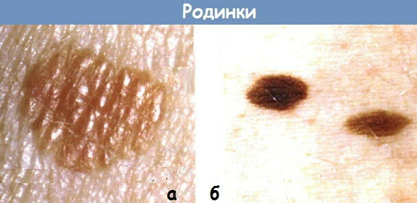
Factors of birthmarks:
- the influence of ultraviolet rays;
- heredity;
- change in hormonal levels in the form of menopause, pregnancy, the consumption of hormonal agents and puberty.
Types of moles (photos with descriptions are given for informational purposes), as well as their placement on the body, are of significant importance. Regarding the location of the neoplasm, it is often permissible to establish the reason for which it manifested itself.
Moles can be located on the following areas of the body:
- Hemangiomas can appear on any part of the body, since their formation will not depend on exposure to ultraviolet radiation. The vessels that provoke the formation of pathology are located throughout the body.
- If nevi hang in large clusters, often located in the armpits or genitals, they are the result of the influence of the papilloma virus.
- Sometimes moles can form on the mucous membranes and on the mucous membranes of the eyes.
- On the chest, cervical and facial areas, as well as on other areas that are exposed to direct sunlight. As a result, the cause of their appearance is considered to be ultraviolet radiation.
The further actions of the specialist will depend on the site of placement of the neoplasm. He can only control the mole or remove it. The decision will be made regarding individual characteristics and a number of factors. At the same time, the placement site is not considered a significant point, which should be assessed by the doctor.
How can a mole threaten?
Types of moles (a photo with a description of nevi will help to establish the risk of danger) can be both safe and pathological. Reborn melanocytes provoke the development of an aggressive malignant tumor in the form of melanoma. The latter is capable of forming on the mucous membrane and skin. Young people are often exposed to this pathology.
Risk factors:
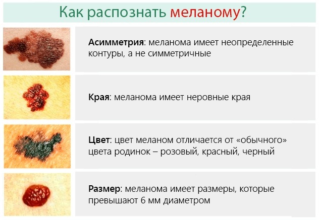
- large pigment spot size (from 6 mm and more);
- hereditary predisposition;
- sunburn;
- traumatic mechanical deformation.
Melanoma can form on its own. However, the risk of developing pathology increases the number, presence and location of nevi.
Skin cancer is not considered the only reason to remove nevi. It is necessary to get rid of them if the neoplasm is regularly susceptible to injury. For example, in the groin area due to regular interaction with underwear, in the cervical area, where the mole is rubbed with the collar, as well as on the head, since it is easily permissible to deform it when combing curls.
It is not recommended to get rid of neoplasms with your own hands. Before removal, an oncologist with a dermatologist should carefully examine the nevus. Based on the results of the analyzes, they can make a decision on removal.
It is permissible to remove neoplasms with:
- laser;
- radio wave coagulation;
- electrical coagulation.
The removal option should be selected exclusively by a specialist. The procedure is often performed under local anesthesia. After surgery, a small scar will remain on the body. The healing phase takes place in a short time period.
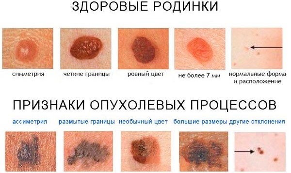
What moles are dangerous?
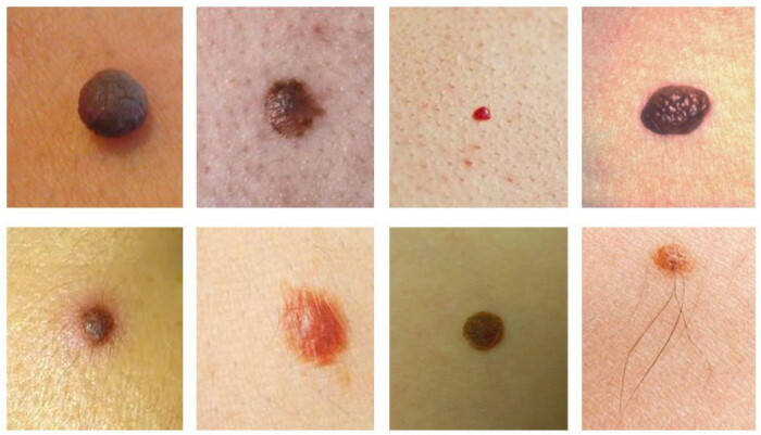
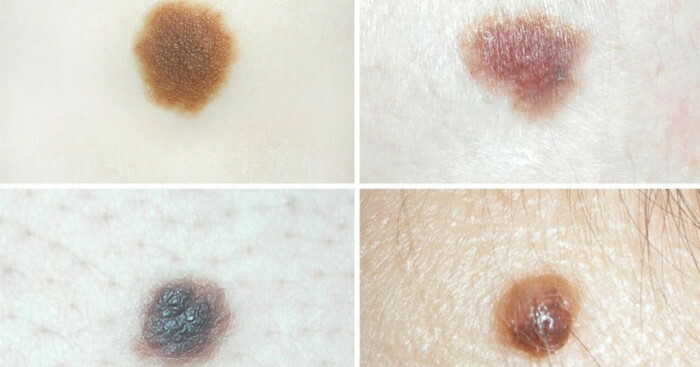
Moles can be very small, convex, or flat. Based on the description and photo, the shade will depend on the volume of melanin: the more there is, the more saturated the color will be. Sometimes there is no pigment, so the nodule will be pinkish or white. If some types of neoplasms that are located in the deep layers of the skin have an intense color, they will have a bluish tint on the surface of the skin.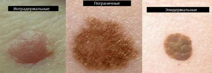
If moles have an irregular shape, uneven shade, spotting or a fuzzy outline, they will be atypical. A large number of such nevi must be monitored, in particular, if there were any malignant skin pathologies in the history of blood relatives.
In rare cases, a mole is confused with a wart, since it looks like a nevus outwardly. Warts are capable of carrying a viral source of origin. However, they are not always cancerous. The exceptions are the mammary glands with the genitals and the perianal zone. At the same time, the formation of papillomas is not a sign of the presence of cancer cells or parasites.
Neoplasms can be safe and pathological. The latter are dangerous and can provoke the development of melanoma.
These include the following types of nevus:
- intradermal pigmented;
- verrucous;
- giant pigmented;
- blue;
- nevus Ota;

- limited precancerous;
- pigmented borderline;
- complex atypical.
Safe varieties include the following:
- "Mongolian spot";
- spindle cell virus;
- halonevus;
- neoplasm from balloon cells;
- papillomatous;
- fibroepithelial.
Any neoplasm needs to be monitored. If it has changed its shade, began to grow, the shape or edges have changed, and itching also occurs, you need to consult a dermatologist.
If a mole is located in an inconvenient place where it is inconvenient to exercise self-control, is regularly deformed or pathological neoplasms have appeared, they must be removed.
Safe moles
Types of moles (a photo with a description will help determine safe types of nevi), which relate to safe neoplasms, after a certain time interval they are able to change the shade with a form. However, this is not dangerous. If the nevus is bulging and located in areas that are potentially traumatic, it must be removed. If a convex mole is ripped off or removed with threads with your own hand, this can provoke the development of oncology.
Safe nevi can be distinguished by a number of signs:
- diameter is not more than 0.5 cm;
- has a slow and measured growth, which is not striking;
- has a type of spot or plaque that is slightly raised above the level of the skin;
- hair grows on the mole;
- has delineated contours;
- the structure of the mole will correspond to the relief of the skin, which is closely located.
A nevus is subject to removal, which:
- located on the pubic area or in the armpit, because there is a risk of injury to the mole with a shaving machine;
- grows very quickly and changes shape;
- when rubbed with tissue matter, it provokes burning, itching and pain;
- brings uncomfortable sensations from the aesthetic side;
- placed in the decollete or neck area, because he is constantly irritated by closed things, a scarf or jewelry.
Safe types of nevus:
| A type | Description |
| Hemangioma | It is formed due to the proliferation of blood vessels, which is of a benign nature. May have pinkish and reddish hues. By the type of growth, the neoplasm can be capillary, venous, arteriovenous or arterial. Often located on the cervical and facial areas. If it begins to grow, it is recommended to get rid of it so as not to provoke injuries.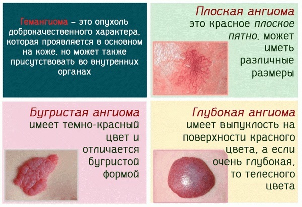
|
| It manifests itself extremely rarely and is presented in the form of a spot or tubercle of brown color, with a thin rim of a yellowish tint. | |
| Lentigo | It is a pigmented, measured flat spot, colored brownish or black. It occurs at the border of the outer and inner layers of the skin. The neoplasm has a darker color than freckles. At the same time, it is not affected by ultraviolet light, and it will not multiply under the rays of the sun. |
| Mongolian spot | It is a birthmark in newborns. It can act as a single or group spot, located on the back, thighs, buttocks or sacrum. It is characterized by a different shade of bluish color. The nevus has a smooth surface and may protrude slightly above the skin. A mole does not need treatment, since it can disappear on its own in childhood. |
| Becker's nevus | The spot on the skin is characterized by high hairiness and hyperpigmentation. It is mainly formed in men. |
| Sebaceous gland nevus | It is a flat convex spot with a rough surface. It can be dominated by different colors of brown. It often manifests itself in children due to the failure of the normal growth of various tissues of the skin. The nevus is formed at the stage of intrauterine formation, appearing on the body 3 months after birth. According to the degree of development of the child, the mole can grow, increase in size and acquire a convex shape. However, it will not develop into a cancerous pathology. This type of nevus can be removed when the child's age reaches puberty. |
| Sotton's neoplasm | It is able to manifest itself and disappear on its own. There is an unpainted ring on the skin around the mole.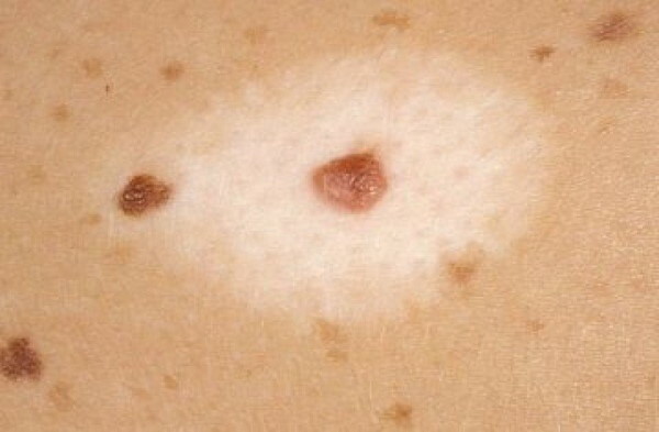
|
| Papillomatous | Outwardly, it looks like a cauliflower or several accrete warts. It has an irregular shape, a bumpy surface, and also rises above the skin. |
| Pink melanocytic | It is a simple epidermal formation. It mainly affects people with fair skin types. Therefore, melanocytes produce a pinkish pigment. |
| Fibroepithelial | The structure is characterized by a large number of connective tissue elements. The nevus has a round, convex shape, as well as a brownish, pinkish or reddish tint. He is able to grow throughout his life and not go into oncology. |
| Epidermal-dermal | It can be colored black, brownish or yellowish. It is presented in the form of a flat mole, which can rise above the skin. Melanin-producing cells are mainly located on the surface. The nevus reaches about 0.5 cm. Often located in intimate places, on the palms and feet. |
Pathological moles
Types of moles (a photo with a description will tell you how to distinguish pathological nevi from safe ones), which are of a pathological nature, can provoke an anxiety state in a person. This is due to the fact that they become a factor in the formation of skin cancer.
These types of nevus must be removed, but this procedure should be carried out by a surgeon or oncologist, drawing up the next therapeutic course of treatment. It is undesirable to remove melanoma-prone neoplasms in cosmetology centers or clinics. An experienced plastic surgeon should prescribe a series of tests if there is a risk of having a malignant growth.
The main symptoms of pathological education:
- active growth, reaching 1 cm in diameter;
- in the area of the mole, swelling with hyperemia appears;
- bleeding occurs;
- intensive change in structure with shape;
- new nodes begin to develop on the surface;
- manifestations are formed on the surface;
- a burning sensation with itching appears on the site of the nevus;
- depigmented zones are formed, and the shade changes partially or completely;
- moisture is separated with exudate;
- the surface peels off and a crust forms;
- the surface is rough or shiny, the skin pattern disappears from the hill;
- asymmetry appears;
- daughter nodules appear;
- hair falls out completely or partially from the surface;
- the texture becomes thin;
- formation at a more mature age.
Varieties of pathological nevi:
-
Verrucous. It is presented in the form of numerous nodules, which are built in a line and are divided by zones of unchanged skin. The shade can range from lighter to darker. Mainly protrudes above the surface. Able to form one nevus or have a systemic shape, looking like a stretching garland.

- Giant pigmented. It acts as a darkish pigmented congenital spot, which increases according to the degree of human growth. The shade can be black, brownish or grayish.
- Blue. A mole can be painted in a bluish or bluish tint, reaching 1 or 3 cm in diameter. Outwardly, it looks like a compacted knot, which grows very slowly.
- Dysplastic. It is considered a pigmented mole, which is presented in the form of a single or group spots that are close to each other. Separate zones may rise above the surface of the skin. At the same time, an uneven tone with uneven contours prevails. The shade can be reddish or reddish. This type of nevus is transmitted at the genetic level.
- Intradermal pigmented. The shade can range from light brown to darker tones. The cells that reproduce melanin begin to accumulate in the deep layers of the skin. A convex neoplasm can be warty, smooth, or hairy. Nevi without hair can progress to the stage of melanoma. These types of moles require more care. The doctor must constantly examine the condition of the nevus, because a non-hairy formation provokes the risk of developing oncology.
- Melanosis Dubreya. It acts as a pigmented flat single spot, in which the uneven contours look like a geographical map. Often focuses on the skin of the facial area, affecting the cheeks with the nose and forehead. It is characterized by a brownish tint. According to the degree of growth of the nevus, the color begins to change. As a result, the birthmark is dominated by an uneven shade, ranging from brown to black, on which there are bluish or grayish areas.
- Non-cellular. It is considered a type of intradermal mole, outwardly resembling a convex small oval. Often manifests itself in adolescents on the cervical or facial area. A nevus is not considered hazardous to health, but it can turn into a malignant stage if it is injured or exposed to prolonged exposure to ultraviolet light.
-
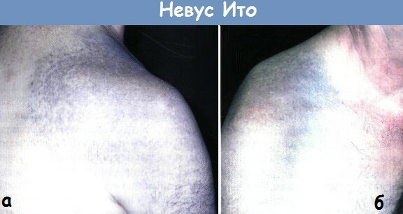
Ito's nevus. Often located on the scapular and supraclavicular area. It acts as a precancerous condition, because it is able to degenerate into oncology.
- Nevus Ota. Focuses on the facial area, located on the cheekbones or along the edge of the eye sockets. It can also manifest itself on the mucous membrane of the pharynx, larynx or oral cavity. Bluish-black color predominates. It is considered a precancerous pathology, because it can turn into oncology.
- Borderline. It has a uniform shade, oval or round type, delineated contours and a smooth surface. Able to rise slightly above skin level. Often, brown, grayish or brownish color predominates.
- Epithelioid. The structure looks like melanoma. It is characterized by a convex, round shape and a brownish-red tint. Has a bumpy or smooth surface. With this type of mole, the risk of developing skin cancer is very high.
After reading the descriptions and photographs of different types of moles, you can protect yourself and your loved ones from a dangerous problem. If you identify such a pathology in time, you can quickly and efficiently deal with it, without harming your health.
Moles video
How to Distinguish Dangerous Moles from Safe Moles: