Right ventricular hypertrophy is a pathological process, accompanied by the growth of the muscle layer of the myocardium. Often the disease is detected during ECG studies and requires surgical treatment.
Record content:
- 1 The mechanism of development of pathology
- 2 Types of hypertrophy
- 3 Causes
- 4 Symptoms and clinical manifestations
- 5 What minor deviations are considered normal?
- 6 Diagnostics
-
7 ECG signs
- 7.1 Emphysematous (s-type)
- 7.2 Blockade (rSR ')
- 7.3 Hypertrophic (qR)
- 7.4 Moderately hypertrophic
- 7.5 RV hypertrophy and dilatation
- 7.6 The predominance and enhancement of the potentials of the right ventricle
-
8 Treatment features
- 8.1 Drugs
- 8.2 Traditional methods
- 8.3 Diet
- 9 Complications
- 10 Forecast
- 11 Video about ECG for ventricular hypertrophy
The mechanism of development of pathology
The mechanism of development of the disease is associated with an increase in blood pressure, which, in turn, causes changes in the bloodstream. The process involves connective tissue, which enters the chambers of the heart with great force. As a result, there is an effect on the mitral valve and the right ventricle, which causes vasoconstriction and leads to an increase in resistance.
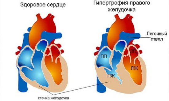
Prolonged stay in this state forces the heart to build up muscle tissue, due to which the number of cardiomyocytes increases. This ultimately leads to the formation of hypertrophic changes.
With the defeat of a separate part, the functional characteristics do not change. With an excessive supply of blood, a dilating state develops, which leads to an increase in the size of the myocardium, vasodilation. A similar phenomenon is often found among those who are involved in active sports or among workers doing hard work.
Types of hypertrophy
The disease is divided into 2 main forms, depending on the mechanism of their development:
-
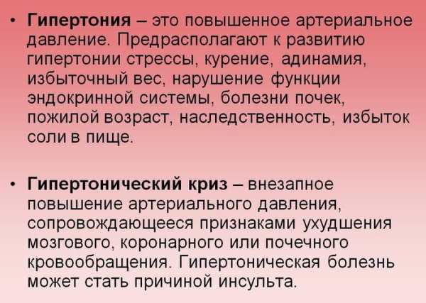
Hypertensive. This type of pathology is associated with an increase in blood pressure, narrowing of blood vessels and arteries. This leads to the fact that blood stops flowing in sufficient quantities and an increase in pressure is required to push it.
- Dilation. The mechanism of development of this type of disease is associated with an increase in the volume of the right chamber of the heart and blood overflow. The process leads to an increase in blood pressure. In the future, the pathology develops in a similar way, as in the first case.
Both in the first and in the second case, the disease requires compulsory treatment.
Causes
Right ventricular hypertrophy (on ECG diagnostics, the pathology is confirmed in the vast majority of cases) can develop for various reasons:
- Pneumonia. Hypertrophy develops against the background of lung damage, which leads to respiratory failure, which provokes an increase in pressure. The process affects all vessels and arteries of the small circle, which causes an increase in pressure in the right ventricle.
- COPD (chronic obstructive pulmonary disease). The mechanism of damage to the right ventricle in this disease is identical to the development of pathology in pneumonia.
-
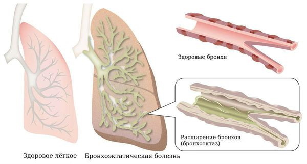
Bronchiectasis. Hypertrophic changes occur against the background of tissue defects formed during the underlying disease.
- Emphysema. The disease causes the expansion of the alveoli and their further rupture, resulting in the formation of inactive cavities.
- Bronchial asthma. Right ventricular hypertrophy develops as a result of respiratory failure, leading to an increase in pressure in the arteries of the lungs and vessels of the small circle. This leads to the proliferation of tissue in the right ventricle.
- Cystic fibrosis. With pathology, blood pressure increases, changes in the structure of cells are observed.
- Congenital heart defects. It is considered the most dangerous condition that can lead to death.
To establish the exact cause, an examination by a specialist and instrumental diagnostics are required.
Symptoms and clinical manifestations
The pathology has a characteristic clinical picture, by which it is possible to determine the presence of any abnormalities in the work of the heart.
Symptoms:
- frequent pressing burning pains in the chest area with minimal discomfort;
- an increase in heart rate up to 150 beats per minute, in rare cases accompanied by the development of arrhythmias;
- drops in blood pressure;
- dry cough, occasionally bloody discharge (possibly the development of tuberculosis);

- shortness of breath that occurs when walking, physical activity, and even at rest;
- general body fatigue - weakness, fatigue, in the absence of proper treatment, the process can lead to disability.
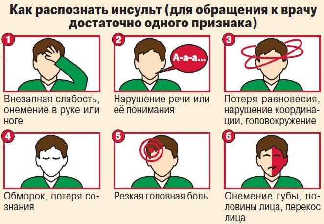
Symptoms may be accompanied by thickening of the fingers or nails, blue skin, headaches, impaired coordination, and fainting. Ultimately, the pathology can lead to stroke or ischemic brain damage.
What minor deviations are considered normal?
Right ventricular hypertrophy (only a doctor can determine the presence of pathology on an ECG) may not always indicate severe disorders that require compulsory therapy. Minor deviations can be observed in 15-25% of all patients. These deviations are associated with the individual characteristics of the structure of nerve fibers and endings.
In most cases, minor disorders occur in athletes, children and in asthenic conditions.
Deviations considered to be the norm:
- time interval before impulses in the right ventricle;
- partial blockade of Purkinje fibers;
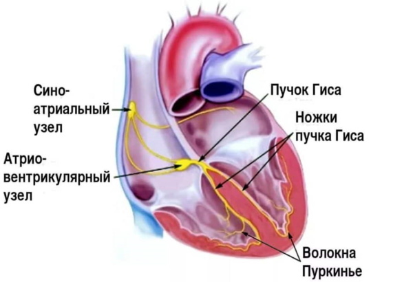
- change in the position of the axis of the heart;
- positive T in combination with high rates of R and V1;
- decrease in the number of contractions to 28 mV (in the first lead).
These violations do not cause concern and do not cause the development of pathological processes.
Diagnostics
For an accurate diagnosis, a visual examination is not enough, therefore, various additional types of diagnostics are prescribed.
Research:
- Measurement of pressure, heart rate, pulse.
- Daily mounting (Holter ECG).
- Electrocardiography - allows you to determine the presence of arrhythmia.
- Spirography is the definition of respiratory function.
- Auscultation - the procedure allows you to listen to the heart sound.

- Chest X-ray.
- MRI of the chest area. Often performed in conjunction with CT.
If necessary, additional (laboratory) diagnostic methods can be prescribed - general or biochemical blood tests, hormone tests.
ECG signs
ECG diagnostics will help determine the presence of hypertrophic disorders in the right ventricle only in moderate and severe forms. It is possible to identify the disease in the early stages by comparing the results of ECG and echocardiography.
Emphysematous (s-type)
This disorder occurs in 23% of patients. It occurs as a result of cor pulmonale. Pathology is characterized by a change in the position of the heart (lowering down). In this case, the upper part of the organ is turned back.
Signs of the disease:
- reduced value of electric current at the ventricular wave in leads V;
- the S wave is deep, while T has a positive value in leads V1 to V6;
- transition region V3-V4 is shifted to region V
Only a specially trained specialist can identify deviations in the emphysematous type.
Blockade (rSR ')
This type of pathology occurs in 18% of cases. Violation occurs with asynchronous excitation of the ventricles.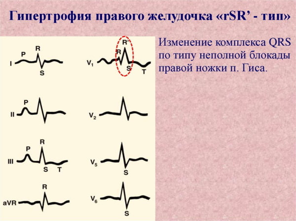
Diseases can be identified by the following features:
- Q wave formation in leads V1-V2;
- S-wave - deep, combined with deep R, located in V6;
- change in the cardiac axis at the very beginning - to the left, at the very end - to the left.
Pathology is detected with heart block.
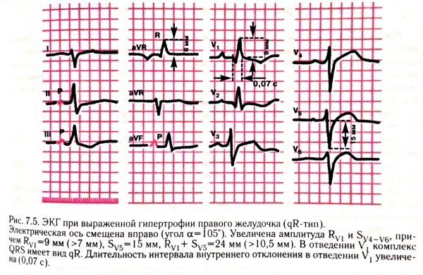
Hypertrophic (qR)
This type of disorder is the most common and occurs in 45% of patients. Pathology is characterized by an increase in pressure in the lungs and an increase in the size of the large chamber of the heart.
ECG signs:
- an increase in the volume of the QRS waves over 12 ms;
- the electric current indicators of the R wave increase by more than 8 mm;
- S wave - deep, while the amplitude of this wave increases to V1-V6;
- ST indices decrease below the isoline;
- T wave - negative.
A negative T wave is observed only in standard and right leads.
Moderately hypertrophic
The pathological process develops in approximately 10-13% of patients. It is characterized by the same ratio of the sizes of the right and left ventricles.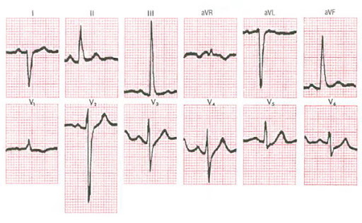
Signs:
- the axis of the heart is shifted to the right side by more than 1000;
- ST indices are below the isoline;
- indicators T - a negative value is observed in the standard and right leads;
- complex ventricular complexes have the form rSR ';
- the electric current of the R 'wave is more than 7mm.
The pathology is also characterized by a moderate overload of the right side of the heart.
RV hypertrophy and dilatation
The disease is accompanied by stretching of all cavities of the heart chambers. At the same time, there is a gradual thinning of the walls of the chambers. In both cases, there is an increase in the size of the ventricles. With the development of the first type, a thickening of the heart muscle occurs, while in the second case, the heart muscle is stretched.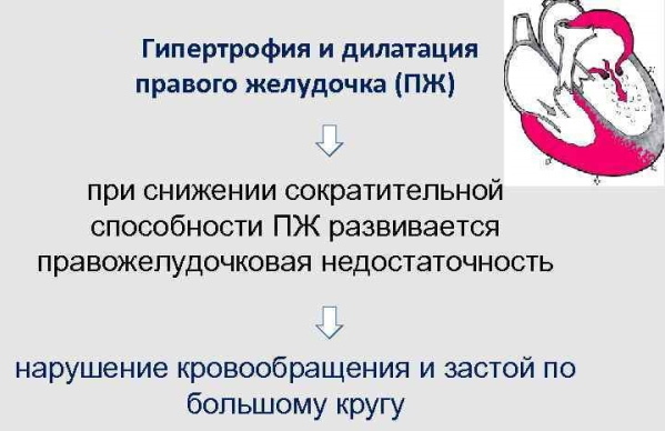
Signs of pathologies on the ECG:
- change in the position of the axis of the heart to the right by more than 300;
- Q - deep, T - negative (in lead V3);
- ST - lengthened in leads V1-V6;
- complete or partial blockade of the right Purkinje fibers.
Both types of pathology in most cases develop simultaneously.
The predominance and enhancement of the potentials of the right ventricle
The process is caused by the predominance of electrical impulses of the right ventricle directed towards the left ventricle. The condition is typical for children aged 8 to 10 years.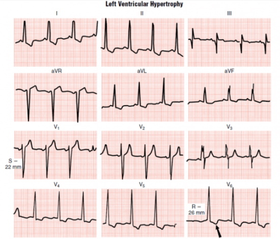
Pathology can also develop in the following cases:
- partial blockade of Purkinje's right fibers;
- agitated emotional state of the patient during diagnosis;
- incorrect position of the heart (the organ is located vertically).
In the absence of any symptoms, the condition is satisfactory and does not pose a health hazard.
Treatment features
Right ventricular hypertrophy (ECG patients are sent only for moderate and severe forms pathology) requires an integrated approach to treatment, including taking medications, adherence to a diet. In rare cases, surgical interventions can be performed.
Drugs
Drug therapy is mandatory and is carried out for a long time.
| Group of drugs | Action | Name of funds |
| Diuretic | This group of funds helps to remove excess fluid from the body. At the same time, cardiac activity for pumping blood is facilitated and restored. | Indapamide, Furosemide, Veroshpiron |
| ACE inhibitors | The action of this group of medicines is aimed at reducing hypertrophic processes and slowing down the progression of defective changes in the heart muscle. | Enam, Diroton, Prestarium |
| Nitroglycerin | These medicines help to reduce the tone of the veins located in the lungs. Thanks to this action, there is a decrease in the load on the myocardium. | Monocinque, Nitrosorbide |
| Calcium antagonists | The drugs of this group have a relaxing effect on the myocardium and help to reduce the heart rate, restoring the contractility of the organ. | Verapamil, Amlodipine |
The dosage and duration of the course are determined by the attending specialist, taking into account the nature and type of the pathological process.
Traditional methods
There are many different recipes from traditional medicine that will help eliminate symptoms and prevent further progression of the disease.
Recipes:
-
Drops based on lily of the valley flowers. To prepare drops, you need to take a small handful of flowers and put them in a dark glass dish. Then the contents should be poured with good quality vodka (no more than 0.2 l). The resulting mixture must be placed in a dark, cool place for 2 weeks. After this time, the resulting infusion should be filtered and taken 15 drops 3 times a day. Can be diluted with a little water. The duration of the course is at least 2 months. If necessary, the course is extended for another 1 month.

- Garlic and honey remedy. To prepare the infusion, you need to take a few cloves of garlic and grind it until a gruel is formed. Then honey should be added to the resulting mixture in proportions 1: 1. After that, the gruel must be placed in a dark place for 1.5 weeks and shaken periodically. Use the remedy for 1 tbsp. l. 3 times a day. The duration of therapy is 24 months with short interruptions.
-
Herbal collection. To prepare the herbal infusion, it is necessary to mix the flowers of hawthorn, grass of the mountaineer bird and horsetail in equal quantities. Then take 0.3 liters of water and prepare the infusion. Use the product 5-6 times a day in small portions.

Before using folk remedies, it is recommended that you first consult with your doctor.
Diet
Hypertrophy of the right ventricle (on the ECG, the disease is not detected in all cases) also requires adherence to dietary rules.
Basic principles of dietary nutrition:
- rejection of fatty meats, fish, flour and confectionery products;
- elimination of salt and salty foods from the diet;
- refusal from canned foods;
- daily intake of fresh fruits and vegetables;
- adding seafood, dried fruits, nuts to the diet.
In case of illness, it is recommended to consume the following foods:
- low-fat varieties of meat, fish;
- fermented milk products;
- cereal;
- plant food;
- fresh vegetables and fruits;
- fresh natural juices, fruit drinks, compotes.

The following types of foods should be limited:
- food of animal origin;
- fresh bread;
- spicy, fried, salty, smoked;
- confectionery and flour products.
You should also limit salt intake and high fluid intake.
Nutrition rules: food is eaten in small portions 5-6 times a day. The interval between doses should be at least 2-3 hours. It is recommended to refrain from eating in the evening and at night.
Complications
In the absence of timely treatment, pathology can lead to serious consequences and impairments.
Complications:
- the formation of swelling in the lungs;
- bronchial asthma;
- dementia of a vascular origin;
- myocardial infarction or ischemic heart disease arising on its background;
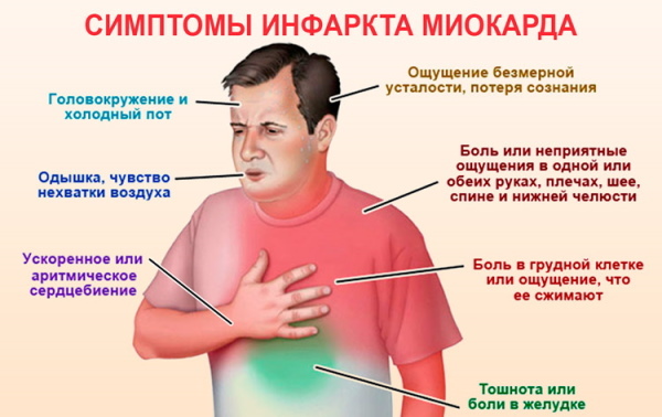
- stroke, up to death;
- heart failure.
It is possible to avoid the development of complications only with prompt referral to a specialist and the appointment of effective treatment.
Forecast
The prognosis of recovery depends on the form and cause of the development of the disease.. In most cases, the pathology does not lend itself to complete clinical cure. However, the disease can be controlled. The lethal outcome in complicated forms is about 7%.
With the development of heart failure, the risks of death increase significantly and range from 20 to 25%. At the same time, the accompanying negative factors are taken into account - age, the presence of concomitant diseases, bad habits, and the ecological situation.
The prognosis may slightly increase with major surgical interventions.
Hypertrophic changes in the right ventricle area require constant monitoring by the attending physician and diagnostic ECG procedures. In this connection, it is recommended to undergo systematic preventive examinations with a cardiologist and to treat in time for any violations of the cardiovascular system.
Video about ECG for ventricular hypertrophy
ECG interpretation for ventricular hypertrophy: