1 Features of examination
By its localization the abdominal cavity is a part of the trunk located in the abdominal region, which is bounded from above by the diaphragm;back - with dorsal muscles, vertebral column and fiber;in front - abdominal muscles;from below - pelvic bone and muscle tissues. The inner surface of the cavity is covered with the peritoneum - parietal and visceral parts. What organs can be attributed to the organs of the abdominal cavity? The liver, spleen, gall bladder, part of the stomach and pancreas are organs located in the abdominal cavity and covered with the visceral part of the peritoneum. The pancreas, the duodenum, the small and large intestines are the organs of the abdominal cavity, partially covered by the peritoneum. Kidneys, adrenal glands, ureters, abdominal aorta, branches of the abdominal aorta, lower hollow vein with inflows are organs of the retroperitoneal space, and the bladder is the organ of the preperitoneal space.

We recommend that you read the
- Preparing for EGD of the stomach
- What can pregnant women with heartburn?
- What is urine diastase?
- Effective agent for gastritis and gastric ulcer
Ultrasound examination( US) is considered a safe non-invasive way of diagnosing diseases with high informative ability. When the ultrasound of the internal organs is performed in the abdominal cavity, the following elements are checked and looked at:
- Liver: study of organ structure changes, fatty degeneration, hepatosis, hepatitis of various types, cirrhosis, tumor formations and other pathologies.
- Gallbladder and ducts: determination of the biliary system;detection of stones, polyps, cholecystitis;assessment of patency of the biliary tract.
- Pancreas: diagnosis of pancreatitis and pancreatic necrosis, incl.at an early stage.
- Spleen: Structural changes and increase in size are determined, pathologies are revealed.
- Stomach: ultrasound is made in different projections, which can partially replace the endoscopy of the gastrointestinal tract;
- Intestine: all parts of the intestine and duodenum can be examined for peptic ulcer disease, tumors, polyps, etc.
- Kidney: The study includes examination for stones, inflammation, tumors and other pathologies.
- Ureters: the study is difficult and requires special training.
- Bladder: the general condition of the body is determined, by its condition, the patency of the ureter is indirectly assessed.
- Prostate: inflammatory reactions and tumors can be identified at an early stage.
- Vessels: the state of the vessels is assessed when combined with duplex scanning.
- Uterus: it is possible to assess the change in structure, size, identify inflammatory processes on the mucosa, the tumors are determined.
- Lymph nodes: changes in the size of the lymph nodes of the peritoneum are very easily detected on an ultrasound image.
-
 IMPORTANT TO KNOW! Gastritis? Ulcer? To have a stomach ulcer not turned into cancer, drink a glass. ..Read the article & gt; & gt;
2 The need for preliminary preparation of
In general, ultrasound is based on transmission of an organ by an ultrasonic wave with a frequency of at least 2.5-3 MHz, which are modulated by special generators. Any structure has its own echogenicity, i.e.different reflectivity. The reflected wave is fixed by sensitive sensors that convert the signal into an electrical signal and send it to a computer monitor, where an ultrasound image is formed. It can determine the size, boundary parameters, uniformity and density of structure, the presence of tumors, the state of blood vessels and lymph nodes, the presence of other pathologies. Modern systems allow you to obtain two-dimensional or three-dimensional data of internal organs in real time.
-
 Gastroenterologist VAZHENOV: "I beg you, if you began to worry about abdominal pain, heartburn, nausea, do not in any way do gases. .."Read more & gt; & gt;
Naturally, the presence of any foreign substances can distort the resulting picture. Study of the abdominal cavity faces additional complexity. The first serious hindrance is the presence of gas in the intestine. Already the gas itself leads to disturbance of the wave movement, and besides, it inflates the intestinal loops, closing access to other organs under investigation. The accumulation of gas, as is known, depends on the intake of food, especially the use of products that provoke active gas production.

In view of this circumstance, a requirement arises: before making ultrasound of the abdominal cavity, it is necessary to remove the gas background. Great interference is created by fat components of food, which are slowly processed and stored for a long time in the digestive tract, which is especially characteristic of obese people. In addition, the question of how to properly prepare for ultrasound, requires the elimination of other random factors. For example, smoking can cause spasms, which can lead to incorrect interpretation of the results. A similar influence is exerted by stresses and other influences.
ADVICE FROM THE MAIN GASTROENTEROLOGIST
Korotov SV: "I can recommend only one remedy for the rapid treatment of Ulcer and Gastritis, which is now recommended by the Ministry of Health. .." Read testimonials & gt; & gt;
3 The purpose of diagnosis
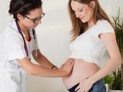
ultrasound allows you to determine the various pathologies of the abdominal organs, while ultrasound can be done for pregnant women and children of the earliest age. Such a procedure can be subjected to an unlimited number of times, as necessary and at the doctor's prescription. The study of the abdominal cavity is directed with the following symptoms: a feeling of heaviness in the stomach;nausea and vomiting;frequent pain syndrome in the abdomen, lower back, hypochondrium;chronic flatulence and bloating;violation of urination and pain when urinating;signs of inflammatory and infectious processes;bitter taste in the mouth. For chronic diseases of the abdominal cavity organs, it is recommended to conduct ultrasound every six months;in general, any person is recommended to conduct preventive studies annually.

4 Compliance with the
diet Before starting the research, it is necessary to decide how to prepare for ultrasound of the abdominal cavity in order to avoid interference. The most serious cause is the appearance of gas in the intestine. To eliminate this factor, preparation for ultrasound of the abdominal organs implies compliance with the diet and cleaning of the gastrointestinal tract.

5 Recommendations for feeding
To ensure that the gas does not accumulate in the intestinal loops and does not interfere with the procedure, preparation of the patient begins 3-4 days before the research. During this period, you should follow a diet designed to reduce gas formation in the intestines. Recommended to eat: buckwheat, barley, linseed porridge and oats on the water;poultry meat and low-fat beef;fish of low-fat varieties( baked or boiled);a stiff egg( not more than 1 piece per day);cheese with minimal fat content. Nutrition is recommended frequent( 5 times a day), but in small portions. The drinking regime is provided by water or loose tea in a volume of up to 1.6 liters per day.
The last time before ultrasound is in the evening in the form of a light meal if the procedure is in the morning or in the morning in the form of a light breakfast if the event is scheduled after 14 hours.
The diet before the ultrasound of the abdominal cavity categorically excludes the following products from use( especially forday before the procedure): fresh fruit and vegetables;leguminous crops;bakery products, muffins, cookies, cakes, cakes;milk and dairy products;fatty meat and fish;sugar and sweets;strong coffee;juices;drinks with gas;spirits of all kinds.

6 Bowel cleansing
The most accurate results are obtained if you clean the intestines before the ultrasound. In former years and sometimes in our time, cleaning procedures are performed with the help of an enema. However, medicines are most often used. The most common method is taking laxatives. Recommended drugs such as Senadé, Senadixin, Fortrans, whose dose is determined from the condition: 1 tablet for every 20 kg of the patient's weight. It is allowed to apply Normase, Dufalac, Prelaxan( strictly according to the instructions).
In the presence of increased gas formation, even during the period of dieting, you can take funds to normalize digestion( Festal, Mezim).To reduce gas formation, Smecta, Enterosgel, Simethicone are appointed. One day before the procedure, Espumizan is an effective way to eliminate gas formation. For chronic constipation, Senadé or Bisacodyl( in the form of a candle) should be used. Directly on the day of ultrasound, you can use sorbents. The most common means is activated charcoal. Good and enterosorbents( Polysorb, Polyphepan, white coal).
7 Conducting the
study How are ultrasound of the abdominal cavity and kidneys done? Usually the procedure is carried out in the morning on an empty stomach or after 14 hours, but in any case, completely exclude food and drink 4-5 hours before the research( except for ultrasound of the kidneys and bladder, where the training regime is different).The procedure is carried out only if the preparation for ultrasound of the abdominal cavity is fully completed, and the intestine is sufficiently cleaned. Otherwise, for 1-1.5 hours before the event, a cleansing enema is put.
How does the procedure work? The patient is lying on his back. If you need to get a picture in different projections, you can use the position on the left or right side. The contrast gel is applied to the area under investigation, and the sensor is located in the abdominal region. The ultrasound picture is displayed on the monitor screen.

8 Reason for distortion of
results In addition to the preparatory measures for ensuring the objectivity of research results, one should also take into account the possibility of their distortion under the influence of other factors. In order to exclude any suspicion of the possibility of receiving false data, the following recommendations should be adhered to:
- The last cigarette can be smoked no later than 2.5-3 hours before the procedure begins.
- You should not use chewing gum and suck lollipops later than 3-4 hours before ultrasound.
- It is necessary to exclude the influence of psychological factors: stress, anxiety.
- In the study of the kidneys and bladder, on the contrary, liquid filling of the body is used.
- When carrying out the radiography on the eve of the examination, this fact should be reported to the doctor conducting the study( it is better to take a break for 3-4 days).
- It is not recommended to take spasmolytic drugs such as No-shpa, Spasmalgon, Papaverin, Dibasol, Papazol before the examination.
When conducting ultrasound examinations of children, the following preparatory measures are recommended:
- In infants( up to 1 year), one feeding should be allowed, i.e.to sustain without feeding 2,5-3 h and not to drink 1 h before the examination.
- Babies at the age of 1-3 years do not need to feed 3.5-4.5 hours and drink 1-1.5 hours.
- Children over 3 years old have an empty stomach in the morning( without food 5-7 hours) and without water 1-1.5 h
Ultrasound examination of the abdominal cavity has a great informative ability, which allows early diagnosis of many diseases. The problem of research is the possibility of distortion of results as a result of gassing in the intestine. To avoid false results, it is necessary to conduct special training before conducting ultrasound.
- 1 Survey features
- 2 Need for preliminary preparation
- 3 Purpose of diagnosis
- 4 Compliance with diet
- 5 Recommendations on nutrition
- 6 Bowel cleansing
- 7 Conducting the study
- 8 Reason for the distortion of results
When an ultrasound of the abdominal cavity is prescribed, preparation for the procedure is important, as it ensures the reliability of the results. The presence of food debris in the organs or the products of its decay cause additional distortions of the wave, so questions such as "can I drink and what can I eat?" Are quite appropriate. When diagnosing diseases of the abdominal cavity, how to prepare for ultrasound is considered to be an important moment determining the accuracy of diagnosis. Ultrasound is sensitive enough to the presence of interference, and this fact must be carefully monitored when preparing the patient for ultrasound.(Figure 1)
Do you have gastritis?
GALINA SAVINA: "How easy is it to cure gastritis at home for 1 month. A proven method is to write down a recipe. ..!"Read more & gt; & gt;