
Located in the masticatory-parotid salivary gland zone is a paired exocrine organ that plays an important role in digestive activity. In an adult, it weighs 20-30 g and has an indefinite elongated-oblong geometric shape.
Record content:
- 1 Parotid anatomy, structural features
- 2 Blood supply and lymphatic drainage
- 3 Innervation
- 4 Embryology
- 5 X-ray anatomy
- 6 Histology
- 7 Research methods
-
8 Diseases of the parotid glands
- 8.1 Parotitis
- 8.2 Granuloma
- 8.3 Stones
- 8.4 Pleomorphic adenoma
- 8.5 Cyst
- 8.6 Tumors
- 9 Hardware video
Parotid anatomy, structural features
The organ is surrounded by a thick fascia capsule that performs a barrier-protective function. The parotid salivary gland is located just under the skin and is not separated from it by soft tissues or elastic muscle fibers.
The organ is limited by the following anatomical structures:
- zygomatic arch in the upper plane;
- mandibular angle;
- the mastoid process of the temporo-cranial bone in the region of the posterior surface;
- adjacent chewing muscles in the anterior segment.
The parotid salivary gland of a person has a lobular anatomical structure, which is given to it by sensitive capsule processes, deeply immersed in the thickness of the organ. The vertical dimension of the exocrine serous element of an adult is 40-65 mm.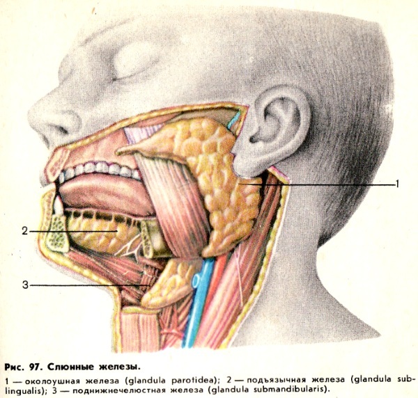
The horizontal value reaches 20-38 mm, and the sagittal (dividing) line extends by 30-50 mm. The peculiarity of the anatomical structure of the digestive organ is the unevenness of the tissue structure with dense and loose areas. The former occupy most of the specific gravity of the gland.
The bed of the functional element is a space bounded by the leaves of the parotid chewing capsule. The inner part of the gland, which does not have a dense fascia cover, is adjacent to the peri-pharyngeal subcutaneous lipid tissue. The organ is dotted with secretory ducts.
Blood supply and lymphatic drainage
The parotid salivary gland is penetrated by a branched vascular network. The functional purpose of such channels is the blood supply to the organ. Another type of vessels is lymphatic.
They carry out a drainage function. Most of the blood vessels and lymphatic channels are located deep in the organ. There are also surface capillaries.
The largest blood vessel in the parotid gland is the carotid artery, tightly fused with the parenchyma, a functionally active epithelial layer.
Here, the main element of the blood supply to the digestive organ is divided into the following final branches:
- posterior;
- superficial temporal;
- cross-facial;
- jaw.
The jugular vein, which is involved in the systemic circulation, is adjacent to the outer surface of the carotid artery. The posterior and transverse-facial vessels flow into it. They play an active role in the blood supply to the salivary gland.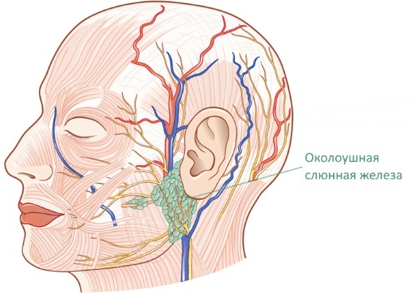
Interstitial veins and capillaries are located in the interlobular septa. The arterial outflow is carried out into the pterygoid node and the bloodstream of the lower jaw. The lymphatic vessels of the glandular body are connected to the superficial and deep-lying nodal plexuses involved in drainage.
Innervation
For the conduction of impulses to the brain, sympathetic and parasympathetic conductive tissues are responsible. The afferent innervation of the exocrine digestive organ is performed by the temporo-auricular neural process. It encircles the salivary gland and the auditory element.
Postganglionic fibers of the autonomic nervous system play a leading role in the innervation mechanism. Sympathetic tissue is drawn to the gland from the plexus of the carotid artery and its branches.
The innervation mechanism is physiologically realized with the help of neurons that give rise to axons and are associated with preganglionic fibers located in the lateral branches of the spinal structure.
Together with the choroid plexus that accompanies the carotid artery, the nerve-conducting tissues reach the parotid gland. Sympathetic innervation regulates the secretion of saliva by narrowing or dilating the lymphatic and blood vessels.
Irritation of the cranial fibers, namely the drum string, leads to the activation of secretion production. The parasympathetic nerves slightly increase the secretion of fluid, but increase its density due to the saturation of organic compounds involved in the breakdown of food.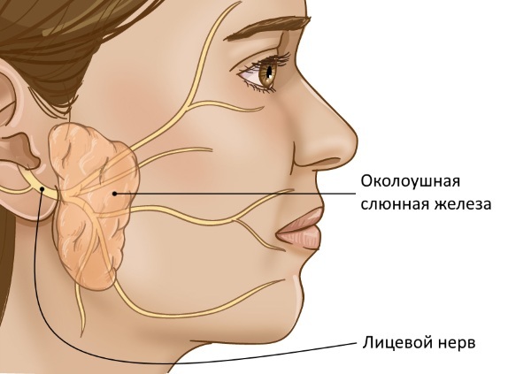
The parotid salivary gland of a person with increased innervation consumes much more oxygen and emits carbon dioxide than in a calm state. This physiological mechanism is focused not only on the regulation of the volume of secreted secretion, but also on the filtration of lymphatic fluid.
Embryology
The parotid gland is formed by the epithelial cells of the dental cavity. It is characterized by a stepwise development in the embryonic period. The organ kidney appears at the 6th week of the intrauterine phase. The zone of its initial localization is the deep layers of the tissue groove.
This elongated indentation separates the inner cheek from the loose gingival fibers. The salivary gland of the parotid cavity at the stage of formation is an epithelial cord. It gradually extends towards the auditory organ.
At the 2nd month of embryonic development, the rounded distal end of the epithelial cord branches, forming the excretory ducts of the secretory sections of the salivary gland. In this phase of formation, the digestive organ takes on a recognizable shape.
Closer to the 12th week of embryonic development, gaps appear in the anlages of the drainage canals. The epithelial lining acquires a two-layer structure, thickening in the area of the salivary ducts due to the increase in the number of layers. Cellular differentiation begins later than in other digestive organs.
X-ray anatomy
The sialographic projection of the glandular body shows shading in the anterior segment of the organ, stretching towards the outer part of the cartilaginous branch of the mandibular anatomical structure.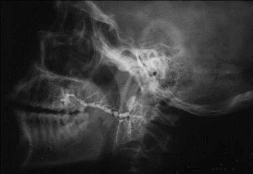
This reflects the geometric configuration and structure of the exocrine element. With a side view of the sialogram, the shadow of the organ is superimposed on the projection of the mandibular cavity.
The largest excretory duct runs diagonally from bottom to top. The inner glandular part of the canal with epithelial lining crosses the posterior process of the mandibular branches on the border itself or superimposed on the projection of the rounded corner of the functional and anatomical constructions.
Sialographic examination allows you to see on a section of the cartilaginous branch of the lower jaw, a large lymphatic canal emerging from the body of the organ, surrounded by small capillaries.
The interlobar excretory tubules merge into one channel, which is involved in the secretory function of the digestive element. They emerge from the deep tissue layers of the organ at an angle and have a different number of branches.
Depending on the X-ray anatomical structure, the excretory canals are divided into multi-, slightly- and moderately branched. Together, they form a single secretory complex.
Histology
The parotid gland is divided by connecting membranes into small segments, which form several large lobular parts. A complex organ is classified as an alveolar organ. The lobar structure is formed by the adjacent excretory sections at the end of the digestive organ.
The parotid gland has acini and internal excretory canals. The body of the salivary organ is speckled with insertion and striated tubular elements. An adult has more than 10 of them. The end sections that perform the secretory function are formed by epithelial serocyte cells.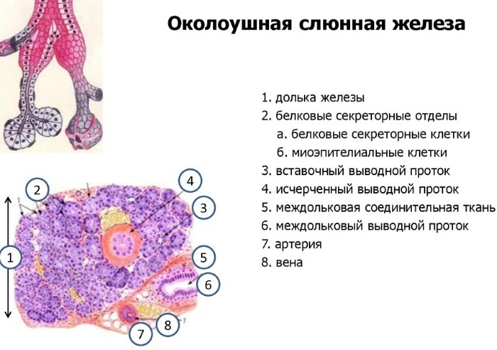
They are located on the surface of the basal membrane covered with reticular tissue. The serocyte cell has a pyramidal shape. Its cytoplasm contains the nucleus of the basal arrangement and the branched endoplasmic reticulum with multiple microscopic reservoirs, plates and excretory granules.
In the end segments of the parotid glandular body, small secretory ducts are located between the serocytic cells. They have a tubular structure and have no walls of their own. Such elements are characteristic of all excretory organs that synthesize protein compounds.
The end sections are connected to narrow insertion canals lined with cubic epithelium, and striated ducts or salivary tubes covered with a single-layer cylindrical mucous membrane.
The apical pole of serocytic cells facing them contains sensitive microscopic hairy receptors. The degree of the characteristic basal striation of the mucous membrane depends on the concentration of mitochondria located between the folded depressions of the plasmolemma (cell membrane).
Such channels are presumably involved in changing the saturation of the primary salivary fluid with organic substances. Between the serocytes and the basal lining of the excretory elements of the gland, in the area of the intercalated and striated canals, there are myoepithelial cells, called basket cells.
These are special contractile functional elements that ensure the production of salivary fluid and maintain the normal tone of the flow tubes. They lie in the layers of connective tissues and are actively involved in the secretory function of the organ.
Research methods
Diagnosis of diseases of the parotid gland involves the initial examination and questioning of an adult patient by a dentist, and a child by a pediatrician. To clarify the clinical picture, laboratory tests are prescribed and a hardware study of the digestive organ is used.
Methods for diagnosing the parotid salivary gland:
| Inspection method | Description and purpose |
| Palpation | It is carried out by a doctor during the initial examination of the patient. Allows you to detect a tumor, suggest the nature of the pathology. |
| Probing of the parotid ducts | It is used to determine the patency and the presence of clogging dense foreign fractions in the channel. |
| Cytological examination of salivary secretion | The biological material is obtained by the method of catheterization from the duct of the exocrine organ. It is placed on a glass slide and stained by the Romanovsky-Giemsa method. Next, a microscopic examination is performed using an immersin optical system. |
| Cytological analysis and biopsy | They help to detect morphological changes in the structure of the organ. Research is carried out using special reagents and microscopic methods. |
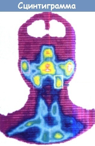
| Sialography | The technology of radio-opaque or radioisotope scanning of the salivary glands. It is intended to study the secretory ability of the organ. Based on the ability of the parenchyma to accumulate and release radioisotopes with liquid. |
| Ultrasonic dowsing | A method for diagnosing tumor neoplasms and inflammatory processes. Allows to draw a conclusion about the biochemical and physiological characteristics of the pathological process, to determine the size of the organ and the degree of degenerative changes in the parenchyma. |
| Thermovisiography | A method for recording infrared radiation, which allows you to set the temperature of an organ. Its increase is a clear symptom of the inflammatory process. |
The parotid salivary gland of a person is often examined by tomographic methods, which make it possible to obtain layer-by-layer pictures of the internal structure of tissues. Panoramic scanning, called pantomography, makes it possible to perform a comparative analysis of the right and left parts of an organ.
Diseases of the parotid glands
The pathogenesis is diverse and includes both typical dental disorders, as well as histological and gastroenterological ones. The salivary glands are susceptible to bacterial infection, seasonal viruses and pathogenic microflora of the oral cavity.
Defects of embryonic development of the exocrine organ are extremely rare.
Of the anatomical disorders of this etiology, the following are described:
- dystopia (displacement) of the glandular body to the chewing muscles or the buccal surface;
- congenital hypertrophy;
- aplastic destruction with preservation of flow channels or their degeneration.
The organ is susceptible to inflammation and formations of the salivary fistula. In the latter case, the restoration of the functionality of the secretory channels by surgical methods is often performed.
Parotitis
This is the most common disease of the salivary glands. Mumps is infectious and epidemic in nature. The pathogenesis is characterized by non-purulent damage to the tissues of the organ and secretory ducts. The causative agent is paramyxovirus. Most often observed in children 3-15 years old during the period of deterioration of the epidemiological situation.
The infection clogs the excretory ducts of one or both glands. Inflammation develops and severe tissue edema is observed. Mumps is capable of causing some types of bacterial agents and benign epithelial hyperplasias.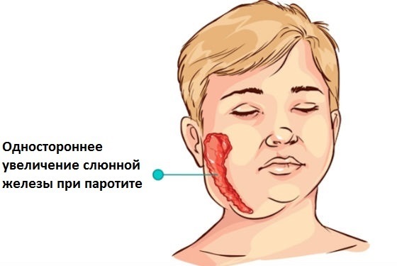
Such a disease is often a symptom of Mikulich's or Sjogren's syndrome. The first is a rare chronic disease of unknown etiology, characterized by an increase in the size of all glands.
Sjogren's syndrome is an autoimmune pathology, as a result of which connective tissues are deformed. Mumps are treated with antibiotic therapy, rinsing the mouth with antiseptic solutions.
Granuloma
An inflammatory disease of the connective tissue resulting from bacterial infection. The formation of such foci in the parotid gland provoke tuberculosis, syphilis, viral encephalitis. Granuloma can occur for non-infectious reasons.
Such a disease of the salivary glands is provoked by some medicines and foreign bodies trapped in the cavity of the secretory canals. Clinical manifestations and methods of therapy depend on the type of granulomatous inflammation.
Both surgical intervention and conservative treatment options are used. Pre-biopsy and sampling of salivary fluid for laboratory analysis to verify the benign nature of multiple neoplasms.
Stones
Sialolithiasis is a chronic pathology in which calculus clogs the excretory ducts. The parotid gland is affected less often than the submandibular gland. Pathology is common among men and is practically not recorded in children.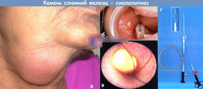
The parotid salivary gland of an adult is susceptible to bad habits, in particular, smoking and the carcinogens contained in cigarettes. This is the main reason for the formation of sialolithiasis.
Secretory dysfunction of the glands leads to stagnation of salivary fluid, which contributes to the concentration of sodium sediment, which forms the basis of stones. Therapy consists in the use of drugs that enhance the outflow, decongestants and anti-inflammatory drugs.
Pleomorphic adenoma
Tumor pathology affects the epithelial coating. It has a grayish tint and a loose structure. Sometimes pleomorphic adenoma contains cartilaginous inclusions. There are necrotic areas and sites of hemorrhage. Such a tumor has a brownish-gray tint.
According to the degree of influence, the causes of the development of pathology are distributed as follows:
- Smoking. Carcinogenic and toxic substances contained in cigarettes provoke not only sialolithiasis, but also tumor or inflammatory processes.
- Unbalanced diet. The consumption of large amounts of salt with microcracks in the oral cavity can cause the formation of a pleomorphic adenoma.
- Hypovitaminosis. Deficiency of vitamins of groups A and B often causes inflammation of the parotid salivary gland.
- Traumatic lesions of the facial surface. Regular shock loads, for example, in boxers, lead to this pathology.
- Exposure to oncogenic viral agents. These include herpes, cytomegalovirus infection, and others.
Such an adenoma is eliminated exclusively by surgical intervention.
Cyst
The parotid gland is characterized by formations of the retention type. The cyst is formed as a result of complete or partial cessation of the outflow of salivary fluid, which is caused by a violation of the patency of the secretory ducts.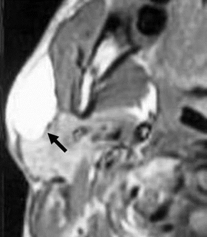
The disease is characterized by an often asymptomatic course. Elimination of pathology does not provide for conservative methods. Surgical intervention is used to remove the neoplasm of the adjacent destructive buccal tissues. After the operation, stitches made of biodegradable material are applied.
Tumors
Hyperplasia of the salivary glands is mainly of a benign nature. Cancer hyperplasia is unusual for this digestive organ. They are recorded in 1% of cases.
Despite the rarity of occurrence, malignant hyperplasias of the salivary glands pose a considerable danger, since at the initial stage of the pathology it is asymptomatic and is not diagnosed.
Benign tumors of the parotid salivary glands most often affect women after 50 years. In this category of patients, hyperplasias have a more pronounced tendency to malignancy and metastasis.
As the neoplasm grows, there is swelling, a feeling of fullness in the mouth and a slight pain that borders on discomfort. Treatment of a neoplasm is exclusively operative, followed by radiation and chemotherapy.
The role of the parotid salivary gland is important in the mechanism of homeostasis of the dental zone. In a smoking adult, salivation helps to clear the cavity of plant tar and nicotine plaque. In children, the production of a secret prevents the development of caries.
Hardware video
About the parotid gland:


