
Muscles and nerve fibers, which are responsible for the mechanism of muscle contraction, allow a person to perform many movements, and internal organs to function. There are more than 600 muscles in the human body, and they all differ in structure and physiological properties.
Record content:
- 1 Definition
-
2 Types
- 2.1 Isotonic mode
- 2.2 Isometric mode
- 2.3 Auxotonic mode
-
3 Views
- 3.1 Single
- 3.2 Tetanic
-
4 The contractile proteins myosin and actin
- 4.1 Myosin
- 4.2 Actin
- 4.3 Tropomyosin
- 4.4 Troponin
-
5 Transformations
- 5.1 Electrochemical
- 5.2 Chemomechanical
-
6 Reduction stages
- 6.1 First
- 6.2 Second
- 6.3 The third
- 6.4 Fourth
- 6.5 Fifth
- 6.6 Sixth
- 7 Muscle Contraction Videos
Definition
Muscle contraction is the mechanical work that a muscle produces as a result of the process of shortening or developing tension.
During muscle contraction, the change in length is caused by the thin fibers being pulled along the thick fibers. Although the length of the overlap between thick and thin filaments (sarcomere) varies, the length of the filaments themselves remains the same.
Excitation of skeletal muscles, their voluntary contraction is stimulated by electrical impulses carried along the nerves. The contractions of the heart muscle cells are stimulated by the heart muscles.
There are also involuntary contractile muscle movements. They are characteristic of the stomach, intestines, bladder, blood vessels, where smooth muscle contraction causes peristalsis (wave-like contraction).
Types
The mechanism of muscle contraction, the physiology of which is the same for all types of muscles, consists of the transition of a muscle from a "high tension state" to a "low tension state". 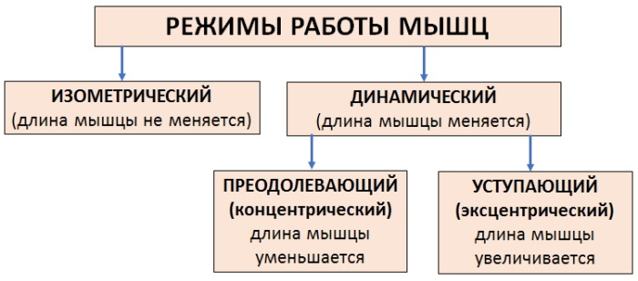 In other words, the muscle tenses and then relaxes again.
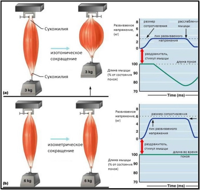
Isotonic mode
With this type of contraction, the muscle changes length, while the tension or resistance remains the same. The result is movement of a part of the body.
There are two types of isotonic contraction:
- Concentric. This is a standard contraction and lift movement in which a muscle shortens and its two points of connection move closer together. In doing so, the movement acts against gravity and allows the muscle to pull and lift.
- Eccentric the contraction uses the same muscle, but acts on the descending part of the movement. As the muscle lengthens, the muscle's two junction points diverge further, but the muscle continues to contract and still exerts pressure on the weight. It acts as a kind of brake mechanism, slowing down the descent by gravity.
 Eccentric contractions are the opposite of concentric contractions and occur when a muscle lengthens as it contracts. This can be seen, for example, when lowering a dumbbell down in a biceps curl exercise. The muscle is still contracting to support the full weight, but the biceps muscle lengthens.
Isometric mode
The mechanism of muscle contraction (physiology of the isometric type) in this case occurs without a change in muscle length. Isometric contractions differ from the other two types in that they are not associated with either lengthening or contraction of the muscles. Rather, it is a type of activation in which a muscle is deliberately tense, but the joints associated with it do not move.
Examples include carrying an object in front of you without moving, squatting against a wall without moving, or holding the body in a balanced position for a minute or two. In each case, the muscle is activated without movement.
This is typical of the muscles in the forearms and hands. For example, when the hand squeezes an object, the muscles of the forearms and hands work, but the joints of the hand do not move, and the muscles create sufficient force to prevent the object from falling.
Another example is when a person is holding something in his hand, there is no movement in the joints, but the muscles contract to provide sufficient strength to hold the object.
Auxotonic mode
The type of mixed mode, in which the elements of isotonic and isometric contraction are combined, is of the auxotonic type. This type is characterized by a change in muscle tone and length, and has another name - dynamic mode.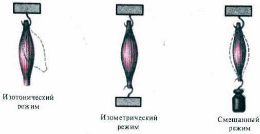
To measure this type of contraction, special equipment is required, an isokinetic dynamometer. Isokinetic contractions in everyday life and in sports are rare. This is best observed in breaststroke mode, where the water provides constant, uniform resistance to the adduction movement.
Views
The nature of muscle contractions depends on the order of contractions and the frequency of the arising nerve stimuli.
Single
In the case of direct irritation of the muscle or through the nerve that innervates it, a single muscle contraction occurs.
This mechanism is divided into 3 phases:
- the latent period, from the beginning of the arrival of the impulse to the response of the muscle, during which the action potential (AP) develops;
- the period of contraction or shortening of the muscle;
- relaxation period.
Muscle excitability in this case changes in accordance with certain phases. But in life, single impulses do not enter a person, but follow each other in series, at certain intervals. The muscle responds to these impulses with a prolonged contraction.
Tetanic
When an ongoing muscle contraction is maintained continuously without relaxation, it is called a tetanic contraction. Muscles can shorten, lengthen, or remain of constant length.
In this case, the movement is stimulated by multiple impulses with a sufficiently high frequency. The impulse coinciding with the relaxation phase contributes to the emergence of a whole series of sequential single contractile movements.
If the frequency of the impulses increases, then the relaxation phase of the previous cycle may coincide with the new incoming impulse.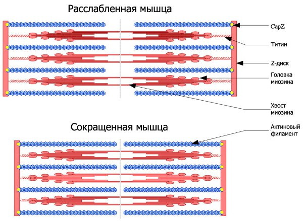
In this case, they speak of dentate tetanus, in which a prolonged contraction occurs, interrupted by periods of incomplete relaxation. Another type of tetanic contraction will be smooth tetanus, when prolonged muscle contraction is not interrupted by periods of relaxation.
Mild theetanic contraction is normal and occurs as a normal physiological process during activities such as lifting weights. In severe cases, with inflammatory and infectious conditions, tonic convulsions of symmetrical muscle groups occur.
The contractile proteins myosin and actin
The individual muscle cells are called muscle fibers. They are long, cylindrical, and contain several nuclei. These muscle fibers are surrounded by a cell membrane called sarcolemma. Inside each muscle fiber, there are tiny rods called myofibrils that are surrounded by sarcoplasm.
Myofibrils are made up of repeating segments called sarcomeres, which are tiny units responsible for skeletal muscle contraction. Each sarcomere contains myofilaments, protein structures of actin and myosin. Thick myofilaments are composed of myosin, while thin ones are composed of actin, troponin, and tropomyosin.
Looking at the structure of the sarcomere, you can see the zigzag areas that mark the end point of each sarcomere. These are called Z-lines, and are involved in the attachment of thin (actin) filaments.
Myosin
Protein, a thick filament, has a structural role as a building block for thick fibers and a functional one by catalyzing the breakdown of ATP during contraction and interaction with actin. 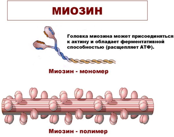 A single myosin molecule is about 160 nm long, asymmetric, and contains 2 main protein chains.
Actin
Actin is the main component of fine fibers in muscles and makes up about 25% of myofilament protein. A single actin cell is a single coiled protein chain.
In the muscle, actin is twisted in the form of 2 long strands of bead-like molecules. The thin actin filament consists of alternating bundles intertwined with bundles of thick myosin filaments.
Tropomyosin
Tropomyosin is a long, coiled protein that is structurally similar to the tail of the myosin molecule. Each molecule is in contact with 7 units of actin, covering most of the actin filament. The molecule is a regulator of muscle contraction, preventing myosin from sticking (and therefore preventing contraction). Within each tropomyosin protein are troponin complexes.
Troponin
Troponin is composed of a complex of protein subunits. One part of the protein complex is associated with the tropomyosin molecule, and the other with Ca2 + ions. Troponin, its molecule, is located along the filament every 40 nm.
Both troponin and tropomyosin are regulators of contracting and relaxing muscle movements through Ca2 + binding.
Transformations
Physiologically, a person is a biochemical engine, because food is a fuel that is broken down into its constituents: glucose (the smallest building blocks of carbohydrates), amino acids and fats. The mechanism of muscle contraction occurs through the use of glucose and energy provided by ATP.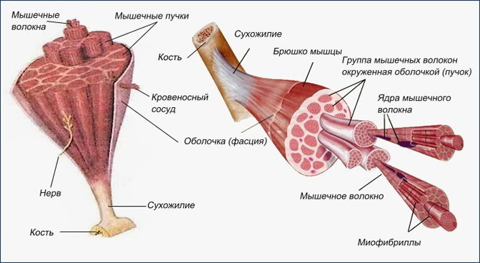
Skeletal muscle converts chemical energy into mechanical energy. Thus, the process of muscle contraction occurs under the influence of a 2-stage molecular transformation: electromechanical and chemomechanical.
Electrochemical
All living cells have membranes with proteins embedded in it. The membrane serves as a diffusion barrier and insulator for the advancement of positively and negatively charged ions.
Neurons and muscle cells across their membranes control the movement of charged ions by selectively opening and closing ion channels. In this way, electric currents and electrical signals are generated. Although the currents created by the ions moving through the channel proteins are very small, they form the basis of both neural signaling and muscle contraction.
Nerve impulses are electrically excitable, therefore, they are able to generate an action potential (AP) - a special type electrical signal that travels along the cell membrane in the form of a wave, quickly and accurately transmitting the signal to large distance.
This first stage of muscle contraction occurs due to the excitation of the muscle fiber membrane, along which the action potential circulates. This property is called excitability, when PD generates neurons and muscle cells.
Chemomechanical
The contractile work of muscles occurs due to the energy released during the oxidation of carbohydrates or lipids.
In the process of a mechanochemical reaction, a gradual transformation of chemical energy into mechanical energy occurs:
- first, the process of binding of troponin with Ca2 + ions is going on;
- the next step is the interaction of the head of the myosin heavy chain with actin;
- actin and myosin slide relative to each other,
Reduction stages
Whether a person runs, rides a bicycle or lifts weights, all movements are part of a complex process of coordinating muscle contractions.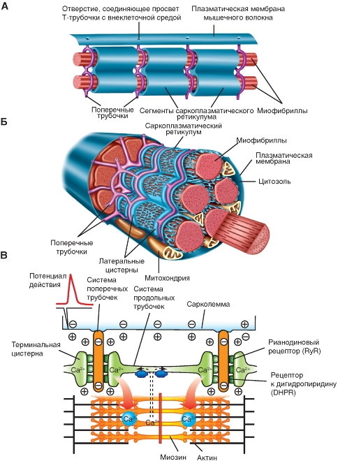
At rest, Ca2 + ions involved in the process of muscle contractions are stored in the cytoplasm of the cell, or the sarcoplasmic reticulum. In this case, an excitation-contraction connection does not arise, which converts the electrical signal of the neuron through acetylcholine into an electrical signal on the muscle membrane.
The mechanism of muscle contraction, the physiology of which depends on the properties of skeletal muscles, occurs only with a certain sequence of events.
Contractile work begins when the sarcomeres are shortened, thick and thin fibers slide past each other, according to the principle of sliding threads. At the same time, proteins responsible for muscle sensitivity to Ca2 + ions, such as troponin and tropomyosin, control the formation of transverse bridges that appear during the interaction of actin and myosin.
| Stages of muscle contraction | Actions |
| I | The process of adding calcium to troponin takes place. |
| II | The adhesion of the myosin bridge to actin begins. |
| III | There is a cyclic movement of the cross bridge. |
| IV | Dissociation and the formation of a complex between myosin and actin occurs. |
| V | Rotation of the head of the myosin transverse bridge. |
| VI | Completion of the cycle with the decay of ATP. |
The process of muscle contraction lasts as long as the necessary reserves of Ca2 + ions and energy (ATP) are available. As soon as the impulse ceases, the ions return back to the sarcoplasmic reticulum, actin goes into a state of rest, and the muscles lengthen and relax.
First
The signal for muscle contraction comes from neurons, is transmitted to the point of contact of the motor nerve with the muscle. Or, in other words, an electrical impulse travels from the motor nerve cell body in the spinal cord along the nerve axon to its destination, the neuromuscular junction.
Second
An electrical impulse causing depolarization and AP in the sarcolemma triggers the release of calcium ions from the sarcoplasmic reticulum. Then, calcium ions interact with the troponin complex on the actin myofilament. This gives rise to a displacement of the troponin and tropomyosin complex, which leads to the unblocking of the site of binding of myosin to actin molecules.
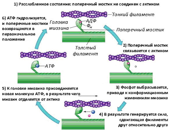
At the moment when the signal for contraction is sent along the nerve to the muscle cell, proteins, actin and myosin are activated. Myosin begins to work as an engine, releasing ATP energy, which facilitates the sliding of the myosin filament along the actin filament.
Each actin molecule has an active center, which is blocked by proteins, troponin and tropomyosin. They do not allow actin to act with myosin ahead of time.
As soon as the myosin binding site is exposed, the head of this molecule forms a connection with the actin filament. In this case, it is said that the actin myofilament is in the “on” stage.
The third
After actin is “turned on”, the myosin head bends in the hinge regions, attaching itself to the adjacent actin filament. This forms an active cross bridge. It works as an enzyme (myosin-ATP) that breaks down stored adenosine triphosphate (ATP), retained in the myosin head, into adenosine diphosphate (ADP) and inorganic phosphate, releasing at this energy.
Fourth
The energy released during hydrolysis is used to move the myosin head relative to the actin filament. The head of the myosin heavy chain tilts and begins to pull the actin filament, while the thick and thin filaments move relative to each other.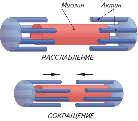 The opposite ends of the actin myofilaments inside the sarcomere begin an oncoming movement, which leads to contractile muscle movement.
Fifth
The mechanism of muscle contraction (the physiology of a complex process is multi-step) at this stage is that the head part of the myosin chain, using the energy of ATP hydrolysis, attaches to the site of action actin. The release of inorganic phosphate enhances the binding interaction between myosin and actin and subsequently triggers a "force hit".
Power punch is a key step in power generation utilized by myosin motor proteins. Forces are generated on the actin filament when the myosin protein returns to its original form. The actin filaments are pulled out. It is at this stage that the muscle contracts.
Sixth
When myosin regains its original shape or conformation, ADP is released, but the myosin head remains tightly bound to the filament in a new location, thereby returning the cycle to the beginning. The breakdown of ATP from myosin completes the cycle, and the entire actin-myosin complex remains in a fixed state.
The myosin head continues to position itself at a 45 ° angle to the thin and thick filaments. Under these conditions, a large number of ADP-free myosin heads remain bound to actin until a new ATP molecule binds to it and thus starts a new contractile cycle.
One neuronal impact only results in about 1% shortening of the entire muscle. Therefore, to achieve an overall reduction of up to 35%, the entire process must be repeated many times. It is believed that while half of the cross bridges are active in pulling actin through myosin, the other half is looking for their next binding pathway.
The living system is characterized by the mechanism of muscle contraction due to the interaction of proteins. Contractility is a property of cells in any tissue. But only muscle ones have a complex physiological structure, which leads to a variety of movements.
Muscle Contraction Videos
Physiology. Mechanisms of muscle contraction:


