Content
- What is tooth decay?
- Black classification of cavities
- Black caries classes
- 1 class
- Grade 2
- Grade 3
- 4th grade
- Grade 5
- 6th grade
- Types of caries according to Black according to the depth of the lesion
- Black preparation steps
- 1st grade pathology
- 2nd grade pathology
- Pathology of the 3rd and 4th grades
- Video about tooth decay by Black
In dental practice, different classifications of caries are used to determine the stage development of a destructive process. The most common is according to Black. The American dentist developed his own gradation in 1896. The pathology ranking system has been expanded, supplemented and modernized over time.
What is tooth decay?
Disease of the elements of the jaw rows is a process of the formation of pathological cavities in dentin due to demineralization of hard tissues and destruction of the morphological structure of the tooth.
The resulting cavities are inhabited by pathogenic and opportunistic microflora. Typical provocateurs of pathological changes are cariogenic bacteria, which include streptococcal agents forming an acidic environment.
Such microorganisms are characterized by anaerobic fermentation, which causes inflammation, edema, hyperemia of the mucous membranes. The disease progresses actively with dysfunctions of the salivary glands, which produce antibacterial compounds. Black's classification of caries does not take into account the causes of the development of the pathological process and the predisposition to the disease due to the anatomical features of the jaw structure.
The grading scale is based only on the degree of damage to the dentin tissue. The rate and extent of dentin destruction depends on the provoking factor or their combination.
These include:
- unbalanced diet;
- hereditary predisposition;
- systemic pathologies;
- gastrointestinal diseases;
- weakened local immune defense;
- violation of the basic rules of oral hygiene.
A sharp increase in the sensitivity of the teeth to tactile and temperature exposure, the appearance of dark pigmentation on the enamel coating, intense pain syndrome.
Each stage of the disease has its own set of clinical signs. Caries is considered not a separate nosological unit, but a pathogenetic link in the general physiological state of the body.
Black classification of cavities
The process and stage of destruction of units of the jaw row are described by the depth of the lesion, the nature of complications, and the dynamics of progression.
Black's classification system divides caries into the following types:
- Fiisurnaya. The most common variant of the disease in children. Fissure caries is difficult to detect at an early stage of development.
- Interdental. The process of destruction of dentinal tissues captures the anterior teeth with incisors. Demineralization proceeds rapidly.
- Cervical. This type of disease is characterized by the formation of a white-matte pigmentation on the enamel coating. Cervical caries is easily detected at the initial stage by visual inspection of the oral cavity.
- Atypical. The destruction begins with noticeable damage to the cutting edges of the chewing teeth. Carious cavities are localized on the lateral surfaces, penetrate into the root and subgingival part of the jaw unit.
The scale developed by the American dentist takes into account the depth of tooth damage. According to this grading system of carious cavities, destruction can be initial, superficial or deep.
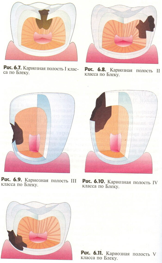
Black caries classes
Initially, the scale contained 5 degrees of damage, later a 6 was introduced into it, additionally specifying. The classes of caries according to the system of the American dentist Black are presented in the table.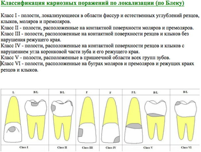
| Class | Defeat option |
| 1st | Fissure caries |
| 2nd | Demineralization of the contact planes of the teeth |
| 3rd | Damage to cutters with canines without destruction of the cutting edge |
| 4th | Lesion of the chewing surface |
| 5th | Formation of pathological cavities in the cervical area with necrotization of adjacent gingival tissues |
| 6th | Extensive lesion of units of the jaw row |
Belonging to a certain class of the observed clinical picture is determined by the established symptoms, localization, and the depth of damage to dentinal tissues.
1 class
Carious cavities are formed in the depressions of the chewing surfaces of the units of the jaw row or on the fissures - natural anatomical grooves. Incisors with canines are most susceptible to such caries.
Less commonly, the spread of the destructive process to molars is observed. Class 1 caries can be treated with a filling. A high-strength composite material is used.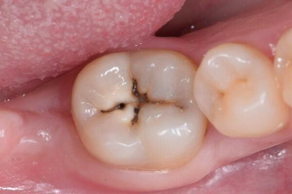
The edges of such a filling do not break off and withstand significant mechanical stress when chewing. The dentist forms a special bevel angle of the enamel surface. A cone-shaped bur is placed in the 1st class carious cavity to make the cavity look like a natural fissure.
To close the pathological cavity, a special chemical composite is used, which is applied to the damaged plane. To eliminate caries of the 1st class, photopolymer filling material is often used.
UV-hardened, it provides adequate strength. The composite is applied in beveled layers, which reduces the risk of the edges of the filling breaking off.
The Black classification of caries allows dentists to choose the optimal treatment tactics. For each variant of the destructive process, its own therapeutic model has been developed and a protocol approved by WHO is in effect.
Grade 2
Such caries affects the contact planes and chewing edges of adjacent teeth of the same jaw row. Molars with premolars are most susceptible to demineralization.
When treating caries of the 2nd class, the following therapeutic algorithm is used:
- Opening of the pathological cavity.
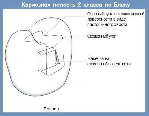
- Cleaning from microbial plaque and necrotic tissue.
- Formation of the surface for the application of filling material.
- Composite filling.
- Finishing leveling of the surface.
For a tight seal, the dentist uses a special matrix. Wedges provide access to the lateral surfaces of the affected tooth. To fix the unit of the jaw row, severely damaged by the pathological process, an adhesive composition is used.
Grade 3
Assigned to carious cavities when destructive changes are localized on the front surface of the incisors or canines. The angular plane of the coronal part of the units of the jaw row is not destroyed. The upper edge of the incisors is not damaged by caries of the 3rd class.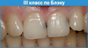
Pathology manifests itself synchronously on the distal and medial surfaces. Tooth decay changes the aesthetic appearance of the face when smiling. For treatment, do not use standard dental cement, amalgam, or cast liners.
The therapeutic procedure involves the removal of necrotic particles of dentin tissue. A filling material of the corresponding shade is selected according to a special table. White or transparent composite fillings are installed.
4th grade
Such caries affects the proximal planes of the teeth. The anterior units of the jaw row are most susceptible to class 4 pathology. The demineralisation process destroys the coronal angle and the incisal edge of the teeth. In the 4th class, caries passes with the development of the previous stage of the disease, mechanical damage to the enamel coating, trauma to dentinal tissues.
Treatment includes:
- etiotropic measures aimed at eliminating the cause of tooth decay;
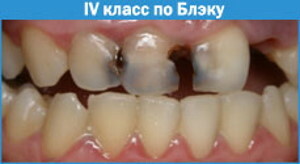
- removal of necrotic tissue and bacterial plaque in carious cavities;
- grinding the surface with a drill;
- installation of a composite filling of a suitable shade;
- restoration of the aesthetically pleasing appearance of the enamel coating.
If less than 1/3 of the tooth is destroyed, a composite restoration is performed, half or more - the installation of a crown. From dental materials used porcelain, cermets, zirconium dioxide.
Grade 5
The carious process affects the cervical segments of the teeth. The lingual and vestibular surfaces of the units of the jaw row are especially susceptible to demineralization. Carious cavities often form on the roots of molars.
Leads to grade 5 pathology:
- reproduction of pathogenic microorganisms;
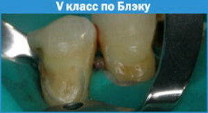
- erosive processes in hard tissues;
- hypoplasia of tooth enamel;
- mechanical damage to the cutting edge.
Classification of caries according to the scheme proposed by the American dentist J. Black, suggests performing a procedure for correcting the angular edges of soft tissues with the installation of a temporary filling to protect the pulp.
From dental materials, a cermet or photopolymer composite is used. To restore the aesthetic properties of the jaw line, veneers or lumineers are installed.
With an extensive lesion surface, a unit of the jaw row is increased using a special isomeric-composite composition. Destruction of the enamel coating in the smile zone is eliminated with a photopolymerizable gel-based material.
6th grade
This form of dental disease is characterized by the formation of deep pathological cavities in the coronal part of the units of the jaw row with lesions of fissures. Caries captures the dentinal tissue of the canines, premolars, incisors.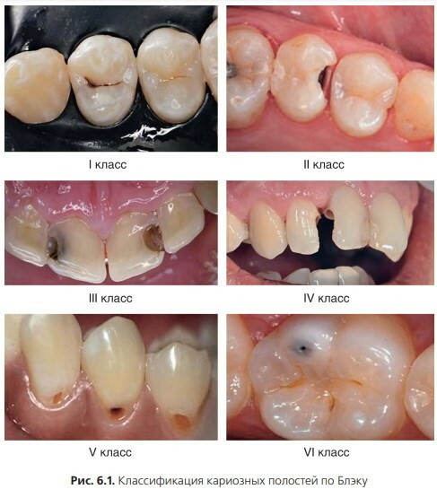
Defective changes are caused by a violation of the mineral composition of the tooth enamel, which leads to its rapid erasure and exposure of hard fibers. For the 6th class of carious pathology, a change in the bite and chewing ability of the units of the jaw row is characteristic.
Types of caries according to Black according to the depth of the lesion
The pathology grading scheme proposed by the American dentist was later refined and supplemented. The current interpretation of the topographic system takes into account the depth of the damage to the teeth.
She divides caries into the following types:
- The stage of formation of a pigmented spot behind the tooth enamel. The lesion has a whitish tint and is visually different from the surrounding tissues.
- Surface demineralization. The pathological process is characterized by a slight involvement of dentin tissues. Painful reactions to tactile and temperature effects occur.
- Average degree. The pathological process completed the destruction of the enamel coating in the rapidly growing focus of carious lesions. The pathogenic microflora makes structural changes in the hard fibers of the tooth.
- Deep defeat. At this stage of the pathological process, there is a rapid destruction of dentin. There is a risk of pulp damage and the development of periodontitis.
Deep caries is fraught with complications with irreparable damage to the jaw unit. There is a risk of tooth root destruction. In the gingival zone, stony deposits are formed, foci of infection with cariogenic microflora.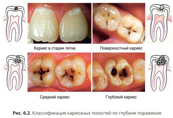
Such bacteria colonize mucous membranes with a high probability of colonizing the structure of the gastrointestinal tract and other functional systems of the body. Deep caries causes inflammatory and purulent-septic processes in the oral cavity, affects the salivary glands.
Black preparation steps
The procedure involves the opening of carious cavities, followed by cleaning the surface and installing a composite or photopolymer filling. In the course of therapeutic measures, the space is expanded, necrotic elements and bacterial plaque are removed.
1st grade pathology
Dissection is performed in stages.
Class 1 caries treatment includes:
- the use of local injection anesthesia, which stops the painful reactions of nerve fibers in the focus of therapeutic manipulation;
- instrumental opening of the carious cavity;
- cleaning the enamel layer and the dentin located under it from dead elements;
- preventive expansion of the prepared surface;
- preparation of the plane for filling;
- applying aesthetic gloss in a chosen way;
- the supply of ultraviolet radiation for solidification of the material when using a photopolymer composition.
The Black classification of caries makes it easier for dentists to choose the preparation method. When installing a permanent filling, the topographic scheme ensures that the final height of the processed jaw unit is adjusted to the adjacent teeth for bite correction.
In class 1 pathology, which is characterized by a shallow depth and insignificant extent of carious lesions, anesthesia is used at the request of the patient. Anesthesia is recommended for hypersensitivity of the enamel coating.
2nd grade pathology
Dissection of such a carious lesion involves the reconstruction of the natural interval between adjacent teeth, strong adhesion of the photopolymer or composite filling compound to dentin fibers.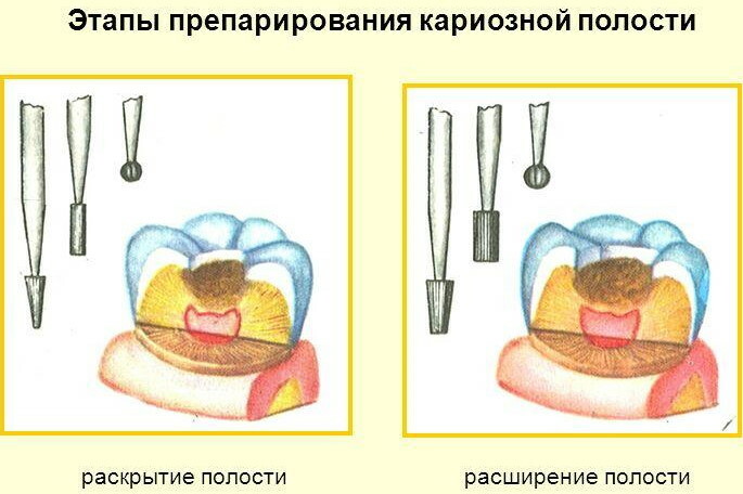
Reliable adhesion is only possible with tooth enamel. When placing a seal on the hard tissues of a unit of the jaw row in pathology of the 2nd class, difficulties often arise. To ensure reliable fixation, a special bonding material with increased adhesive properties is used.
The step-by-step procedure includes the following sequence of therapeutic manipulations:
- Anesthesia of the focus of medical intervention.
- Opening of the carious cavity to the entire depth of the lesion.
- Cleaning and correcting the gingival margin as needed.
- Restoration of the destroyed enamel coating by artificial spraying.
- Filling with composite material.
- Giving the dental surface an aesthetic gloss.
For pathology of the 2nd class, it is important to select the tone of the filling material in accordance with the individual natural a shade of bone tissue, which is due to the localization of carious cavities on the frontal surface of the teeth, called a zone smiles.
The peculiarity of the treatment of the disease at this stage is the involvement of the mucous membranes of the dental space in the inflammatory process. A general antiseptic is performed and when the filling is installed, the overhang of its edge over the gums is avoided.
Pathology of the 3rd and 4th grades
Pre-clean the tooth surface from microbial plaque. Just as in the previous case, the exact selection of the shade of the filling material is important. With pathology of the 3rd and 4th classes, there is a significant area of destruction of the tooth surface.
After injection anesthesia, a thorough cleaning of the deep carious cavity is performed, the gums are corrected with the removal of stones, and a filling is installed. The procedure ends with a final polishing of the affected area with adjacent tissues that are affected by the demineralization process.
When performing therapeutic manipulations with respect to caries of the 3rd or 4th class, gingival correction with suppression of inflammation of the mucous membrane is considered mandatory. Black classification of the pathological process facilitates the diagnosis and selection of filling material.
Video about tooth decay by Black
Preparation of carious cavities according to Black: