Content
- Definition of disease
- Classification and stages of development
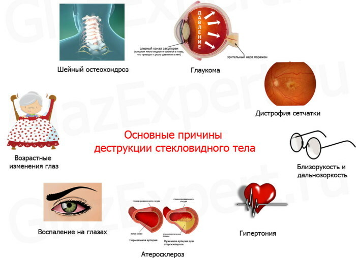
- Causes
- Complications
- Diagnostics
- Treatment of destruction of the vitreous body
- Medicinal effects
- Eye microsurgery
- Drops
- Vitamins
- Physiotherapy treatment
- Traditional treatment
- Forecast
- Video about the destruction of the vitreous body
Age-related causes, systemic or ophthalmological pathologies, hereditary predisposition cause a violation of the physicochemical properties and density of the vitreous body of the visual analyzer. To eliminate destruction, surgical intervention is used in combination with treatment with conservative methods.
Definition of disease
The morphological structure undergoes a gel-like vitreous filler that serves as a natural biological lens and is designed to refract light radiation.
Destructive transformations clinically manifest in outbreaks, floating opacities, dark objects of various shapes in the field of view. The pathology is common among elderly patients.
At a young age, the destruction of the vitreous body (DST) is caused by mechanical damage to the visual organ, progressive myopia, and other factors. Functional impairment is diagnosed with the same frequency in men and women.
There is no pronounced gender differentiation. Destructive changes in the biological lens change the shape of the organ and stretch the retina behind it, which leads to detachment.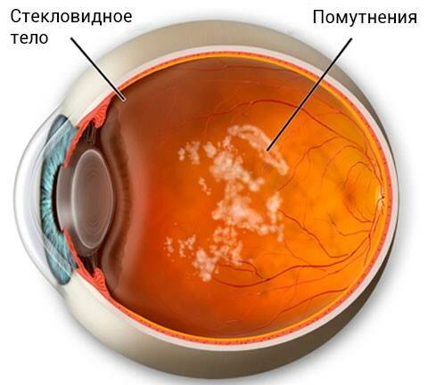
This form of the disease is called ZOST. Ophthalmologists do not make diagnostic and therapeutic differences between pathological processes due to the similarity of pathogenetic and clinical characteristics.
Sclerotic changes lead to structural disorders in fibrils - special transparent filamentous elements that form the basis of the gel substance of the optical lens of the eye and give it its integrity.
Destruction of the vitreous body (treatment with modern microsurgical methods allows restore the natural structure of the organ and visual function) is found in 25% of patients in aged 50-59.
Classification and stages of development
DST can be full or partial. More often, sclerotic changes affect the central portion of the inner space of the vitreous body. At an early stage of the development of visual pathology, a cavity is formed in the colloidal filler, filled with liquid and a coagulated mass of collagen substance.
With the progression of a destructive disease, structural changes are observed in fibrillar proteins that make up the molecular basis of filamentous formations. Coagulation of such substances causes an increase in volume and growth of organic matter.
As a result, the gelatinous mass is liquefied, which is filled with the space between the lens element and the reticular membrane. A pathological film coating and strands of various origins are formed.
Such structures attach to the fundus, provoking its wrinkling and causing the formation of adhesions of the sclerotic type. The progressing process leads to a decrease in the volume occupied by the vitreous body, deformational changes.
At the next stage of DST development, there is an excessive tension of vitreoretinal bundles attached to the retina, which begins to exfoliate. According to the forms, destructive transformations in the vitreous body are classified into filamentary, granular and crystalline.
Ophthalmologists call the trigger of the first type of systemic atherosclerosis or progressive myopia. Granular destruction of the vitreous body develops under the influence of inflammation in the retinal layer of the visual analyzer.
The forming foci with a changed morphological structure damage the surrounding colloidal substance. A rare form of DST in clinical practice is crystalline. The natural structure of the biological lens is damaged by cholesterol and tyrosine deposits.
The final stage of the development of the pathological process is the exfoliation of the posterior surface of the vitreous body from the sagittal plane of the eyeball.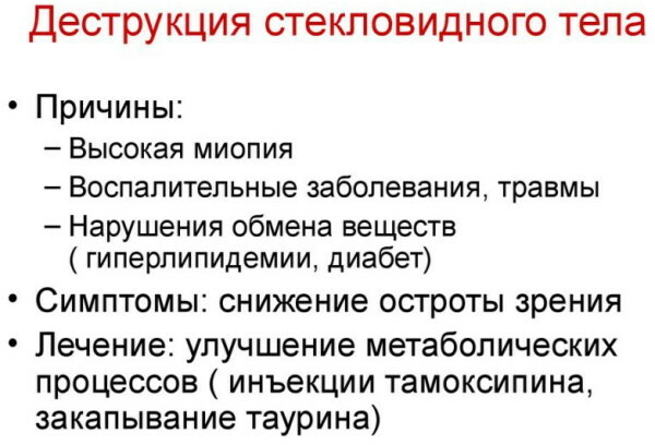
Happened:
- death of ciliary and pigment cells;
- penetration of blood cells into a gel-like substance from destroyed capillaries;
- free wandering of particles of membrane capsules of necrotic cytological units;
- inflammation of the retina.
Complete destruction encompasses the entire internal space of the organic lens. A partial type of pathology is characterized by the localization of sclerotic changes in the central segment of the vitreous body or on the periphery.
Causes
The etiological factors of CTD are numerous and varied.
Common causes of an ophthalmic disorder:
- Mechanical damage. Structural changes in the vitreous body cause contusions, craniocerebral trauma, penetrating wounds of the orbit.
- Local inflammatory processes. These reasons for the destruction of the vitreous body include chorioretinitis, uveitis, keratitis.
- Expressed myopia. Myopia with optical indicators of at least -6 diopters characterizes the stretching of the eyeball in the sagittal plane.
- Peripheral dystrophic processes in the retina or its breaks.
- Diabetes. Endocrine-metabolic pathology of the 1st and 2nd types, decompensated form, increases vascular and capillary permeability. Erythrocytes, leukocytes and other formed elements penetrated into the gel contents of the vitreous body damage the structure of the fibrils.
- Cardiological disorders. DST often develops against the background of tachyarrhythmias or bradycardia, stenosis of the coronary arteries.
- Hemodynamic disorders. Violation of the blood supply to the cerebral regions and the optic organ causes structural changes in the vitreous body.
- Systemic intoxication. Common causes of DST are infectious and purulent-septic processes, toxoplasmosis.
Destruction of the vitreous body, the treatment of which requires the use of radical methods of eye microsurgery, develops under the influence of acute vitamin deficiency, chronic micronutrient deficiency, amino acid deficit.
Such violations increase the fragility of the vascular walls. The etiological factor is intraocular neoplasia of benign or malignant nature. Less commonly, DST is caused by parasitic invasions. The larvae of some worms are able to penetrate into the structure of the vitreous body by hematological or lymphatic pathways.
Complications
Consequences of morphological transformations of a destructive nature in the organic lens of the visual analyzer are fraught with a destructive effect on the retina, leading to its detachment and loss of physiological functionality.
Damage to the central area that focuses light radiation causes a rupture of the macula, called vitreomacular traction syndrome. The resulting pathological crack leads to the formation of cystic edema.
This condition significantly reduces visual acuity. With the loss of central optical perception, the ability of spatial orientation is retained. Complete DST leads to blindness.
Diagnostics
The severity and specificity of the symptoms of ophthalmic dysfunction simplifies the determination of the destruction of the vitreous body. Questioning and physical examination of the patient quickly clarifies the clinical picture.
To confirm the diagnosis, set the stage and form of the disease, a set of hardware examinations is prescribed. Standard instrumental techniques for detecting ophthalmic pathology are presented in the table.
| Hardware technology | Purpose and essence of the survey |
| Ophthalmoscopy | Used to study the fundus, assess the transparency of light-sensitive media. The method detects signs of sclerotic changes, congenital or acquired developmental anomalies of the optical analyzer. The technology allows to identify inflammatory foci, destructive dystrophic areas, traumatic damage to the retina, vitreous body, optic nerve head. |
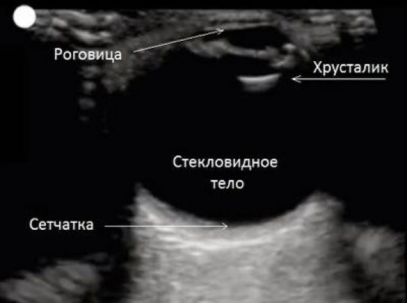
| Eyeball ultrasound | Ultrasound examination is intended for a detailed study of the structure of the optic organ, the state of the vascular network, muscle fibers and nerve tissues. Included in the standard diagnostic package for any ophthalmic diseases. |
| Biomicroscopy | It is used for intravital study of cells of the conjunctival sac, the anterior chamber of the visual analyzer, and the lens element. With DST, attention is focused on the structure of the vitreous body, reticular, corneal and iris. |
| Optical coherence tomography | Non-invasive method for studying the structural and morphological state of the visual organ. The technology is intended for probing retinal tissues with near-optical spectrum radiation. |
| Visometry | Hardware technology is used to determine the maximum visual acuity of distant objects. Allows you to establish the stage of destructive changes in the vitreous body. |
 Additionally, according to the indications, a perimetric examination is prescribed, which consists in determining the field of view. To measure intraocular pressure, the tonometric method is used.
The destruction of the internal structure and outer membrane of the vitreous body, provoked by vascular-cardiac or hemodynamic causes, leads to a hypertensive effect. Differential diagnosis using tonometry is needed to select an adequate treatment regimen for visual dysfunction.
With a partial form of pathology, the boundary membrane does not have specific changes, which is determined by the method of ophthalmoscopy. For complete destruction, the formation of a single hollow space with fragments of exfoliated fibrils is characteristic, which is detected by biomicroscopy or ultrasound examination.
Treatment of destruction of the vitreous body
Specific methods for eliminating sclerotic changes in the structure of the visual analyzer have not been developed. The complex of therapeutic measures combines surgical intervention with the use of drugs.
The therapeutic tactics are selected on the basis of the observed clinical picture, the degree of structural changes in the colloidal gel filler of the vitreous body. Therapeutic measures are aimed at restoring the natural shape of the retina and the natural lens to improve optical function.
Medicinal effects
Correction of the initial stage of destructive changes allows conservative methods of therapy that eliminate symptoms and prevent further development of the pathological process.
It is necessary to change the lifestyle and diet, observe the sleep and wakefulness regime. As part of drug therapy, local preparations are prescribed containing potassium iodide, which has absorbing properties.
Antioxidants are used to improve blood microcirculation in the eye vessels and capillaries. Vinpocetine and Cinnarizine are taken orally. The first belongs to the pharmacological category of cerebral circulation correctors.
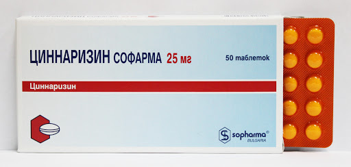
Cinnarizine is a selective calcium channel blocker. Systemic angioprotectors are used as part of complex drug therapy of the initial stage of pathology or partial CTD.
Combined drugs are prescribed to improve metabolic processes and metabolic reactions. Generalized drug therapy is focused on suppressing symptoms or has etiotropic significance, affecting the cause of destructive changes in the vitreous body and retina.
Eye microsurgery
With pronounced and deep sclerotic transformations in a natural biological lens, surgical intervention using vitreolysis technology is used. The technique provides targeted grinding of large collagen fragments.
Complete destruction is considered an indication for vitrectomy. The operation is performed under local anesthesia or systemic anesthesia. The vitreous element is removed with microsurgical technique, followed by plugging with silicone. Restoration of optical function takes 2-3 months.
Drops
It is impossible to eliminate the pathology with such pharmaceuticals. Ophthalmic drops are prescribed exclusively as part of complex symptomatic therapy or are used to accelerate postoperative recovery.
Depending on the etiological factor, the following types of sterile water-based or oil-based drip solutions are used: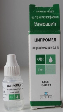
- Anti-infective. This pharmaceutical group includes Albucid, Uniflox, Tsipromed. Such eye drops suppress bacterial microflora, reduce the impact of parasitic infections.
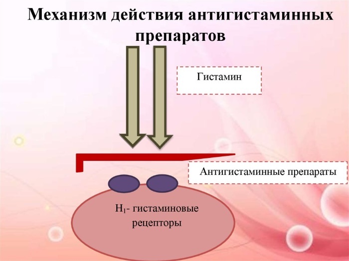
- Antiallergic. In the complex treatment of destructive changes in the vitreous body, provoked by systemic anaphylaxis, prescribe drugs from the pharmaceutical group of histamine receptor inhibitors, intermediates, glucocorticosteroids. These drops include Lekrolin and Allergodil.
- Anti-inflammatory. Such drugs are used to suppress aseptic inflammatory reactions and traumatic injuries that caused the destruction of the vitreous body. Non-narcotic analgesics and non-steroidal drugs are used.
- Regenerating. Effective for damage to the cornea or vitreous body of various origins. Regenerating drops are prescribed to speed up postoperative recovery.
- Moisturizers. This category includes artificial tear drops. Such preparations are saturated with hyaluronic acid. The most famous and widely used drops with moisturizing properties in ophthalmic practice are Hilo-Komod.
Destruction of the vitreous body, (treatment with sterile solutions is possible only in the initial stage with insignificance of sclerotic changes) requires a complex and systemic therapeutic impact.
Vitamins
Such pharmaceuticals are used for prophylactic purposes, in the framework of maintenance or rehabilitation therapy. With minor destructive changes, vitamin-mineral complexes can improve vision, prevent further development of the pathological process.
The composition of such drugs includes anthocyanins, carotenoids, and other physiologically active substances. The first ones prevent the development of tumor neoplasms and have a wide range of biological effects.
Among the proven beneficial properties of anticyanins are:
- antispasmodic;
- adaptogenic;
- stimulating metabolic reactions;
- mild antibacterial;
- decongestants;
- antihistamines.
Carotenoids are a class of natural plant pigments. They are synthesized by algae and some microorganisms. In the composition of vitamin preparations, such compounds are needed to enhance local immunity, prevent the destruction of cell membranes, and strengthen the vascular walls.
The combination of physiologically active substances of groups A, B, E reduces eye fatigue, normalizes the functions of the visual analyzer. For the prevention of destructive changes in the vitreous body, Vitrum Vision is prescribed, saturated with ascorbic acid, copper and zinc oxide.
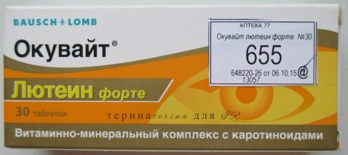
Ocuwaite Lutein Forte effectively compensates for the lack of vitamins.
The complex serves as a source:
- carotenoids;
- ascorbic acid, known for its anti-inflammatory and antibacterial properties;
- tocopherol;
- zinc;
- Selena.
The vitamin complex protects the visual analyzer from negative external influences, improves ocular hemodynamics, and strengthens the vascular walls. Elderly patients with diagnosed DST "Ocuwaite Lutein Forte" are prescribed to slow down age-related changes.
Physiotherapy treatment
Special eye gymnastics in combination with ophthalmic massage procedures are used for temporary suspension of the development of the destructive process, the displacement of collagen fragments from the central part of the focusing lens to periphery.
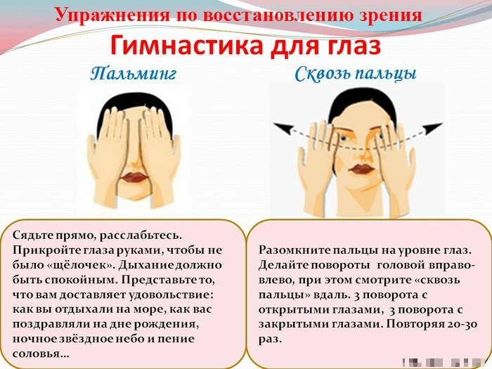
The physiotherapeutic methods of Bates, Zhdanov and Norbekov are effective. The first developed the technology of palming - exercises for relaxing the eye muscles.
Execution algorithm:
- Sit at the table with an emphasis on your elbows.
- Cover the eye sockets tightly with your palms for 5-10 minutes.
- Move the eyeballs in different directions without moving your hands.
- Make several circular movements with the eyeballs.
The destruction of the vitreous body (physiotherapy treatment is prohibited with retinal detachment) cannot be eliminated by gymnastic exercises. Such methods are used exclusively for preventive purposes with the approval of an ophthalmologist.
The massage procedure involves gently kneading the eyelids and the corners of the eye sockets with your fingers. The technique improves the condition of the skin of the periorbital zone and has a moisturizing effect by stimulating the lacrimal glands, but is unable to affect destructive changes in the vitreous body.
Traditional treatment
Unconventional recipes are only of secondary importance. Alternative treatment cannot serve as an alternative to drug therapy or surgery.
Such methods can only be used after a complete examination and consultation with an ophthalmologist. Folk recipes cannot eliminate destructive changes in the vitreous body and suppress clinical symptoms.
However, herbal remedies can enhance the therapeutic effect of pharmaceutical preparations, relieve eye tension, vascular and muscle spasms.
When using DST:
- honey-based eye instillation solutions;
- aloe tincture;
- propolis;
- cloves;
- drinking liquids with medicinal chamomile.
It is recommended to bury the eyes with herbal extracts or decoctions no more than 2-3 times a day. Special compresses are used based on starlets - a flower of the carnation family.
Forecast
With timely and adequate treatment, destructive changes in the vitreous body can be stopped. The prognosis of the course of the disease depends on the etiological factor that caused the sclerotic changes in the natural biological lens.
The situation can be significantly improved by maintaining a healthy lifestyle, changing the diet, and giving up bad habits. Patients with CTD are advised to undergo regular ophthalmological examinations to monitor the dynamics of optical impairment.
Favorable prognosis is ensured by microsurgical operation using a YAG laser and constant use within maintenance therapy of drugs based on L-lysine aescinat, which improves blood circulation in the capillaries of the visual organ.
To slow down the destruction of the gel-like filler of the vitreous body, it is recommended to perform daily massage of the periorbital zone and eyelids. The effectiveness of treatment and the favorable prognosis increase the use of fortified foods, the rejection of foods saturated with animal fats.
Video about the destruction of the vitreous body
Destruction of the vitreous body. "Flies" before the eyes: