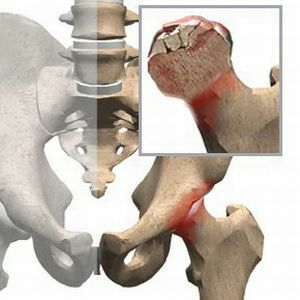
 Disease, called aseptic necrosis of the head of the femur, occurs quite often. Women suffer from them several times less often than men. It is characteristic that 2/3 of the patients are young people 20-45 years old. It is a rapidly progressing disease.
In the absence of proper treatment, it threatens to disrupt the function of the joint and, as a result, disability.
That's why it's important not to miss the first symptoms of the disease.
Contents of the article
- The main causes of the disease
- The main causes of the disease
- Vascular causes
- About the mechanical theory
- Exchange disorders and pathological conditions
- Symptomatics and diagnosis of the disease
- Diagnostic methods
- Treatment and pain relief with conservative methods
- Medication therapy
- Therapeutic gymnastics and massage
- Orthopedic rules
- Surgical treatmentdiseases
- Decompression of the femoral head
- Autograft transplantation from maloebe
- Endoprosthetics of the hip
- Video: What systemic diseases can provoke the development of necrosis of the GDK
- The main causes of the disease
The main causes of the disease
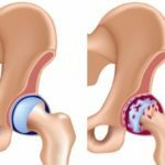
The head of the femur is a closed compartment that is sensitive to circulatory disorders that alter the architectonics of the bone.
Blood supply to the head is accomplished through three small arteries. When one of them stops( violates) the supply of blood, necrosis( ischemia, necrosis) occurs in the area of the head that was supplied by the damaged artery.
The essence of asthenic necrosis is a violation of microcirculation and further necrosis of the bone tissue in the head of the hip bone. As a result, the integrity of the cartilage covering this area is disturbed and a secondary deforming arthrosis develops.
Vascular causes of
Common causes of discontinuance of artery supply with blood from the head of the hip bone:
- compression or twisting of the artery in trauma,
- its occlusion by small thrombus,
- venous congestion,
- prolonged vasospasm,
- increase in blood viscosity,
- disturbed venous outflow.
Vascular disorders increase intraosseous pressure, resulting in mechanical destruction of bone tissue.
About the mechanical theory of
The vascular theory of causes is supplemented by a "mechanical" theory. According to her, the head of the femur undergoes "fatigue".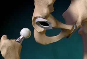
Impulses about this are sent to the cerebral cortex.
Feedbacks lead to vasospasm or blood congestion, disruption of metabolic processes, accumulation of decomposition substances in bone.
As a result, the physico-chemical and structural properties of the bone change, which gradually breaks down with difficulty in local circulation.
Exchange disorders and pathological conditions
Among them leads as the causes of the :
- ; long-term use of alcoholic beverages;
- long-term use of corticosteroids in large doses;patients with arthritis or bronchial asthma take corticosteroid hormones for a long time( metipred, prednisolone, etc.);
- chronic pancreatitis;
- large doses of radiation irradiation;
- caisson disease;
- osteomyelitis;
- sickle cell disease and other diseases,
- trauma( bruised thigh, hip dislocation, hip bone fracture, etc.).
One of the causes of the disease is a congenital defect in the form of a hip dislocation( hip dysplasia).
Symptoms and Diagnosis of the Disease
stages of aseptic necrosis of the head of the hip joint with distinctive symptoms:
- Initial. Pain serves as a starting clinical manifestation. It grows to the maximum and becomes intolerable in the first two or three days. Usually appears in the groin, less often in the thigh, knee joint, lower back. The joint fully retains its mobility.
- The second is an impression fracture. At the patient constant strong pains in a joint even in rest. During the period from several days to six months, vascular disorders develop. Possible atrophy of the muscles of the thigh. The aching leg seems to decrease in volume. Movement is limited. Lightness is noted in the gait.
- The third is a secondary arthrosis. For 6-8 months, the bone beams are destroyed, the femoral head is deformed. In the joint, severe pain is noted. Movement is limited in three directions. When walking, there is a starting pain, an average limp, a desire for support.
- Fourth .When the disease lasts more than 8 months, there is complete destruction of the head. Constant pains in the joints of the hip and knee, in the lower back. Movement is severely limited. Atrophy of the muscles of the buttocks and thighs is strongly pronounced. The aching leg becomes shorter, according to a more severe variant, it lengthens.
Diagnostic methods
The widely used methods for diagnosis of include:
- MRI .The early stage is detected by magnetic resonance imaging or computed tomography. This method of diagnosis almost 100% reveals the disease, when the X-ray does not "see" it. Therefore, during the first weeks of the disease, diagnosis with MRI is a priority.
- Radiography .Aseptic necrosis on the X-ray is made visible only in 2-3 stages of the disease. When the disease has an "experience" of more than a year, its signs are very clearly manifested in the pictures. At this stage, the tomogram is not needed.
- Radioisotope scanning .This method shows the unequal absorption of the radioactive preparation by pathological and normal bone tissues. The administered dose of the drug serves as a "mark" for the abnormal zone in the bone. The result is a two-dimensional image where the affected areas of the bone are visible.
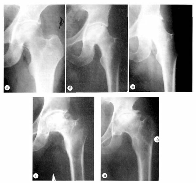
X-ray patterns of patients with different stages of aseptic necrosis of the femoral head: from a - the initial stage, to d - complete destruction of the bone.
Treatment and pain relief with conservative methods
Drug therapy
The main groups of medicines used to treat the disease include:
- Anti-inflammatory non-steroid preparations , for example, diclofenac, indometacin, piroxicam, butadione, etc. They contribute to reducing pain in the thighand groin. This group of drugs does not cure the disease. But due to anesthetic action, prevention of reflex spasm of muscles occurs with pain. These drugs are especially effective in the first six months of the disease.
- Vessel dilatation drugs , for example, trental, theonikor. They eliminate stagnation in the circulation. As a result, the arterial blood flow is activated and the spasms of small vessels are removed. Decreased vascular night pains in the affected joint. Effective in the first 6-8 months of ailment.
- Bone Restorers .To stimulate the recovery process help with vitamin D( calcium D3 fort, oxide oxidation, napokal D3, etc.).These drugs contribute to the accumulation of calcium in the head of the affected bone of the thigh.
- Calcitonins effectively stimulate bone formation and eliminate bone pain. These include miacalcic, sibacalcin, alostin, etc.
- Chondroprotectors ( chondroitin sulfate and glucosamine) provide cartilage tissue and restore the structure of the destroyed cartilage. Treatment gives an effect in the period of the disease from 8 months.
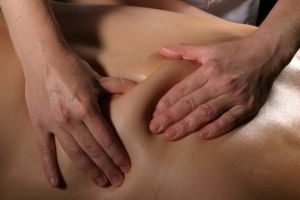
Therapeutic gymnastics and massage
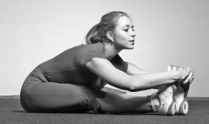 One of the most important methods of treatment of necrosis of the head of the femur is therapeutic gymnastics .Without it, it is impossible to overcome the progressive deterioration of blood circulation in the region of the femoral head and the growing atrophy of the hip muscles.
You need to choose exercises to strengthen the muscles and ligaments of the sore leg. And there should be no pressure on the head of the hip bone without active flexion-extension of the legs.
An example of a static exercise is a small rise of a straight leg in a position on the back of a prone. The leg is supported by weight. There will be fatigue, although the joints do not work. A set of exercises should be carefully thought through with the doctor.
Therapeutic massage is used as an additional method of treatment. But if you do it correctly, without rough pressure, it will bring real benefits. With massage of the femoral muscles and back, blood circulation improves.
 Pains in the hip joint can talk about the development of arthrosis. How to properly treat coxarthrosis read in our stelye. Chto do, if you have a suspicion of deforming arthrosis of the ankle joint? The measures that need to be taken first look here.
 Orthopedic rules
In their opinion, this threatens:
- progressive muscle hypertrophy,
- by the formation of pain resistant syndrome,
- violation of motor stereotypes.
For to facilitate the flow and reduce the duration of the disease, the following:
- walking up to 20 min.average pace,
- walking up the steps,
- swimming,
- exercises on the stationary bike,
- using canes in the first weeks and with long walks,
- fighting overweight.
It is necessary to exclude inertial loads on the joint in the form of lifting of weights, jumps, running.
Surgical treatment of
disease Surgery is resorted to when conservative drugs are ineffective.
Femoral head decompression
The operational decompression method consists of drilling a channel into the femoral head region with no blood flow. The drill passes along a large trochanter and neck of the hip bone.
Decompression objectives :
- increased blood supply of this site due to the growth of new vessels in the formed channel( puncture),
- decrease in intraosseous pressure in the head of the thigh.
By reducing the pressure in 70% of patients reduce pain.
Transplant of autograft from fibula
Unlike decompression, a fragment of fibula, located on the vascular pedicle, is transplanted into the drilled cavity. Such a transplant from your own body gives an improvement in blood flow and strengthening of the neck of the thigh.
Endoprosthetics of the hip joint
It consists in the complete replacement of the damaged hip joint by an artificial one. A titanium pin( or zirconium) with an artificial head on the edge of the joint is inserted into the formed cavity of the hip bone and fixed.
Simultaneously, the second articulating part of the joint is operated, inserting a concave bed to rotate a new head in it. Correctly done surgery removes pain and restores joint mobility.
In another part of patients, the condition stabilizes, which does not lead them to surgical measures.