1 What are they looking at?
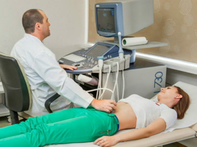
The dimensions of the internal organs of a person allow us to consider them as thoroughly as possible using a sensor, check the structure, the thickness of the walls, and determine the echogenicity.
OBP( organs of the abdominal region), which explores ultrasound:
Do you have gastritis?
GALINA SAVINA: "How easy is it to cure gastritis at home for 1 month. A proven method - write down a recipe. ..!"Read more & gt; & gt;
- The pancreas is located in the retroperitoneal space and is protected by ribs.
- The liver, located mainly on the right side, is one of the largest human organs.
- Spleen, closed by ribs on the left side.
- Kidneys are not located in the lower parts of the spine, but immediately below the ribs.
It is recommended to read
- What is the diffusely heterogeneous structure of the pancreas
- When to do MRI of the abdominal cavity and retroperitoneal space
- What is carried out and what does the biochemical blood test show in adults, children
- Effective agent for gastritis and stomach ulcer
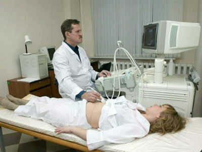
What does ultrasound show? First of all, the expert evaluates the location of the organ, checks whether there is any displacement. After this, measurements are made, the form and the sharpness of the boundaries are established. The doctor on ultrasound will check if there are any lesions, will determine the presence or absence of fluid in the abdominal space. Based on the findings, the doctor makes his assumptions about the patient's health, makes a diagnosis.
The attending physician will help to interpret the results of the ultrasonic sensor, because for the most part the conclusion of this procedure consists of medical terms and numerical measurements.
Ultrasound of the abdominal cavity, the decipherment of which is performed by a doctor, should be carried out with special preparation. On the eve of the echography should be observed diet, directed against the accumulation of gases. Often, gas accumulations are taken by the ultrasound doctor as neoplasms, and then an incorrect diagnosis is made. It is necessary to exclude from the diet legumes, fatty varieties of fish and meat, bread, fruit, carbonated drinks, sweets.
The attending physician may prescribe a drug aimed at getting rid of flatulence - Espumizan - or ordinary Activated Carbon. Medications are taken the day before the procedure. If the patient is suffering from constipation, then an enema is made to clean the intestines of the contents.
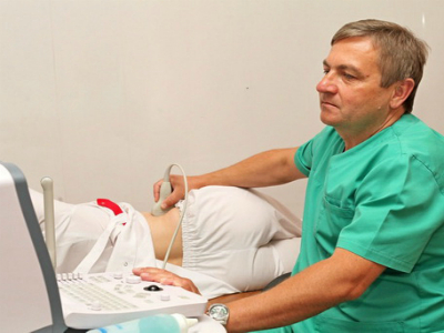
The procedure of ultrasound of the abdominal organs lasts from 15 to 30 minutes, then the patient gets results in the hands of the patient. They can be partially deciphered independently, but the final diagnosis is made by the attending physician.
2 Diagnosis of the pancreas
The pancreas is an important organ of the digestive system, its main functions include digestion of food and the establishment of carbohydrate metabolism in the body.
First of all, the doctor evaluates the shape of the organ, in unchanged form the pancreas resembles the letter S. If the ultrasound shows another form, it will be considered a deviation from the norm.
-
 IMPORTANT TO KNOW! Gastritis? Ulcer? To have a stomach ulcer not turned into cancer, drink a glass. ..Read the article & gt; & gt;
Defective pancreas occurs as follows:
- In the form of a ring.
- Spiral-shaped.
- With doubling one of its parts.
- Double - split.
Deviation in the form of this organ can be both congenital and pathological, acquired. Often this defect has no effect on the quality of life of the patient.
Once the shape of the pancreas is established, the doctor makes measurements.
The regulatory parameters are as follows:
- The size of the gland itself is from 15 to 22 cm.
- Weight varies from 70 to 80 g.
- Head dimensions, including hook-shaped process, from 2.5 to 3 cm.
- Tail 1-2 cm.
- Body insideglands 1.5-1.7 cm
An error of up to 2-3 mm is permissible. For children, these indicators are reduced, their rate is set when compared with the growth of the child.
Normally, the outline of the pancreas should be clear, even, without any peculiarities.
One of the most common diagnoses that is put on the basis of ultrasound is pancreatitis. To confirm the disease, the treating doctor must also be assigned other tests, since echography is an additional study that can both confirm and refute the diagnosis.
-
 Gastroenterologist. IMPORTANT: "I beg you, if you began to worry about abdominal pain, heartburn, nausea, do not in any way do gases. .."Read more & gt; & gt;
Pancreatitis - inflammation of the mucosa of the pancreas, often accompanied by severe pain. Occurs for many reasons - infections, alcohol, endocrine gland diseases, trauma, bacteria. In the case of this diagnosis, ultrasound will record a number of the following symptoms:
- Body size exceeds normal values.
- Darkening is traced.
- The structure of the gland is not uniform.
- Fluid is detected.
- The main duct of the gland reaches 2 mm.
If there is a suspicion of a tumor, the following signs have been revealed by the specialist:
- In most cases, the boundaries of the tumor are well traced and have a clear contour.
- The nearest lymph nodes in size exceed the norm.
- Protocols extended.
- The pancreas has uneven borders.
- Metastases can be found in neighboring organs, for example, in the liver.
3 The norm and pathology of liver research
The liver takes on many functions - it is participation in digestion, and the body's defense against bacteria, and the filtration of harmful substances. Before visiting a doctor, you need to learn as much as possible about ultrasound of the abdominal cavity: what the echography shows, what standards for the liver exist.
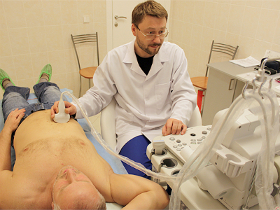
A properly formed liver has 4 parts: right, left, tail and square. The largest sizes are the right and left lobes, while the other two are much smaller.
TIP FROM THE MAIN GASTROENTEROLOGIST
Korotov SV: "I can recommend only one remedy for the rapid treatment of Ulcer and Gastritis, which is now recommended by the Ministry of Health. .." Read the reviews & gt; & gt;
Norms of liver lobe sizes:
- The thickness of the left lobe varies from 5 to 8 cm, and the height is 8-10 cm.
- The right side is even larger, the thickness is 9-12 cm, the height is 8-13 cm, the vertical oblique size is up to 14-16 cm.
- The hvostataya share should not exceed 7 cm in length and 2 cm in width.
- The length of the whole organ is 13-18 cm, the cross section is up to 23 cm. The weight of the liver in men reaches 1500 g, in women the norm is up to1300 g.
The specialist measures the sources of the organ blood flow - it is a portal vein, its norm is up to 15 mm, and its own artery, up to 6 mm.
Fibrosis of the liver is an overgrown connective tissue, it appears on the liver tissue sites. The disease has 4 stages, the latter grows into cirrhosis of the liver.
Confirm or refute the disease will ultrasound of the abdominal cavity, which once again confirms the need for this procedure. Ultrasound diagnosis does not reveal the stage of the disease, this is the responsibility of the attending physician, studying the results of other analyzes.
Signs of fibrosis:
- Surface of the liver in the mounds and waves.
- The structure of the organ is uniform, grainy interspersed.
- Increased blood pressure in the veins of the liver.
- The walls of the vessels are pronounced.
Hematomas inside the liver are also detected by ultrasound. They appear because of physical trauma or surgical intervention.
WE RECOMMEND!
For prevention and treatment of gastrointestinal diseases our readers advise Monastic tea. This unique remedy consists of 9 medicinal herbs useful for digestion, which not only supplement, but also strengthen each other's actions. Monastic tea will not only eliminate all symptoms of the gastrointestinal tract and digestive system, but will also permanently eliminate the cause of its occurrence.
Opinion of doctors. .. "
The hematoma is the damaged vessels. It looks like an oval formation with a liquid inside. At the initial stages has vague boundaries. If the number of damaged vessels grows and the bleeding continues, the ultrasound will fix it.
The study diagnoses cancers only if the tumor is more than 2-3 cm, with smaller dimensions it is difficult to distinguish the neoplasm.
Symptoms of oncology:
- Increased liver size.
- Neophytes have fuzzy, often hummocky borders.
- Increased blood supply.
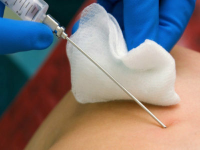
Complete diagnosis of the disease involves puncture of the liver.
4 Inspection of the spleen
The ultrasound of the abdominal organs, namely the spleen, will show the already existing defects, in addition, will warn possible deviations. For a long time, the functions of the spleen were incomprehensible to doctors, they are still not fully understood, it is known that the body produces antibodies, removes defective red blood cells and bacteria.
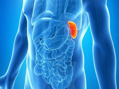
Spleen size norm:
- Length is 10-13 cm.
- Width from 7 to 9 cm.
- Thickness not more than 4-6 cm.
The artery leads to the spleen, its diameter is 1.5 mm, and the vein has a diameter up to6-8 mm. The organ resembles the shape of a half moon, located under the ribs, between the chest and stomach.
Echogenicity - a reflection of the sound wave - should be average, a small-sized vascular reticulum on the organ is allowed.
On the pathological changes in the background of tumor formations, the following signs indicate:
- Increased size and thickness of the spleen.
- Pointed edges of the organ.
- Increased size of the proximal lymph nodes.
- Rough surface.
Tumor grows quickly, in the future, its size is controlled by ultrasound. Often the oncology of this organ is confused with pathological increases in the size of the spleen, diagnosis of cancer in the early stages is difficult. In time, the detected disease is cured in 80% of cases.
Based on only the results of this study, the diagnosis is not posed, additional tests are needed.

On suppurative inflammations testify:
- Spleen cyst.
- The pathology of the echostructure is mixed or hypoechoic.
Abscess - the onset of a purulent sac in the spleen. It can appear because of previous infections of internal organs, mechanical damage, against the background of scarlet fever or malaria. If untimely medical care is provided, a lethal outcome is possible.
In the case of an abscess on ultrasound, you can see:
- Fuzzy boundaries.
- Fluid in the abdominal cavity.
- Pathology of echostructure.
5 Possible kidney diseases
Kidneys are the main filter in the human body. Because of the heavy load, they are susceptible to many diseases.
The normal size of the kidneys:
- The width does not exceed 5-6 cm.
- The length of the kidney reaches 10-12 cm.
- Thickness 4-5 cm.
- Tissue cover - parenchyma - has a thickness of 1.7-2.2 cm. Normvaries depending on the age of the patient.
The shape of the organ resembles a bean - it is indicated in the doctor's opinion, the kidneys are not on the same straight line - the right one is slightly lower than the left one. Contours equal, well traced, clear.
The doctor checks and adrenal glands, sometimes they can not be visualized - this is the norm option. The right adrenal gland has the shape of a triangle, and the left one resembles half the moon.
Normally, organ pelvis is not visualized. If the doctor saw them, then it indicates the diseases of the urinary tract, the presence of stones, neoplasms.
Cancer can be diagnosed on ultrasound in 96% of cases. In conclusion, this diagnosis may sound like an echo-positive education.
Symptoms of kidney cancer:
- Uneven edges.
- Places increased density of the body.
- Possible areas of necrosis.
The renal cyst has similar characteristics, but the organ does not change its contours - it is well visualized, has clear boundaries.
Stones can not always be diagnosed on ultrasound, if they are detected, then they are listed as hyperechoic formations. During the study, they can move inside the organ.
ultrasound reveals acute inflammatory processes of the kidneys.
Symptoms of pyelonephritis:
- Swelling of the tissues of the organ, possibly a hemorrhage.
- Extensions and contractions of the sinus are traced.
- Shadows with obscure outlines.
- With severe disease, abscesses are visualized.
ultrasound detects complicated forms of pyelonephritis, for example, emphysema. It is characterized by a large number of harmful bacteria living inside the kidney and emitting gases that are tracked on the study.
After the ultrasound examination of the abdominal cavity, the interpretation, the norm of the indices can be interpreted by the patient himself, but the statement of the exact diagnosis and the appointment of the treatment is the doctor's business.
Ultrasound diagnosis today is an auxiliary research method, other tests are needed to confirm the diseases.
- 1 What does the survey look like?
- 2 Pancreatic Diagnosis
- 3 Liver Research Norm and Pathology
- 4 Spleen Inspection
- 5 Possible Kidney Disease
When the doctor prescribes an ultrasound of the abdominal cavity, the decoding, the patient's norm of indices will be provided quickly. Internal organs and systems check by this method does not always give a clear picture of their usefulness, therefore ultrasound is included in the complex study of the patient.

