 In the human body, bones can be articulated in both a movable and a fixed way.
The main type of joint is the joints, the mobile joints, which include the articulating parts of the bones, the joint cavity, the capsule and the retaining ligament. In a smaller amount, there are syndesmoses.
These are inactive joints of bones through an interlayer or strand of dense connective tissue, they are present between the bones of the skull, shin, forearm, and spinous processes of the vertebrae.
In the form of a layer of connective tissue, syndesmoses can look like a membrane( membrane), a seam or "piercing".The membrane is connected to the tibia with fibula, elbow bone with radial, transverse surfaces of the vertebrae and their spinous processes.
In the form of seams, syndesmises are present between the cranial bones. They are subdivided into scaly, flat and serrated. The term "piercing" refers to the connection between the dental root and the inner surface of the alveoli.
Article Contents
- Features injury
- gap tibiofibular membrane
- clinical picture
- Conservative therapy
- surgery method also
- Features ankle injury
- reasons
- injury Treatment after injury
- injury prevention
Features
injury most often damaged the interosseous membrane between the tibia in the lower third, anterior and posterior syndesmosis, which form part of the ankle joint and are involved in providingits stability.
In general, injuries occur in athletes when jumping or running, in ballerinas, circus performers. It looks like isolated stretching or rupture of ligament cords, and also as a combination with fractures of bones or detachment of the ligament fragment together with bone fragment.
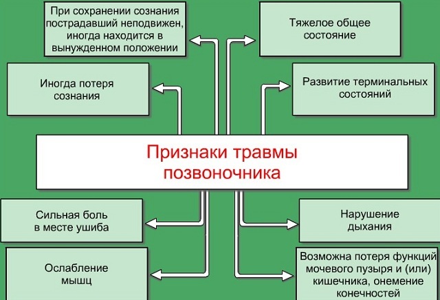
Damage to cranial or vertebral syndesmoses always accompanies craniocerebral or vertebral injuries.
In particular, with birth trauma in newborns, the interosseous membrane of the skull may be ruptured or have hemorrhages.
In a compression fracture of the spine, when one vertebra is pressed into the other, interstitial and transverse syndesmoses are often not completely ruptured, but can be stretched or have partial damage to fibers with hemorrhages.
Interstice membrane rupture
Interstitial syndesmosis is represented as a sedentary connective tissue membrane connecting the medial surfaces of the 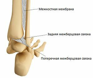 of the tibia and peroneal in the entire length.
The main part of the membrane is called the interosseous membrane, only its lower part is called the actual interdental syndesmosis.
Normally, the width of the intercellular cleft is no more than 3 mm. Fibrous fibers covering it, are crossed or parallel to each other and go into several layers, of which the inner ones are more durable, and the outer ones are more likely to be subjected to stretching and tearing.
This explains the possibility of a partial rupture of the distal interbody syndesmosis.
Almost all injuries of the lower leg, especially in the lower third, are accompanied by injuries of the intercostal joint, and 10% of the ankle joints are "upper", that is, they are accented in the syndesmosis.
None of the injuries of the intercellular membrane are insured, although features of some professions( ballet, circus art, choreography) increase their risk.
Tears and sprains of the intercostal joint are involved in combined injuries in many car accidents and accidents, they can be obtained by falling on a slippery road or from a low altitude.
High heels also increase the possibility of injury. The most common is the rupture of the intercellular membrane with fracture and dislocation of the ankles, with strong pronation( turning outward) of the foot and simultaneous rotation of the foot( turning the toe inward).
With this mechanism, the rupture occurs along the line of attachment of the fibers to the tibia.
Clinical picture of
Symptoms in the trauma of syndesmosis consist of severe pain, which increases with palpation and changes in the position of the foot, and swelling that increases with every minute.
The stop takes a forced, unnatural position( more often turned outwards), the area of the lesion is hyperemic, there are hemorrhages.
Radiography in such cases is mandatory. In pictures taken in two projections and with a load on certain ligaments, the widening of the intercellular fissure, the line of rupture, the presence of fractures are clearly defined.
With the help of the radiograph, stretching of ligaments, partial rupture of the membrane joint, and clarifying the treatment tactics can be avoided.
Conservative therapy
Uncomplicated partial and complete rupture of the intercostal articulation is treated with conservative methods.
 Novocain blockade is used for anesthesia. The main task is to completely immobilize the injury site, compress the enlarged intercellular cleft, allow time for the ligaments to recover independently.
For this, an ankle joint is applied with gypsum in the form of a boot for 5-6 weeks. Then it is removed, replaced by a removable tire for another 2 weeks. At the same time they begin to conduct physiotherapy, massage, and exercise gymnastics.
Conservative treatment is long-term and not always effective at 100%.For another 6 months, a decrease in functionality and soreness may be of concern.
Surgery is also the method of
With advanced or complicated forms of tearing of the intercostal articulation( failure with conservative attempts to close the gap between the shin bones, improperly fused ankle fractures), therapy consists in applying surgical methods.
The two methods are traditionally used:
- The first is a tandoplasty, that is, the transplantation of a part of the wide fascia of the thigh, the canned tendon or the tape from the lavsan to the place of the ruptured syndesmosis, its complete renewal. A new ligament is implanted in canals drilled in the tibia. The percentage of complete recovery is 92, this is a very good result.
- The second surgical method gives the greatest strength to the ankle fork and consists in the use of a bolt-screed or compression screw. Its essence is to install a reliable tightening mechanism from a special metal alloy, which fixes the bones of the shin at a certain distance from each other, preventing them from fusing or moving, and hinders the contraction of the ankle joint.
Tears of the intercellular membrane are accompanied by vascular disorders. Sometimes thrombosis of venous vessels is possible. To prevent such complications, anticoagulants and angiotropic agents are prescribed, which accelerates recovery.
Features of ankle injury
Syndhesmosis of the ankle joint is formed by the interosseous membrane, as well as transverse, anterior and posterior intercellular ligaments.
It firmly fixes the ankle joint, not allowing its components to move relative to each other. Injuries of syndesmosis occur quite often: 20% of all damage to the support apparatus.
Of these, up to 12% falls on partial and complete ruptures of the ligamentous apparatus.
Causes of injury
Causes of injuries - the effect of lateral or direct force on the joint. 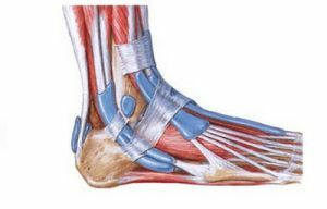
Various strokes, collisions, slip or fall with podtytyvaniem feet - the most common etiological factors.
Symptomatic of stretching and tearing of ankle syndesmosis is similar: sharp pain, hyperemia and growing swelling of the joint, bruises, unnatural and sparing position of the foot, its deformation.
Often the injury is combined with ankle fractures, with and without bias. To carry out differential diagnostics will help radiography.
The images in several projections clearly differentiate the condition of all ligaments and bone components of the joint.
Treatment after trauma
Therapy of fresh( up to 20 days) ankle syndesmosis injuries is conservative.
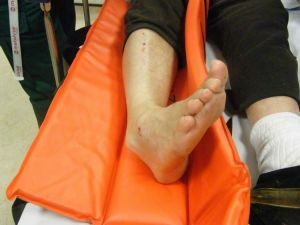
It consists of local anesthesia, reposition and fixation of the foot in a normal position, the imposition of a plaster bandage and complete rest.
Older breaks( more than 20 days) are best immediately treated surgically. If possible, the ligaments are sewn or produced by their plastic, while in the presence of fractures, they fix the fragments with the help of metal structures.
Prevention of injury
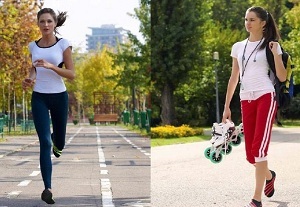 Measures to prevent the trauma of syndesmosis consist in wearing comfortable shoes with a wide and low heel, caution on ice, running and jumping.
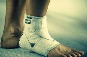
It is necessary to strengthen the ligament apparatus with a full-fledged diet, exercise by feasible kinds of sports. Professional athletes use special fixative bandages.
Therapy for syndesmosis injuries should be started on time, as neglected cases are treated longer and more difficult. Correct and competent treatment restores all the functions of the joint in full.