 Forearm injuries are the most common injuries. The forearm consists of the elbow and radius bones. At the top they are directed to the elbow, below - to the wrist. The ulnar bone approaches the little finger, and the radial bone - to the thumb of the hand.
Fracture of the arm beam is the following the fall on the outstretched arm of the .
Injuries associated with fracture of radius:
- fracture of ulnar bone;
- dislocation near the located bones;
- ligament tears.
These injuries account for a quarter of the total number of fractures of the bones of the hands and 90% of the fractures of the bones of the forearm. In women, fractures of the radius in a "typical place" occur 2 times more often than in men. The reason for this is the lower density of bone tissue of the female body.
Contents of the article
- Possible causes of fractures
- Other causes of
- fractures Other types of beam injuries
- Diagnostic methods
- First aid for fracture
- Non-surgical treatment
- Surgical treatment
Possible causes of fractures
Wedde most common causes of fractures of the radius arms recovered are:
- fall on an outstretched hand;
- Osteoporosis - increased brittleness of bones, especially in cases of stress and shock, is typical for people of 60 years of age;
- car accident;
- fall off the bicycle;Injury in the workplace,
- , etc.
This is due to the anatomical structure of the bone, which in some places is more delicate. Accordingly, in these places it is easier to break.
There are 2 types of damage:
- Fracture of the Wheel - a fragment of the radial bone is pushed to the back of the forearm. He bears the name of a surgeon who first described this type of fracture. Such a break is called extensor.
- Smith Fracture is the opposite of the fracture of the Wheel. Displacement occurs in the direction of the palm. For the first time such a case was described by a doctor in 1847.It is called the flexor.
 It is especially important to know the first signs of fracture of the ribs. It is this fracture that most often results in damage to the internal organs. See our article for more details.
Timely first aid for a collarbone fracture determines the overall success of treatment and subsequent rehabilitation. Details you can find here.
Other types of ray injuries
Among other types of fractures are:
- intraarticular - the fracture line covers the wrist joint;
- extraarticular - does not span the joint region;
- open is accompanied by skin damage;
- closed fracture of the radius;
- fracture of the cervix of the radius;
- Splintered - the bone is broken into 3 or more pieces;
- is primarily open - skin damage is observed outside the bone;
- re-opened - skin damage from the inside.
Classification of fractures is important due to the fact that the type of fracture depends on the method of its treatment.
What happens with a fracture
Symptoms of a fracture of the radius: 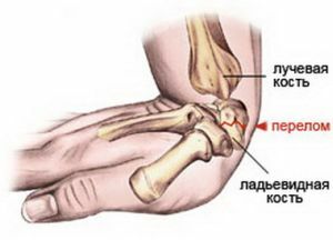
- pain in the joint, which are strengthened when moving by hand;
- forged movements;
- edema;
- hemorrhage in the joint;
- swelling in the area of the shoulder joint.
Diagnostic methods
Such fractures are clinically weakly expressed, therefore the final diagnosis is made after the examination of the X-ray. In addition, you need to consider whether a ray fracture is combined with a fracture of the ulna or dislocation.
Types of diagnostics
The main diagnostic methods are:
- Conventional radiography in 2 projections is the most popular and available method for diagnosing fractures.
- Computed tomography - is relevant for intraarticular fractures to assess the alignment of the joint surface. In the postoperative period gives precise information about bone fusion.
- Magnetic resonance imaging - is used to diagnose complex fractures, combinations of several fractures.
Treatment and first aid
First aid for fracture
Professional first aid and prompt treatment to the doctor is the basis of competent treatment and a prerequisite for restoring all the functions of the hand.
With a closed fracture, you must immobilize the injured limb with a solid tire or other improvised means. The tire is applied from the middle of the shoulder to the base of the fingers.
The arm is bent at right angles and placed in a kerchief tied around the neck. Reduce pain by injecting analgin or applying ice to the site of injury.
With open fracture, it is necessary to stop bleeding, disinfect the wound and apply a clean bandage. To prevent blood loss during arterial bleeding, you need to put a tourniquet on the middle of the shoulder. The fixing bandage is the same as with a closed fracture. Ice will help to remove swelling. Then the patient needs to be hospitalized.
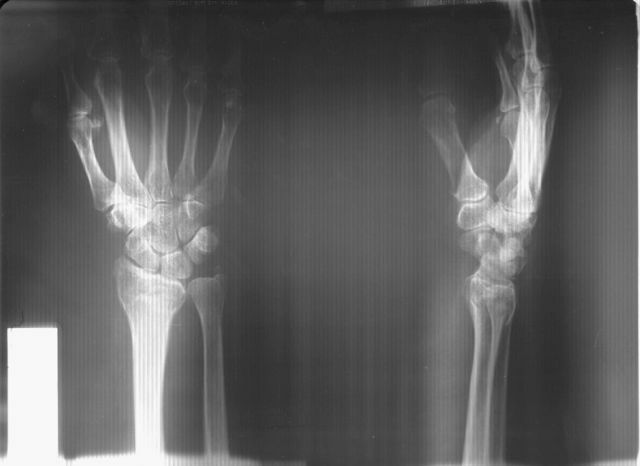
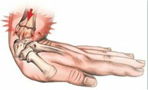
The photo shows a fracture of the radius of the hand
Treatment procedures
In order to correctly treat a fracture, you must first evaluate the nature of the damage, and only then choose the method.
There are many treatment options.
Non-surgical treatment
Fractures of the ray without bias are fixed with gypsum or polymeric bandage. If the fracture of the radial bone with displacement, then the parts of the bone are put in the correct position and fixed until the fusion.
In time, untreated treatment threatens the development of joint arthrosis and loss of mobility of the hand.
The limb will remain stationary for 4-5 weeks.
Then the doctor prescribes a referral to the exercise room, where after a fracture of the radius, the joint undergoes the necessary rehabilitation.
Surgical treatment
The operation for fracture of the radius is applied in case of inability to properly support the bone before adhesion with gypsum. In this case, physicians perform a fixation with their knitting needles through the skin or operation, a closed reposition and fixation with knitting needles through the skin, is the most popular method of international medicine.
First, the doctor closes the displacement, then inserts the knitting needles in certain directions.
Negative points:
- risk of infection of the wound and infection instead of fracture due to the presence of the spokes over the skin;
- prolonged wearing of plaster bandages;
- risk of lack of movement in the joint due to late development.
Open fracture repositioning
An incision is made, the muscles and tendons are moved away, and the fragments are repositioned in the 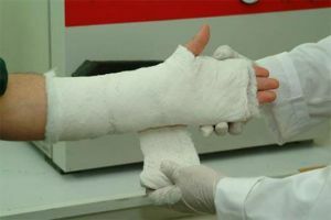 in the correct position. The bones are fixed with metal plates.
In this case, wearing gypsum is not required, becausethe bones are in the correct position due to the plates.
External fixation devices
Are indicated for wearing with contraindications to the use of plates and screws. With all open fractures, the patient should be operated on as soon as possible, thoroughly disinfecting the tissues around the fracture. The wound is sutured and the apparatus is fixed for 4-6 weeks.
Negative moments:
- devices are expensive;
- risk of infection due to rods over the skin;
- uncomfortable dressings and wound treatments;
- risk of lack of dynamics in the joint.
Recovery after fracture
The types of fractures of the radial bone are so different as the ways of their treatment, then the rehabilitation after the fracture of the radius for each patient is selected.
The hand grows after 1.5 - 2 months.
At first, after fracture for the removal of pain and swelling use UHF and ultrasound. Also, after a fracture of the radius, exercises are useful to restore blood flow and prevent muscle damage.
If the patient is operated with a plate, the doctor will prescribe the LFK joint after 7 days after the operation.
After the end of the splicing period, the following restoration procedures are prescribed:
- exercise therapy;
- massage;
- phonophoresis.
After convalescence warm coniferous, coniferous-salt baths are useful.
It all depends on the patient. How much he himself is stubbornly struggling to restore limb mobility.
Possible complications of
For non-surgical treatment with the application of gypsum or a polymeric bandage, it is necessary to observe the brush. To look, whether there is an edema, whether pales fingers, whether sensitivity remains.
Preventive measures
At the heart of prevention of fracture of the ray of the upper limb lies:
- avoiding various types of injuries;
- falls from a height that can lead to this type of damage;
- treatment and prevention of osteoporosis.