1 The essence of magnetic resonance imaging
Magnetic resonance imaging allows you to accurately diagnose various pathological changes in the organ at an early stage. This method reveals abnormalities in work, helps to detect stones, tumors, measure their size, determine the presence of metastases in cancer.

We recommend that you familiarize yourself with
- What is Colonoscopy of the intestine
- How to prepare for colonoscopy of the intestine
- How is computerized tomography of the abdominal cavity
- Effective agent for gastritis and stomach ulcer
This method does not cause any unpleasant sensations, the only drawback pointed to by doctors is the incomplete picture of the conditionintestines.
At what indications can MRI be recommended?
- The assumption of the presence of neoplasms of both benign and malignant nature.
- When diagnosing oncology, the organ is examined for metastases.
- When this method is the most optimal, giving a full picture of what is happening in the intestine.
In order for this method to work, the bowel is filled with water to create a swelling effect. It is strictly forbidden to carry out MRI when there are metal implants, pacemakers, piercings, insulin pumps in the body. Also, it is not possible to conduct research during pregnancy.
There are certain rules that must be observed before the procedure:
- for 10 hours nothing to eat, the stomach during the procedure must be empty;
- on the body there should be no metal objects;
- before going to the doctor's office to turn off the phone.
During the study, the patient should lie still so that the apparatus can diagnose all areas of the intestine and detect pathological changes in the body.
Computed tomography of the intestine is mainly performed in parallel with the MRI.This device allows you to study the body layer by layer and get pictures. All the layers of the organ and lead are seen perpendicular to the body.
The intestinal CT scan can be performed quite often, since this method:
- does not cause discomfort or pain;
- does not need to be anesthetized;
- does not endanger the patient's health;
- can be used in people with chronic diseases of the circulatory system, respiratory failure;
- on the received pictures will be visible not only intestine, but also nearby bodies of a small basin, an abdominal cavity;
- X-rays, which are used in the study, do not carry any harm to human health.
-
 IMPORTANT TO KNOW! Gastritis? Ulcer? To have a stomach ulcer not turned into cancer, drink a glass. ..Read the article & gt; & gt;

2 How is the CT scan performed?
The intestinal CT is examined in this order:
- the patient lies down on a special table, on his side or on his back, depending on what the doctor needs to see on the pictures;
- the patient's body is attached with special straps to the couch, a pillow is placed under the head;
- in the anus is placed a small tube;
- intestine is filled with air using a pear;The
- table enters the scanner, and the patient at the command of the doctor, holds his breath for 15 seconds in order for the scanner to work.
The entire procedure takes no more than 15 minutes, while patients do not experience significant discomfort or severe pain.

intestinal CT requires special preparation:
- , you can not take any solid food 24 hours before the test;
- should be taken within 12 hours and again 1.5 hours before the procedure, laxatives for complete bowel cleansing;
- in the morning, before going to the procedure, a cleansing enema is performed;
- before the procedure you need to remove all the metal products, jewelry, clothes. For the procedure of computed tomography, a special gown is issued.
-
 Gastroenterologist. VAZHENOV: "I beg you, if you began to worry about abdominal pain, heartburn, nausea, do not do gas in any way. .."Read more & gt; & gt;
3 Characteristics Hydro-MRI
Hydro-MRI is an innovative research method that allows to determine pathological changes in the intestine at the initial stages of their inception. During the procedure, a contrast agent is injected into the vein of the patient, which makes it possible to trace the work of the whole organism and the intestine in particular.

This method is very effective for the study of certain areas of the body. It allows you to accurately determine the picture of what is happening in the intestine, rather than using a conventional MRI or computed tomography.
Most often hydro-MRI is used to determine the presence of polyps, incipient tumors, ulcers, or bleeding.
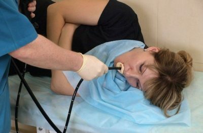
4 Colonoscopy: the essence of the procedure, the advantages of
Colonoscopy is not very fond of patients, as it causes discomfort, in some cases, even painful. But doctors recommend this method, as it allows not only visually inspect the entire intestine, but also take samples of organ tissues. Thanks to this procedure, you can make minimally invasive interventions, remove polyps.
Colonoscopy is recommended when there is a need:
- to eliminate obstruction in the intestine;
- eliminate the foreign body;
- to stop internal bleeding;
- to investigate hemorrhoids in the phase of exacerbation;
- is suspected of having tumors, ulcers, cancer;
- the patient sharply loses weight;
- patient has frequent and prolonged constipation;
- presence of inflammatory processes in the large intestine;
- Crohn's disease( ulcerative colitis);
- for chronic peritonitis;
- with frequent colds( then this method helps to find out the situation with the microflora in the intestine in order to choose the right treatment for the patient);
- is a fulminant form of ischemic colitis.
ADVICE FROM THE MAIN GASTROENTEROLOGIST
Korotov SV: "I can recommend only one remedy for the rapid treatment of Ulcer and Gastritis, which is now recommended by the Ministry of Health. .." Read the reviews & gt; & gt;
Colonoscopy is very often recommended in pregnancy, diabetes or congenital abnormalities in the intestine.
Each of these cases only requires a colonoscopy, since MRI and CT will not produce the results that the doctor needs.

How correctly to prepare for a colonoscopy? This procedure requires special preparation:
- should not eat hard and rough food for 24 hours;
- one hour before the study it is necessary to make a cleansing enema.
During the procedure, an endoscope is inserted into the anus, which is equipped with a micro-camera and a lighting lamp. To fully study the organ, air is introduced into the intestine. If polyps are found during the study, they can be removed immediately. With oncological tumors, tissue biopsy is taken for analysis. Before the procedure, the doctor performs anesthesia, but discomfort during the passage of the endoscope can be.
WE RECOMMEND!
For prevention and treatment of Digestive Gastrointestinal Disorders , our readers advise Monastic tea. This unique remedy consists of 9 medicinal herbs useful for digestion, which not only supplement, but also strengthen each other's actions. Monastic tea will not only eliminate all symptoms of the gastrointestinal tract and digestive system, but will also permanently eliminate the cause of its occurrence.
Opinion of doctors. .. "
To date, doctors reserve the choice of diagnostic methods, but colonoscopy is at the forefront.
Accurate MRI and CT can indicate the size of tumors, their condition, detect metastases, but only the colonoscopy can accurately consider the organ. This is explained by the anatomical structure of the intestine. A large number of fabrics are superimposed on each other and form a loop-like shape.
Doctors and patients can refuse colonoscopy, motivating their choice by the emotional state of the patient, as there have been cases of mental states that can turn into panic attacks and disturbances.
5 What is the virtual colonoscopy?
This method is used extremely rarely due to the lack of special equipment. Those clinics that have a virtual colonoscopy claim that this is a progressive method that allows you to combine all the possibilities of computed tomography, MRI and colonoscopy. The images show a three-dimensional image of the organ, which is convenient for each doctor during the diagnosis.
The main advantage of this method is the possibility of a one-time study, which combines all the necessary diagnostic data.
Despite the fact that this method is innovative, it has its drawbacks:
- high risk of receiving radiation exposure;
- the use of tubes in the intestines can provoke unpleasant sensations;
- does not show cancers up to 10 mm;
- is not possible to take tissue samples for biochemical study.
Only a doctor can choose between diagnostic tests. He clearly understands the whole picture of the intestinal disease, can predict the general condition of the patient and outline the data that he needs in each case. Therefore, it is difficult to talk about the advantages of any of the methods to the patient, everything is decided by the doctor in each specific situation.
- 1 The essence of magnetic resonance imaging
- 2 How is the CT scan performed?
- 3 Characteristics Hydro-MRI
- 4 Colonoscopy: the essence of the procedure, the advantages of
- 5 What is the virtual colonoscopy?
Which diagnosis is optimal? Which is better: an intestinal MRI or a colonoscopy? What does each of the studies show?
MRI is a more modern method, allowing to identify inflammatory processes, pathological changes in the intestine at an early stage of development. Despite this, colonoscopy is also considered a reliable and reliable research method, which has not lost its popularity until today.
The decisive aspect in choosing a colonoscopy or MRI is the informative results that are necessary for the treating physician.
Do you have gastritis?
GALINA SAVINA: "How easy is it to cure gastritis at home for 1 month. A proven method - write down a recipe. ..!"Read more & gt; & gt;