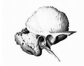
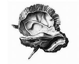
It consists of many elements( channels, furrows, surfaces, tubercles and others) and students of medical academies remember how they studied it in Latin as a terrible dream.
The temporal bone is located on the boundary between the cranial vault and the base of the skull. It is connected with almost all other bones of the skull by different kinds of connections. In it lie the organs of balance( vestibular apparatus) and hearing( inner ear).From the bottom, various muscles of the neck are attached to it, from inside it passes the carotid artery( internal branch), on the outside on its surface there is an auditory opening. This is not all education that has a temporal bone.
The temporal bone channels
In the temporal bone there are several channels and tubules:
- canal;
- tubules are sleepy-drum;
- musculoskeletal canal;
- facial canal;
- drum canaliculus;
- canalicule of a drum string;
- mastoid tubule.
Each channel of the temporal bone contains a certain anatomical formation. Consider the anatomy of these channels in more detail.
 Sleep channel
This channel is named so because it contains the temporal part of the internal carotid artery. The canal canalis( Latin canalis caroticus) originates from below the temporal bone with the outer orifice, passes through its thickness upwards and then, at almost right angles, turns anteriorly and ends in the cavity of the skull. BCA( internal carotid artery) supplies the majority of the brain. A carotid artery in the canal is accompanied by veins and a plexus of the nerve fibers of the sympathetic nervous system.
Sonnoparasian tubules
Latin - canaliculi caroticotympanici - represent two small tubules that branch off from the carotid canal and lead into the tympanum. Contain these canals with sleepy-tympanic nerve fibers.
Muscular tube channel
In Latin - canalis musculotubarius. Begins from the front of the wall of the drum cavity. The entrance to the channel is located near the external auditory aperture. Inside the channel itself there is a horizontal partition, which divides it into two half-channels. In the upper half-channel lies the muscle, which strains the eardrum. It is smaller, relatively lower. The lower channel forms an anatomical connection between the pharyngeal cavity( atmospheric pressure) and the drum cavity to equalize the air pressure on opposite sides of the tympanic membrane. Thanks to this channel, we can always hear the same for different fluctuations in atmospheric pressure. On the other hand, inflammation of the mucosa of this channel can lead to inflammatory processes in the tympanum.
Face channel
The face channel( in Latin canalis facialis) originates in the lower part of the internal auditory meatus and goes horizontally. Inside the temporal bone, it turns at right angles, forming the knee of the facial canal, and exits into the tympanum. Passing through the latter in the direction of the back, it unfolds downward and comes to the surface of the temporal bone, where it ends with an orifice called the stylophyllum, due to the proximity of the styloid and mastoid process near it.
Truncate of the drum string
Latin - canaliculus chordae tympani. It originates from the facial canal near the stylophyllum aperture and ends in the tympanic cavity. The content of this channel is a nerve that innervates the anterior two-thirds of the tongue( taste sensations) and the secretions( sublingual and submandibular).This nerve is called a "drum string".
Drum canalis
In Latin - canaliculus tympanicus. It originates on the surface of the temporal bone( its stony part) and also leads into the tympanum.
Plate canalic
Latin canaliculus mastoideus. It contains the ear branch of the nervus vagus( vagus nerve).It begins in the pit and leads to the tympanum.
Apparently, the temporal bone is literally dug by various canals, tubules, furrows and other anatomical formations. Especially if you consider that its volume( rocky part) is slightly larger than the volume of a matchbox. All this is due to the presence in the temporal bone of hypertonic organs of hearing and co-ordination, which have a rich innervation, as well as blood supply.
Video: Temporal Bone -
Channels
 The temporal bone( from the Latin os temporale) is one of the most complex bones of the human body.


