Perinodular blood flow( vascularization) is a combination of words that is sometimes referred to by a doctor as an endocrinologist in the medical history, and this term is used to describe pathology, during Doppler examination, including in the study of the thyroid nodule.
What is perinodular blood flow
Diagnosis, this phrase can not be named, because it is a description of the picture that the expert sees on the monitor, when carrying out color Doppler mapping( DCC) or energy( EKD).
The formation of the word "perinodular" is derived from the Latin language, namely, from 2 words: peri( around, about) and nodus - meaning "knot".Following the translation, it can be understood that perinodular can be considered vascularization, which is located on the outer part of the tumor, that is - on the periphery. The term itself, can not talk about the nature of the detected thyroid nodule, namely, benign this education or not. Therefore, to rush into panic - it is not necessary.
Also, in the description, after the spent CDC, the term "intranodular blood flow" can be used. In Latin, the word "intra" means "inside" or "through".This means that the use of this phrase, is used to describe the picture seen, when the study shows the vessels inside the node.
Intranodular blood flow in the node, most often appears in malignant course of the disease, but there are cases when with such a blood flow, there may be a benign neoplasm. To clarify the diagnosis, often used thin-needle biopsy.
Why is not enough ultrasound
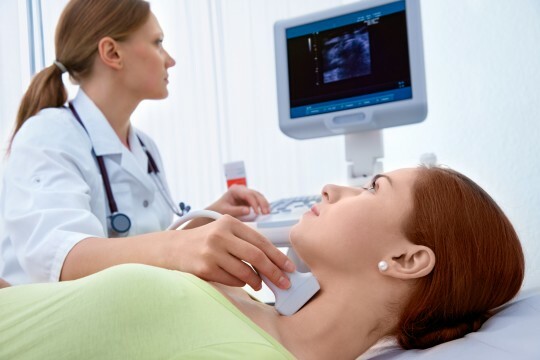
To get a detailed idea of the vascularization in the thyroid gland, ultrasound is not enough. And since the doctor needs more information, he appoints EKD or CDC thyroid. These studies mean the following:
- color dopplerography( CDC).With the help of this study, you can determine the direction in which blood flow in the vessels moves. In this case, the different direction of the particles differs in color. Blue shows particles moving in the same direction. At that time, as red color, the stream of particles in the opposite direction is allocated. With CDC, it is possible to accurately separate fluid formations in the gland, from blood vessels with active blood flow. All these observations can be used by the endocrinologist when diagnosing.
- energy dopplerography( EDC), is able to show the intensity of blood flow in the gland tissues and the intensity of blood filling of the selected site, at the time of the study. On the monitor, you can see an image in red-brown tones or a red-orange picture. A large number of moving particles is shown in bright colors. With a high blood flow in the tissues of the thyroid, we can talk about the presence of an inflammatory process. The more moving particles are smaller, the color on the monitor screen tends more towards brown.
4 types of blood flow
When the diagnosis is established, ultrasound is often used in conjunction with the DCC and EDC.In modern devices, the possibility of using all these modes has already been implemented, which significantly affects the time savings, as well as the means, for the patient.
Both types of dopplerography show the state of the vessels in the thyroid gland, only at the time of the study. If the processes rapidly progress, then it is required to conduct more than one procedure to investigate changes in dynamics.
When conducting CDC and EDC, you can identify 4 types of blood flow in the nodes:
- Vascularization is not detected.
- Perinodular blood flow.
- Intranodular vascularization.
- Peri-intranodular blood flow.
Vascularization not detected
If, in the study, vascularization in the thyroid nodule is not detected, this means that the patient has a benign neoplasm that can remain in the body for a long time.
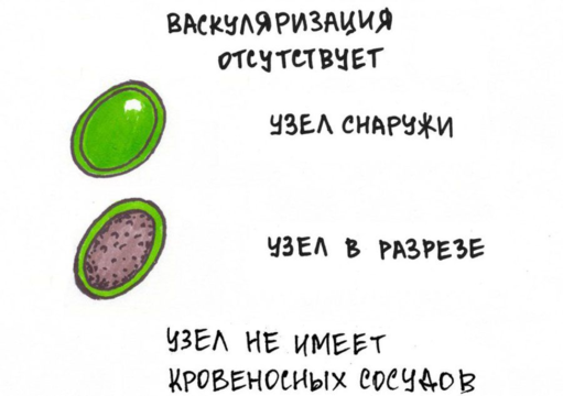
In the absence of vascularization in a cyst or knot, it is observed that the formations do not increase in size and do not contain an inflammatory process. All other cases imply the presence of blood vessels, which feed the neoplasm.
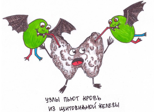
Perinodular blood flow
With this type of vascularization it is found that the walls of the neoplasm have a good blood supply, but inside it, the vessels are not observed.
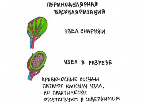
Statistics show that about 85% of detected nodes with peripheral vascularization have benign pathogenesis. Capsule, usually filled with liquid or gel-like content( colloid).
But, there are cases( very rarely) when in the CDC, such a picture can give a malignant tumor, directly - at an early stage of development, when angiogenesis has not yet started. What is noteworthy, both types of formations are hypoechoic, as they contain a liquid filling.
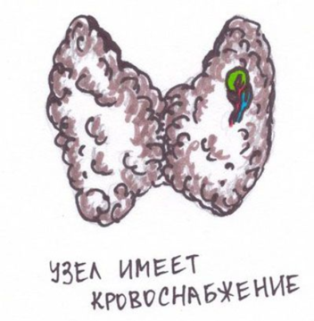
Intranodular vascularization of
With this pathological change, there is a presence of blood vessels inside the tumor, the tissues of which, in this way, receive ample nutrition. In this case, vascularization on the walls may be absent or be negligible.
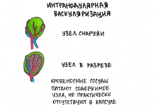
If, again, refer to the statistics, in 20% of cases of detection of this type of blood flow, the picture indicates a malignant neoplasm. If an ultrasound is observed that there is no capsule in the formation, and while it is hypoechoic, the probability that the tumor is malignant increases by 10%.
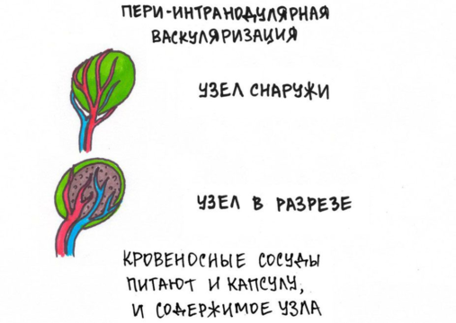
Peri-intranodular blood flow
With CDC, it can be seen on the monitor that the contents of the node or capsule are actively feeding on blood. Such a picture can be observed in nodules and adenomas, of a toxic nature, due to which the synthesis of excessive amounts of thyroid hormones occurs, inevitably falling into the bloodstream.
Such "combined" neoplasms, in 15% of cases, are of a malignant nature. Since there is liquid or jelly-like substance( colloid) inside the formation, it will be hypoechoic in case of ultrasound.
The endocrinologist, in drawing up the conclusion, should rely on the results of ultrasound, as well as the CDC and EDC in the aggregate. But such a diagnosis can be considered superficial, since, until the cellular composition of the tumor is examined, it is impossible to make a conclusion about its nature.
For laboratory cytological examination of the contents of the tumor, TAB is conducted. After the analysis, it is already possible to say exactly what kind of tumor the patient has.
Reasons for the appearance of
nodes The following factors can be the reasons for the appearance of thyroid neoplasms:
- cysts in the gland can form with congenital anomalies, with trauma that caused hemorrhage. Infringement of outflow of a colloid, because of the broken blood flow in a certain part of the gland, in 90% of cases, can provoke the appearance of tumors;
- with prolonged influence of low temperatures, there is a spasm of blood vessels in the thyroid gland. Cells do not receive sufficient nutrition, as a result, local immunity decreases. Such a process is triggered by prolonged emotional overstrain. Spasm of blood vessels, significantly increases the risk of nodal neoplasm in the gland;
- unsatisfactory ecological situation, also provokes the appearance of thyroid gland diseases. In the presence of free radicals and toxic substances in the environment, the structure of thyrocytes is disrupted, as a result of which they begin uncontrolled division. In this process, tumors, both benign and cancerous, can form;
- with iodine deficiency in food, there is a lack of it in the human body. This adversely affects the condition of the thyroid gland. It produces pathological processes, the aggregate of which can cause the appearance of cysts and tumors;
- when exposed to radiation on the human body, the first one, the thyroid gland reacts to it. The cells of the organ undergo mutations, the result of which is predictable;
- in inflammatory processes, for example, with thyroiditis, there may be edema in both lobes of the gland, as a result of which pseudo-nodes very resembling tumors can form;
- autoimmune processes, in which the body attacks its own cells, can provoke inflammation in the gland;
- appearance of hormonal imbalance in the body, with adenoma of the pituitary gland, can provoke the formation of many tumors in the thyroid gland;
- hereditary predisposition, is also of no small importance, and often, is the cause of the appearance of this pathology.
Thus, determining the type of vascularization, namely, the location of blood vessels in the nodes of the thyroid gland, it is possible to establish what kind of neoplasm it is.