 In medicine, there is a common collective term, as "tumor of bone and spine".All types of benign and malignant tumors are meant here.
Statistics show that malignant formation occurs in only 1% of cases among all such bone manifestations. Therefore, when diagnosing - a tumor of the bone - you should not panic or despond.
With timely detection and treatment, almost every growth can be cured.
Content of the article
- Concept and statistics
- Who is at risk?
- Classification of education
- Tumor localization
- Degrees of development
- How does education manifest itself?
- Toolkit for tumor detection
- Treatment: the earlier, the better
- Preventative measures
Concept and statistical data
Chondroma is an enlarged cartilage cell that collectively forms a benign tumor.
Presented neoplasm in medicine refers to potentially malignant manifestations, as the disease has the property to expand and form metastases in the internal organs of a person.
The tumor is prone to slow growth, so it is often diagnosed on a general commission or when another disease is detected, by radiography.
Who is at risk?
Science still does not know all the risk factors for the formation of a benign tumor described. Scientists and eminent doctors, other specialists can not explain the peculiarity of manifestation in girls and boys of adolescence.
During this period, they can not be exposed to harmful factors, which at least somehow could affect the growth of cartilaginous tissue. In addition, the genetic predisposition also has no basis, since its load is still small and can not lead to the formation of this type of tumor.
Classification of education
Aging and isolation of cartilaginous cells can have a distinctive location. Depending on the localization of the chondroma is divided:
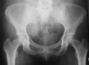
- of the endodroma - is a tumor located inside the bone. As growth grows, a kind of "raspiranie"

On the photo of the chondrome of the ilium bone
bone. In the X-ray image, in this case one can see a pronounced hollow circle with single calcification centers of cartilage.
- Echondroma - is a tumor located on the bone and has a tendency to proliferate towards the soft tissues, where the compaction takes place, and in the x-ray photograph the centers of calcification of the cartilage are visible. In this case, it is impossible to determine the exact boundaries of the neoplasm. This greatly complicates the specification of the size of education.
In the process of growth and modification, the chondroma can be transformed into a malignant growth - the chondrosarcoma.
The localization of a tumor
Chondroma often affects tubular bones - fingers, ribs, but there are also serious formations of the spine, clavicle, sternum, bones of the hands, feet, and also manifest on the cartilages of the larynx and soft tissues.
Sometimes it is possible to diagnose the build-up formation at the base of the skull or paranasal sinuses. A tumor at the base of the skull leads to squeezing the brain, which leads the person to the clinic for examination, which is caused by severe headaches and frequent faints.
Degrees of development of
Approved stages of the onset and development of chondroma do not exist. Here you can only describe the process of the disease itself, what happens to the cells of the cartilage.
At the initial stage, cartilage cells multiply. Their process is slow, so for several years a person will not see any changes in himself.
Increased cell size does not occur, but their continued existence can not be called uniform, because the chondroma consists of the disordered arrangement of mature hyaline cartilage with a low content of elastic fibers.
In the course of its development, it is possible to observe myxomatous changes and foci of ossification, which are common along soft tissues and internal organs. Their formation causes episodes of pain in a person, which pushes him to the examination.
How does education manifest itself?
Symptoms of chondroma are so scarce that sometimes a person for several decades can not guess about his ailment.
The degree of manifestation of the chondroma directly depends on its location:
- The location of the neoplasm in the paranasal sinuses or the tracheal area of the can be felt much earlier. As the tumor grows, anatomical formations are squeezed. As a result, a person will feel a characteristic difficulty in breathing and pain.
- The location of the at the base of the skull of causes headaches, dizziness, fainting, as well as paralysis and convulsions in the limbs. All these symptoms are manifested due to compression of the brain.
- In the articular parts of the tubular bones lead to pain and aches, as a result of which the patient turns to the orthopedist, assuming the presence of the disease - arthritis of the joints.
- Tumor tubular bone brushes or feet does not cause pain even if there is a pronounced tumor and its palpation. Pain syndrome occurs after reaching a large size, thereby squeezing the nerve endings. If a person has found a small seal, which at the moment does not cause any inconvenience, he should immediately consult a specialist.
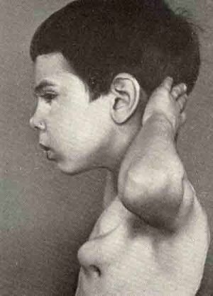
- When the ribs are affected, the tumor does not appear for several years. An exception can be a cosmetic defect - a bulge from the ribs, somewhat resembling a small horn. Even with such a characteristic manifestation, the tumor will not disturb the patient with a pain syndrome. The "internal" chondroma leads to the germination of the periosteum and pleura leaf, which causes pain in the person with a change in position or breathing.
Toolkit for detecting a tumor
 Initial diagnosis is reduced to an X-ray study. At attacks of a pain in the field of nasal sinuses or tracheas use an ultrasonic research.
If the patient complains of headaches, he is sent for examination using an MRI.This method of diagnosis can reveal a tumor of bone in small tubular bones and foci of ossification of cartilage.
When a chondroma is identified for a clinical picture, the patient is often subjected to a biopsy. Experts should make sure that the neoplasm is based on the reproduction and accumulation of hyaline cartilage.
Also using a biopsy, the type of tumor is diagnosed - malignant or benign. Based on the conducted studies, a scheme for the treatment of chondroma is made.
Treatment: the sooner, the better.
The treatment of chondromas is based on the removal of the damaged area, bone tumor. The goal of the treatment is to prevent further growth of the tumor and to exclude its possible repeated manifestation in the same place. Therefore, the treatment of a tumor occurs only through surgical intervention.
There are three methods for removing chondromas:
- Radical operation - resection of bone tissue - removal of the affected bone or only parts with further prosthetics. The complexity of the presented method may consist in carrying out an operation, for example, in the case of a chondroma of the base of the skull. Since the majority of patients are children, prosthetics is supposed to be using a special prosthesis with the possibility of increasing it every 2-3 years as the child grows. With adult patients, such problems do not arise and postoperative recovery lasts only 7 days.
- Radiotherapy - is used in those cases when carrying out a radical operation is impossible or will entail many
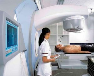 problems. It should also be noted that radiotherapy is used extremely rarely, because it leads to complications. In connection with this fact, radiation therapy is used only to treat the chondromas of the base of the skull.
 - Chemotherapy is the next stage in the treatment of chondromas after radical surgery or radiotherapy. Helps to eliminate the metastases that caused the chondroma. Treating the tumor with chemotherapy is also possible, but not used, because it leads to disruption of the internal organs and other unpleasant consequences.
With timely intervention and thanks to the professionalism of the surgeon, the chondroma is cured completely and does not lead to relapse.
Complications, of course, can occur, but only in the case of partial removal of the affected bone. After the operation, the patient must undergo a regular examination within the first 5 years at least twice a year.
Preventative measures
As far as prevention is concerned, it is not possible to determine the actions that can help prevent the formation of tumors. Take an annual comprehensive examination for the timely detection of the disease.
Treat your health carefully and at the first manifestations of the presented symptoms, consult a specialist.