The most safe and informative method for diagnosing thyroid diseases is ultrasound. With its help, you can determine the condition of the gland, the features of the structure, the structure, identify the characteristic features of the disease in the early stages.
The interpretation of the results of ultrasound of the thyroid gland is carried out exclusively by a specialist conducting research. After all, the correct interpretation of the ultrasound of the thyroid gland plays a decisive role in the resolution of the primary diagnosis. In cases where a more detailed examination is required, computer and magnetic resonance imaging is used. To conduct ultrasound, modern devices are used that are equipped with dopplerographs.
Preparation for inspection
The procedure is completely safe and painless and does not require special preparation. However, there are some recommendations that will help make the examination as accurate and comfortable as possible:
- is not recommended for a few days to take hormonal and iodine-containing drugs;
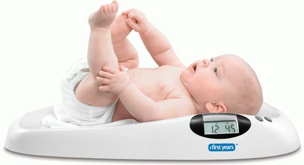
- for a few hours, children and seniors should refrain from eating. To avoid the occurrence of a vomitive reflex, which can appear when pressing on the gland;
- if earlier similar studies have been conducted, you should take their results with you. Comparing the new with the previous results, the diagnostician will be able to determine whether there have been any changes in the state of the thyroid gland.
How the procedure passes
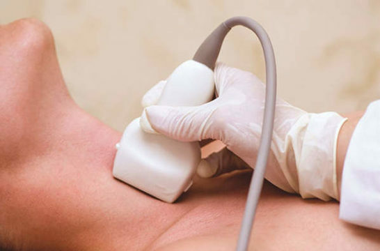
During the study, the patient lies on his back, a towel is placed under his neck. This position is the most comfortable for diagnosis, since there is convenient access to the gland. A special gel is applied to the surface to be examined, which increases the patency of ultrasonic waves. Then the specialist conducts diagnostics at different angles. The procedure takes an average of no more than fifteen minutes. How accurate the results will be depends, first of all, on the experience and qualification of the diagnostician. The sensitivity of the device also influences the accuracy. Usually decryption is given after the examinations are over.
Defined thyroid parameters
- Location
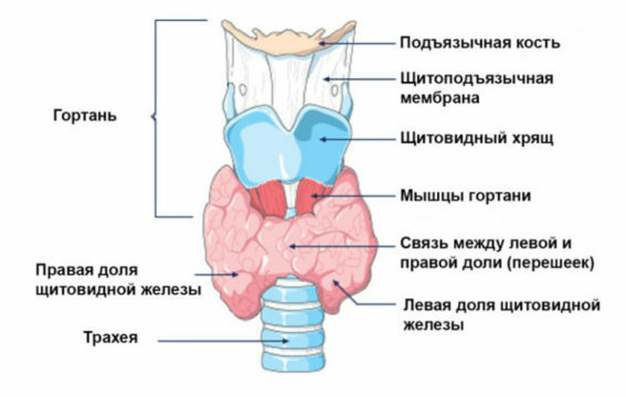
The thyroid gland is located in the middle or lower portion of the anterior part of the neck.
The location is of two kinds:
- Typical( normal).The thyroid gland is within the anatomical norm.
- Aberrant( pathological).The most common example is the location at the root of the language.
- Structure
A healthy gland consists of two sections, which are connected together by a small isthmus. A common deviation is the formation of an additional share and small outgrowths in the lower part. Also, the pathology is a one-sided arrangement, which occurs even during the intrauterine period, during the formation of the organ.
- Dimensions and volume of
As a result of ultrasound, the size of the thyroid gland is accurately indicated. Throughout life they can change. During the examination, the doctor determines the size by measuring the thickness of the isthmus. Correct measurement is necessary from the front to the back.
Dimensions of thyroid gland OK( in cm):
- Length - 2,5-4;
- Width - 1,5-2;
- Thickness - 1-1,5;

The volume of the thyroid gland in children should not exceed the size of more than 15 cc.see The volume of the child is calculated individually, depending on age and sex.
The norms of volume in adults:
- For women - no higher than 18 cu.cm;
- In men - not more than 25 cu.see
The volume may differ markedly from the generally accepted parameters, since the human body weight should be taken into account in the calculation. Parameters taking into account the patient's weight are given in the table:
| Weight ( in kg) | 50 | 60 | 70 | 80 | 90 | 100 or more |
| Volume ( in cm3) | 15,5 | 18,5 | 22 | 25 | 28,5 | 32 |
- Contours
With this evaluation criterion, it is possible to determine the presence or absence of inflammatory processes and tumors. Contours are clear and fuzzy. The presence of fuzzy contours indicates the presence of pathological processes.
- Echogenicity
Determined by the intensity of ultrasound reflection. It is displayed on the screen as a different degree of blackening of the component parts.
- Structure of
When pathologies are absent, the organ structure is uniform, with the presence of granularity. In the presence of inflammatory processes, the structure has the property of losing uniformity.
- Additional parameters of
Also for evaluation, take into account the size and structure of the lymph nodes that are closely adjacent.
Based on the results of thyroid ultrasound, a specialist creates an opinion in which ultrasound signs are painted. The diagnosis is not made only by ultrasound. Do not put the diagnosis alone on ultrasound, you need to seek help from a doctor endocrinologist. For further examination, diagnosis and correct treatment.
thyroid ultrasound norm
Basic key figures:
structure One of the most significant indicators for diagnosis. Happens as a homogeneous, and not a homogeneous structure. Normally, when the gland is healthy, there is only a homogeneous characteristic, which has a special granularity. If there is heterogeneity, then you need to worry. After all, these are signs of pathological processes. Against the background of heterogeneity, inflammatory diseases of an autoimmune nature occur. There are also cases of a moderate heterogeneous structure, which can also occur in healthy people.
- Contours
Have the properties of varying degrees of clarity. Normally, the indicators are absolutely clear. Blurred contours are a deviation from the norm and indicate the presence of inflammation. Also, fuzzy outlines are one of the signs of malignant tumors. Due to the fact that the process went beyond the boundaries of the body and a blurred picture is visible.
- Echogenicity
The thyroid gland is visible on ultrasound in a gray shade. The norm is the correspondence of the parotid salivary to the thyroid gland. If there are inflammatory processes, echogenicity decreases, but in severe forms it can also increase. With a decrease in the tone of the gland is darker than the nearby muscles. Indicators in the norm can slightly change. Usually the organ is compared to the surrounding muscles of a lighter shade.
nodes Normally absent. Admissible deviation from the norm is considered small neoplasms, the size of which is not more than four millimeters. They represent uniformly black formations, which are classified as follicles. Formations more than four millimeters are referred to as nodes. A healthy organ of a homogeneous structure, the presence of nodes is a deviation from the norm.
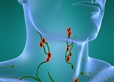
- Regional lymph nodes
Normal lymph nodes of the neck in the examination of the thyroid gland are enlarged. In a normal state they have a smooth, well-defined contour. The length is usually not less than twice the width. The place where the lymphatic vessel enters is clearly pronounced. Blood flow should not be elevated. Cysts are also a deviation from the norm. Very often, impaired indicators are a sign of the presence of a malignant lesion.
- Blood flow
Blood supply disorder can lead to serious complications - atrophy of tissues. At the norm, the screen displays multi-colored signals over the area of the gland.
With inflammatory processes in the body, blood circulation increases. It will be displayed on the screen in the form of a flaming fire. Normally, the image looks stable.
When thyroid ultrasound
is prescribed In most cases, the examination is appointed by the endocrinologist after examination if deviations were detected.
Also appointed in such cases:
- a sharp increase or decrease in body weight;
- abnormalities in the results for hormones;
- pain when swallowing;
- prolonged body temperature increase, without revealing the cause;
- feeling of weakness;
- irritability;
- is a choking;
- when taking hormonal drugs.

Also for the purpose of prevention:
- in occupations that are associated with harmful production;
- for congenital abnormalities of the thyroid gland;
- when planning pregnancy;
Advantages of the test method:
- no contraindications, as the procedure is harmless;
- availability;
- is not invasive;
- low cost.
Common pathologies
- Hypothyroidism
A characteristic feature of this disease is a decrease in the size and volume of the organ. The indices for hormones are also lowered.
- Nodular goiter
When palpation, the tissue is tightened. On ultrasound, there are foci with increased density, which have boundaries and are separated from healthy tissues. Education can be either one or several.
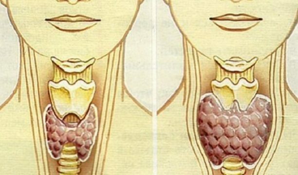
- Diffusive-toxic goiter
The examination shows an increase in the size and volume of the organ above normal. The structure remains homogeneous and has no deviations. To confirm the diagnosis, additional tests for hormones are also prescribed.
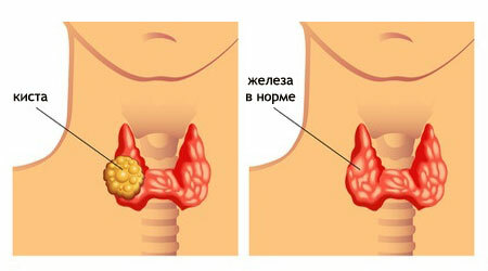
- Cyst
The ultrasound looks like a circular cavity with clear contours, inside it is filled with liquid. Fabrics around are not changed. To exclude malignant pathologies, it is recommended to make a puncture.
- Thyroiditis
Because of edema, an increase in the volume of the thyroid gland occurs. There is a fever, a headache, painful sensations in the thyroid. Ultrasound reveals a high echogenicity and heterogeneous structure.
- Tumors
Benign formations look like foci of compaction with a clear restriction. Malignant - also as foci, but with germination deep into tissues. Enlarged lymph nodes can also be a symptom of malignant formation.
After receiving the results of ultrasound diagnostics, the physician should give a characterization of a large list of indicators. All these parameters are of paramount importance in the diagnosis. A number of common values makes it possible to identify a number of thyroid diseases. In the presence of complaints it is necessary to undergo a survey. Timely visit to the diagnosis is very important. After all, early detection of diseases can prevent possible complications, and begin treatment in the early stages.