 Valgus deformity of the foot is considered almost the most common orthopedic disease. It manifests itself in the form of curvature of the toe and looks like a kind of lump near its base.
As this disease develops slowly, it can be difficult to identify at early stages, which leads to the development of painful sensations and various complications.
By valgus deformity is understood the pathology of the foot, in the process of development of which the metatarsal-phalangeal joint of the big one curves. As a result, his phalanges are at an angle to each other.
As a result, one of them begins to bulge toward the head of the bone. This leads to the formation of cones on the outside of the foot. Sometimes this process is accompanied by pain.
Often the valgus deformity of the thumb of the foot is accompanied by a circulatory disturbance, which provokes arthritis or arthrosis. If you do not provide proper treatment, chronic bursitis may develop, sometimes a traumatic tendovaginitis appears.
Article Contents
- Causes
- Symptoms of the disease
- Diagnostic techniques
- complex therapeutic measures
- Conservative treatment
- Pharmacotherapy
- Physiotherapy
- Surgical treatments
- Rehabilitation after surgery
- Complications
- Preventive measures
- Video: On peculiarities of treatment of hallux valgus foot tells the doctor of the highest category
Causes of
To the development of valgus deformity of the first toesop can result in the following factors:
- flatfoot;
- congenital disorders of the joints of the toes or feet;
- weak or too loose joint;
- destruction of joints due to arthritis or arthrosis;
- traumatic foot damage.
Wearing tight shoes is not the cause of this pathology, but this can speed up the process or complicate its course.
There are diseases that provoke the development of pathology. These include:
- Osteoporosis. This pathology is accompanied by the washing away of calcium from the bones, as a result of which they change their structure. This is what causes the deformation of the foot.
- Flat feet. Almost all patients with valgus deformities of the foot have transverse or longitudinal flat feet.
- Endocrine disorders. These processes often lead to loosening of the ligaments, which ultimately provokes flat feet and deformities of the foot.
 It is necessary to know that with inflammation of the foot joints, treatment should correspond to the disease that this inflammation caused. How to do it read in our article.
What is the plexitis of the shoulder joint and how the pathology itself shows here.
The following are at risk:
- People who are used to wearing uncomfortable shoes. High heels and narrow shoes increase the load on the foot, which leads to faster development of valgus deformation.
- Women. As a rule, this disease is more often diagnosed in women - most likely because they often wear uncomfortable shoes.
- Ballerinas. Representatives of this profession dances a lot of time on their toes, which increases the risk of developing valgus deformation.
- Older people. It is believed that the incidence of this disease increases with age. So, in people 15-30 years, this figure is only 3%, and after 60 years it increases to 16%.
- People with a genetic predisposition. Scientists believe that there is a direct link between the development of valgus deformity and the presence of this disease in close relatives.
Symptoms of the disease
This disease develops rather slowly, and therefore it is difficult to notice. At first, it seems to the person that habitual shoes have become uncomfortable. 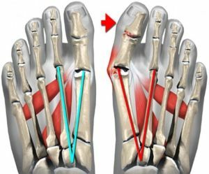
After this, there are painful sensations in the feet.
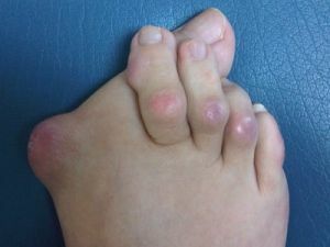
Gradually the appears as the main sign - the deviation of the thumb and the formation of the so-called bone near the base of it. The rest of the fingers are shaped like a hammer. A person may suffer from rapid fatigue or difficulty in choosing shoes. Also, he may have corns that make walking difficult.
The main manifestations of valgus deformity of the foot are as follows:
- appears soft formation, which is accompanied by the fact that the skin in the joint area turns red, there are painful sensations as the joint bag inflames;
- the thumb changes its shape, becoming curved;
- in the region of the first phalanx a solid cone is formed;
- develops a callus, the skin is irritated;
- there are painful sensations while walking;
- the thumb loses its mobility. Over time, motor activity can lose other fingers.
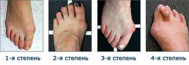
There is a simple classification according to which several stages of development of valgus deformation are distinguished:
- 1 degree. The toe is shifted by less than 20 degrees. In this case, there are no uncomfortable sensations.
- 2 degree. The joint is displaced by 20-30 degrees. In this situation, a person feels minor pain.
- 3 degree. The joint displacement is 30-50 degrees. In this case, there is a constant pain in the area of the big toe. These feelings do not allow a person to perform the usual actions for him in full.
- 4 degree. The joint is displaced more than 50 degrees. This condition is characterized by severe pain, discomfort when walking, difficulty with the choice of shoes, the formation of calluses.
Diagnostic Techniques
In order to make an accurate diagnosis, the physician should examine the patient's medical history for the presence of concomitant diseases and conduct an external examination. During this procedure, the specialist needs to evaluate:
- patient's gait;
- position of the big toe;
- is an outstanding joint position;
- range of thumb movement;
- presence of keratosis;
- the presence of associated deformations.
In addition, if there is a suspicion of valgus deformation of the first toes, additional research may be needed:
- Radiography. This procedure is carried out in three projections and shows changes in the position of the feet.
- Computer Planography. A footprint is placed on a special platform, and its analysis makes it possible to talk about the distribution of the load.
- Computer podmety. With this technique, it is possible to detect changes in the foot at an early stage and prevent the development of the disease.
It is very important to differentiate the diagnosis from other diseases - gout, arthritis, osteoarthritis.
Complex of therapeutic measures

The choice of treatment tactics for valgus deformity of the feet is affected by the degree of its severity and the level of pain syndrome. Of course, the most effective therapy will be at an early stage of the disease, because it helps to prevent joint deformation.
Conservative treatment
 Therapy of valgus deformation of the first toe of the foot usually begins with the choice of comfortable shoes , which does not cause loads or friction. In the early stages of pathology, it is able to stop the development of deformation.
Since pain is caused by pressure from the shoe, the treatment is to eliminate the provoking factor.
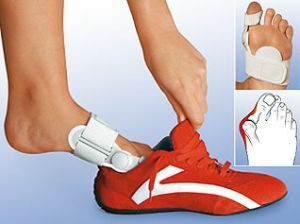
Special gaskets are used to reduce pressure. In addition, there are many devices that change the load distribution.
Medical treatment
During the treatment of the disease, usually prescribe drugs that have an anti-inflammatory effect. A good effect is the local administration of steroids. These include Kenalog, hydrocortisone, diprospan. Usually these drugs are injected directly into the affected joint.
Physiotherapy
For the treatment of valgus deformity, physiotherapeutic procedures - ultrasound or diathermy - can also be used. It should be borne in mind that these measures have a temporary effect.
The most effective is the use of orthopedic products, which are manufactured individually after examining the shape of the foot and the gait of a person.
The use of various insteps, leg adjusters or interdigital rollers at the onset of the disease can stop subsequent deformation. If the process is started, then the use of such products can only slightly reduce pain. With the help of individual insoles, it is possible to correct the violation of the arch of the foot.
Surgical treatment methods
Surgery for valgus deformities of the foot is usually required when conservative therapy does not bring the desired results. The choice of the method is determined by the stage of the disease:
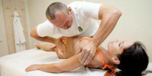
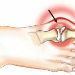
- Deletion of the "built-up" .In moderate cases, during surgery, only the build-up on the joint bag is removed. To do this, perform a small incision and a special chisel remove the build-up.
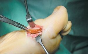
In the photo, an operation to get rid of valgus deformities of the foot
- Distal osteotomy. In this case, the distal end of the bone is cut and moves so as to reduce the angle between metatarsal bones.
- Proximal osteotomy. In this case, the first metatarsal bone is cut at the proximal end of the bone.
The surgeon then performs an osteotomy. With this procedure, it is also possible to reduce the angle between metatarsal bones.
- Operation of Keller-Brandes. In this case, remove the joint of the thumb, and the remaining capsule is sutured into the lesion between the main phalanx and metatarsal bone.
Surgery for valgus deformities of the feet is considered to be the most effective method of correcting valgus deformation and is used in most cases.
Rehabilitation after operation
To ensure that the postoperative period is successful, the following recommendations should be followed:
- wear shoes on wooden soles or use a special bandage;
- use crutches, so as not to emphasize the painful leg;
- undergo a course of physiotherapy, if there is such a need;
- use shoes with a wide toe;
- use orthopedic correctors.
Complications of
It should be noted that surgery can lead to dangerous consequences:
- infection in soft tissues;
- development of osteomyelitis;
- bleeding;
- slow fusion of bones after dissection;
- displacement of bone fragments;
- numbness of the skin in the region of the incision;
- development of arthritis or avascular neurosis after surgery.
Preventative measures
To detect the onset of the disease in time, should be inspected regularly by an orthopedist.
It is also recommended to use orthopedic insoles. If you have to stay on your feet for a long time, you need to adhere to the mode of work and rest.
Valgus deformity of the big toe - this is a rather serious disease, which significantly worsens the quality of human life. However, its treatment is the most effective in the early stages of development of , so do not neglect visits to the orthopedist, especially if you are at risk.