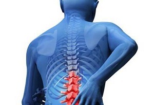
 The formation that occurs at the site of the spinal cord can be both malignant and benign. Such insidious tumors may long remain unnoticed or manifest in the form of some diseases before they grow to substantial sizes. Symptoms of tumors of the spinal cord are very diverse.
It all depends on the location of the tumor, the rate of its development, the variety of education and design features.
The most effective method for diagnosing neoplasms of the spinal cord is tomography using a magnetic resonance device that contributes to the contrast enhancement of painful areas.
The most common method of treating spinal cord cancer is the procedure for surgical removal of tumors. It is also possible to additionally apply chemical or radiotherapy.
Contents
- Classification of spinal cord formations
- About intramedullary and extramedullary formations
- Reasons for the formation of
- Clinical features
- Diagnosis
- Surgery is the only chance
- Possible complications
- Reduce risks in our forces
Classification of spinal cord formations
Unlike brain tumors, neoplasms on the spinal cord are much less common. Most often, the disease affects residents who have reached the age of 30-50 years.
Major varieties of spinal cord tumors:
- intramedullary and extramedular neoplasms;
- subdural and epidural solid-wall tumors;
- formation of cervical, sacral, lumbar, and thoracic spinal cord, tumors also occur on the roots of the bone tail;
- some formations differ in histological structure.
The most common of all are neurinomas, formed from Schwann cells located on the shells of roots of spinal tissue.
The structure of some tumors from time to time resembles the shape of an hourglass, if the formation occurs in the area of the radicular canal. The corresponding symptomatology can be manifested as a result of compression of the spinal tissue by formation in the vertebral canal.
Also the formations are divided into:
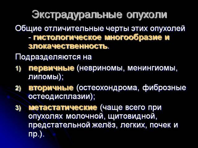
- Primary .In this case, tumor fragments by origin are the result of the transformation of nerve cells or individual elements of the meninges.
- Secondary .In such cases, the formation is located in the region of the spinal cord, and is essentially a metastatic process, which implies the presence of a tumor elsewhere.
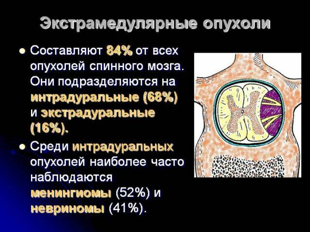
About intramedullary and extramedullary formations
Intramedullary tumors of the spinal cord develop within the spine itself. Formations are formed from directly from the spinal cord located in the region of roots and membranes, as well as from certain fragments of bone walls inside the spinal canal.
In some cases, the formations can grow into channels, passing through the holes between the individual vertebrae.
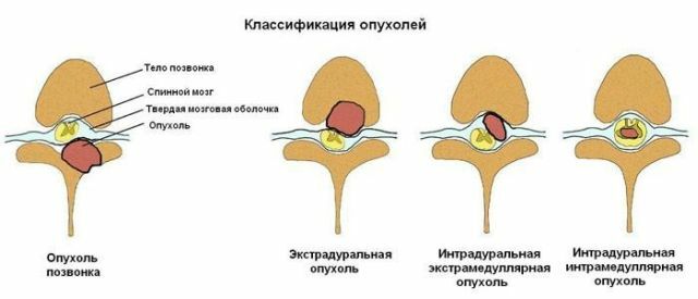
Thus, the formation can develop inside the canal of the spinal cord, as well as beyond it and, accordingly, can be classified as intradural and extradural.
Intradural formations are divided into extra-cerebral( extramedullary, formed in the arachnoid membrane), and intracerebral( intramedullary, growing from glioma).
Intradural localized formations may have an inflammatory origin, for example, as a result of the transfer of a disease such as meningitis. They are also called arachnoid cysts. Education, defined as tuberculoma or even gum, has an extradural nature.
Any intra-vertebral formation increases in size over time and exerts pressure on the entire contents of the spinal canal.
Eccentrically located intramedullary formations primarily exert pressure on the elements placed near the spinal cord. For this reason, there is a violation of blood circulation, as well as normal flow of cerebrospinal fluid.
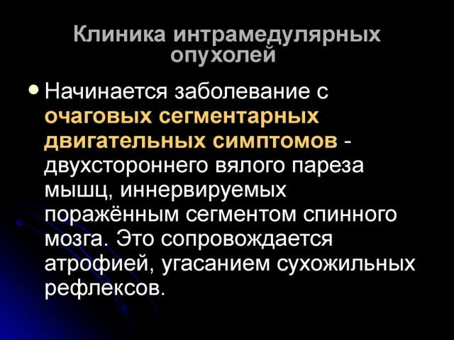
Intramedullary structures contribute to impaired functionality of the spinal cord itself.
The entire fragment of the spinal cord, located below the compressive formation, functions with impaired blood circulation, and also liquor circulation.
In the most malleable vessels the blood is squeezed and stagnant, while the index of intravenous pressure is significantly increased.
In the cerebrospinal fluid, red blood cells and a protein fluid come in excess. As a result of the excessive number of the above-mentioned blood cells, the color of the cerebrospinal fluid changes, which in medical practice is defined as xanthochromia.
Reasons for the formation of
formations To date, the identification of the causes of the formation of spinal tumors is an insufficiently studied field of medicine. 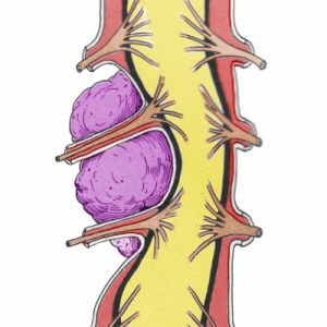
Doctors know that the very essence of the process of formation of tumor tissues is to relax the work of the immune system, which causes a variety of negative factors of internal and external action.
Such diseases always develop at a slow pace and unnoticed, so patients turn to doctors at the advanced stages, when the provision of qualified medical care can be unsuccessful.
If you analyze the lifestyle of patients, you can identify as a cause of cancer of the spinal cord previously suffered trauma, intoxication, as well as various infections, prolonged disorders, psychological, as well as physical overload, transferred stress.
At the same time, there is practically no connection with the beginning of the process of formation of tumor tissues.
Since there is still no clear definition of the causes of tumor tissue formation in medicine, only the main provoking factors can be listed:
- heredity;
- exposure to carcinogens;
- Hippel-Landau disease;
- obstructed circulation of the lymphatic fluid.
Features of clinical picture
The general clinical characteristics of tumors in the region of the spinal cord are represented by only two main types of symptoms. This can be 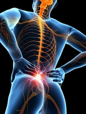 as focal lesion symptoms, which can be caused by compression or deformation of some areas of the brain tissue, as well as cerebral symptoms, the development of which is due to increased pressure.
Each education progresses gradually. At the initial stages, the functionality of the spinal cord is fully preserved. This condition persists until the next stage, when the main signs of decompensation begin to appear, the circulatory disturbance in the problem area of the spine.
The patient may experience localized pain. Often there are convulsive seizures or disruption of the receptors, memory impairment, hallucinations, impaired motor functions. With the development of education until the next stage, there may be sensations of muscle weakness in the limbs.
At the next stage of development, certain neurological symptoms of spinal cord lesions are manifested. Begin to manifest such disorders as general weakness or paralysis of the limbs, a change in sensitivity, a violation of the functions of the genitourinary system, as well as stool retention.
Diagnosis of
Diagnosis of tumors of the spinal cord is carried out in such ways:
- Neurological examinations of are a procedure for examining a patient and checking all of his reflexes and checking for stability in standing.
- X-ray allows you to determine the deformation or displacement of the vertebrae. To diagnose the formation in the region of the spinal cord, the myelography procedure is often used, the essence of which is the introduction of a special substance into the cerebrospinal sub-cauda space during the X-ray imaging.
- Tomography using digital technology , as well as magnetic resonance devices is today considered to be the most modern method for diagnosing such diseases. Special equipment performs layered radiography with subsequent digital processing.
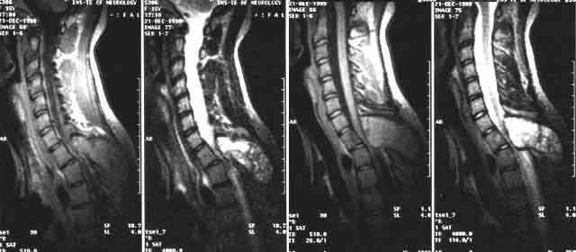
Extramedullary spinal cord tumor on MRI
Surgery is the only chance of
The only method that helps to get rid of the formations in the spinal cord is surgical intervention. This procedure has a 100% effect on benign tumors.
Painful sensations can be reduced, thanks to the use of restorative and anesthetic drugs. Favorable results are always achieved after surgical intervention to address the problem of a benign tumor. The results of this method of treatment in the most successful way can be determined by conducting diagnostics. 
The method of radical removal is also used in malignant formations. The postoperative procedure of radiography can significantly reduce the rate of development of tumor tissue and harbor some neuropathological symptoms. Indications for the use of this technique are serious pain.
The prognosis of the effectiveness of surgical intervention can be determined by the histological nature of the formation, its location and size. Timely surgery often leads to an absolute recovery.
Possible complications of
The most dangerous of possible complications is infection with SKA.The mortality rate resulting from such diseases is too high for the full development of infections.
Since the use of antibiotics, there are fewer than 100 registered cases of abscesses in patients. Complications caused by the development of tumor tissue can progress for years.
To reduce the risks in our forces
To date, medicine does not use proven preventive methods because of the rarity of such diseases.
 Preventive measures in case of such a disease are considered meaningless because the possibility of the formation of cancer cells is caused genetically.
To avoid the emergence of factors that affect their development, it is recommended to get rid of all bad habits, to take timely necessary medications and to treat the emerging inflammatory processes, as well as all kinds of infectious diseases of the human body.
A person who has been diagnosed with spinal cord cancer needs attention and special care. First of all, it is necessary to solve the problem of mitigating the manifested symptoms observed in the development of tumor tissues.
The best preventive measures taken after treatment are basic health care, as well as additional physical activities, a well-balanced diet and regular outdoor walks.
To reduce the risk of development of tumor tissues in the human body, it is necessary to identify the disease at the earliest stages, when from the treatment procedures one can achieve 100% effect and complete recovery. It is always necessary to undergo therapeutic procedures to combat previous diseases.
Often the development of tumor tissue in the human body is an elementary consequence of treatment. To do this, it is recommended to find time for medical examinations. Due to regular visits to the doctor, the risk of harming the body with malignant tumors is significantly reduced.