 The human brain is one of the most complex and poorly studied organs, which is constantly forced to work. For his normal functioning, he needs a full-fledged nutrition and blood supply.
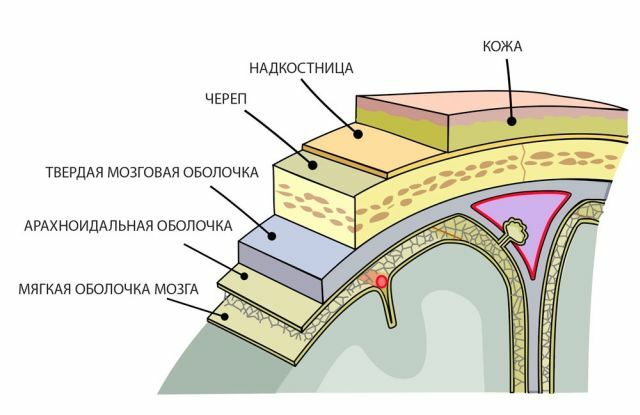
The human brain consists of three membranes: soft, hard and spiny. Subarachnoid space is the space between the soft and arachnoid membrane. The spider web envelops the brain, with other tissues it binds to the sub-paternal junction.
They form the ventricular system of the spinal cord and the brain, consisting of four tanks in which the fluid circulates.
The subarachnoid space is filled with cerebrospinal fluid or cerebrospinal fluid, which is responsible for feeding and protecting the brain. A favorable environment is created for the mutual exchange of useful substances between the blood and brain of man, the movement of nutrients to nerve endings and ventricles.
The final products of tissue metabolism are thrown out and discharged into the liquor. There is its constant circulation in the cerebral cavity.
Up to 140 million cells of CSF should be counted in the subarachnoid space that flows out of the brain through the opening in the fourth ventricle. Its maximum volume is contained in the tanks of space, located above the large cracks and fissures of the brain.
Anatomical Reference - Cases and Spaces of the Brain:
Contents of
- Why subarachnoid space is expanded
- Extent of Extension
- Features of Clinical Painting
- Methods and Objectives of Diagnostic
- Medical Assistance
- Hydrocephalus as a Frequent Complication
- Than it is dangerous?
- Preventive measures
Why subarachnoid space is extended
Crises in the circulation of CSF cause CNS infections, chronic diseases, meningitis, encephalitis, swelling or birth trauma. This leads to a decrease in the amount of gray and white matter in the brain, and as a consequence, the subarachnoid space expands.
The expanded subarachnoid space indicates a failure in the circulation of the CSF, its production occurs excessively and enters the brain cavity, ie hydrocephalus or hydrocephalus develops, and as a result there is increased intracranial pressure.
If a benign local dilatation of subarachnoid spaces occurs, the ventricles are slightly widened or within the normal range, then the disorder itself passes through one or two years and does not harm the baby's health.
But only hope for a favorable outcome of the disease can not, you need to contact a neurologist who will prescribe the necessary treatment.
The expansion of subarachnoid spaces of the brain in adults can cause the following reasons: 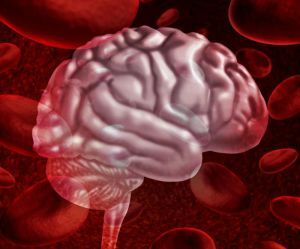
- hemorrhagic or ischemic stroke;
- traumatic brain injury;
- infectious disease of the brain and CNS( meningitis, tuberculosis, encephalitis);
- is a brain tumor.
These factors contribute to the launch of the process of atrophy, the amount of white and gray matter decreases, contributing to the expansion of the subarachnoid space.
Degrees of expansion
The expansion of the subarachnoid space is of three degrees:
- moderate - an increase from 1 to 2 mm;
- average - increase from 3 to 4 mm;
- heavy from 4 mm.
The expansion of the cerebrospinal spaces occurs in proportion to the growth of the head in a newborn and bloating of the fontanel.
The course and outcome of the disease depends on the timely application of medical care and the initiation of treatment. If the treatment is selected correctly, the change in the ventricles remains practically within the normal range.
Features of the clinical picture
To suspect abnormalities in the brain and the expansion of the subarachnoid space in the newborn baby will help such symptoms:
- irritability at moderate or low sounds, noises;
- hypersensitivity to light;
- plentiful regurgitation;
- is broken sleep;
- different in size pupils or strabismus;
- head size increase;
- anxiety for weather change;
- slowly overgrows the fontanel and there is its swelling;
- twitching of limbs and chin.
The fact that subarachnoid spaces are widened in an adult characterize such symptoms:
- headache after morning awakening;

- nausea and vomiting, as a consequence of severe headache, which occurs after vomiting;
- dizziness;
- drowsiness, a dangerous symptom of intracranial pressure, indicating the progression of the disease;
- vision impairment;
- dementia, observed after getting a head injury, sleep disturbed, a person confusing day with the night, memory dips occur;
- apraxia walking, the patient in the reclining position shows how to walk, but with the ascent swings, scuffs, comes with widely spaced legs.
Methods and objectives of diagnosing
Diagnosis of the disease can be only after a comprehensive survey and laboratory tests. After receiving the results of magnetic resonance or computer tomography, the results of blood biochemistry, ultrasound examination of the cerebral hemispheres, assessment of the symptoms and behavior of the patient, the neuropathologist will establish the final diagnosis, the degree of the disease and prescribe medication.
Basic diagnostic methods:
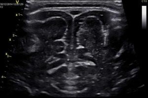
- Neurosonography .It lasts no more than fifteen minutes, is carried out with the help of an ultrasonic sensor through an unclosed fontanel on the head of a newborn. The study can be conducted quite often, without negative consequences for the child. As a rule, neurosonography is done to all newborns in the maternity hospital to reveal pathologies in the development of the brain at the initial stage. Deciphers the survey data of a neurologist or pediatrician. Only by comparing the symptoms and the survey data, the doctor can make a diagnosis.
- Computer and magnetic resonance imaging( ) are very expensive research methods and are conducted when serious abnormalities are detected. As a rule, it is enough for neonates to carry out neurosonography through the fontanel, but adults need more serious methods of diagnosis. Today, these are the most reliable and accurate methods of studying the human body. MRI allows you to see a layered image of the desired area of the brain. Examination of infants is very problematic, as it requires complete fixation and adoption of a stationary state, which is very problematic for young children. If a baby needs this type of examination, he is under anesthesia.
- Cisternography is used to determine the direction of cerebrospinal fluid and clarify the type of hydrocephalus.
- Angiography , is a method of examination when contrast is introduced into the artery and abnormalities in the patency of the blood vessels are revealed.
- Neuropsychological examination - examination and questioning of the patient, the collection of all analyzes and studies together, to identify abnormalities in the functioning and work of the brain.
Medical care
Treatment of an expanded subarachnoid space is aimed at eliminating the causes and factors that triggered the disease. The main therapy includes vitamins, especially groups B and D and the intake of antibacterial drugs, in the presence of infection.
Treatment is long and is assigned to each patient individually.
The main medicines include:
- diuretics , for removing excess fluid from the body( Veroshpiron, Diakarb);
- potassium-containing preparations ( Asparcamp);

- means for improving brain trophism ( Pantogam, Cavinton);
- vitamins B and D ;
- pain relievers after trauma and tumors( eg Ketonal, Nimesil, Ketoprofen, Nimesulide);
- barbiturates ( Nembutal, Phenobarbital, Amitale);
- saluretics ( Acetazolamide, Furosemide, Etacrynic acid);
- glucocorticosteroid preparations ( Prednisylone, Dexamethasone, Betamethasone).
If the disease progresses rapidly and the subarachnoid cavity increases, the main therapy will be to find the cause of the causing disorder, if diuretics are used for hydrocephalus, antibacterial drugs are used to treat infections.
Hydrocephalus as a frequent complication of
In severe disease, when medications and physical procedures failed to produce the desired result, surgical treatment is indicated.
Hydrocephalus is an extremely dangerous disease that can cause blindness or vision loss, speech impairment and a child development gap in 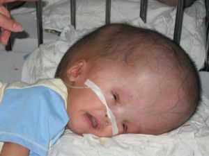 .
.
Principles and methods of treating the disease are carried out continuously and selected individually for each patient. Treatment will depend on the nature, severity and complications. The main task is to restore normal circulation and outflow of cerebrospinal fluid from the over-cerebral region, which will lead to normalization of intracranial pressure, which will enable to improve and restore the metabolism of cells and tissues of the nervous system.
The complex appoints and fiziononevropologicheskie procedures that reduce the symptoms of the disease and accelerate the process of recovery.
Than it is dangerous?
The extended expansion of subarachnoid convectal spaces and untimely treatment of infants in infants can lead to more serious complications:
- manifestation of chronic diseases;
- tumor;
- arachnoiditis;
- hydrocephalus;
- meningitis;
- delay of psychoemotional and physical development of the baby.
Timely diagnosis and treatment will reduce the risk or eliminate complications of the disease, promote the favorable course and outcome of the disease, so that it will not affect the functioning, vital activity and physical development of the child and usually disappears by two years of the child's life.
Preventive measures
Prevention should be handled by a woman( future parents) before conception. Before conception, to conduct a thorough examination of the body  for the detection of chronic and infectious diseases, if they are available to treat them, observe the doctor's recommendations during pregnancy, protect oneself from stress and behave correctly during childbirth.
After the birth of the baby, monitor his behavior, avoid injury.
The widening of the subarachnoid space in adults is diagnosed very rarely, but in order to prevent its appearance, it is necessary to avoid traumatic brain injuries and monitor your health.