 Craniostenosis is understood as the premature infestation of the cranial sutures, which contributes to the limitation of the total volume of the skull, its deformation and provokes the development of intracranial hypertension.
The disease is diagnosed with a frequency of 0.5-1 per 1000 newborns, mostly in boys.
Pathology can occur in isolation or in combination with anomalies in the development of other organs and systems( as a component of the Aperta syndrome, Pfeiffer), often accompanied by a developmental disorder of the brain.
Etiology and pathogenesis of
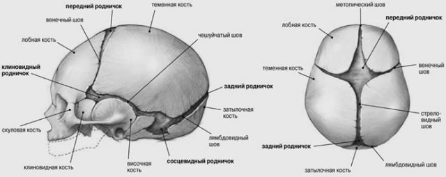
disorders Seams are thin layers of fibrous tissue located between the cranial bones and forming fontanelles( "soft spots") at the confluence. They allow the skull to change its configuration as the child passes through the birth canal, thereby preventing trauma during childbirth.
Normally, the closure of fontanelles occurs by the end of the first year of the child's life. With their premature overgrowth and early ossification of the joints, a picture of craniostenosis is observed, the degree of which depends directly on the onset of the disease.
If the seams are closed in the prenatal period, the deformation of the skull will be significant, if after birth - expressed to a lesser extent.
Pathogenetically the main role in the development of the disease belongs to the impairment of blood circulation and metabolism in bone tissue, which can be triggered by the following factors:
- genetic mutations and hereditary conditionality;
- infections in utero( rubella, influenza, herpes, etc.);
- effect on the fetus of unfavorable environmental factors( bad habits of the mother, household and industrial toxins, radiation, taking certain medications);
- mechanical compression of the fetal head in the uterine cavity;
- giving a wrong position to a newborn, immobility in infancy, limiting motor abilities;
- birth injuries and illnesses suffered in the first months of life.
Species of abnormal development of
Depending on the number involved in the pathological process of stitches, craniostenosis is distinguished:
- simple ( closure of only one seam),
- combined ( multiple infection),
- complex ( all cranial sutures are involved).
In addition, the disease is divided into the following forms:
- Scaphocephaly - premature closure of the sagittal suture, occurs in most cases, mainly in boys.
 It is characterized by: elongation and narrowing of the skull, extension of the forehead, extreme fusion or closure of the large fontanel. With this form of pathology, natural childbirth is often difficult, which is associated with a mismatch between the size of the fetal head and the mother's birth canal.
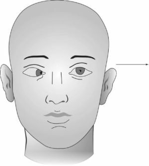
 - Plagiocephaly frontal - frontal stenosis of the skull against the background of frontal and coronal sutures. It occurs more often in girls, the flattening of the forehead is visually determined, with the eye socket raised and the eyebrows on one side, the protrusion of the auricle.
- Platiocephaly occipital - early closure or sclerotherapy of the coronal suture, which arose because of the immobility of the newborn or its incorrect position. In this case, the frontal bone flattenes, and the occipital on the same side of the head bulges out.
- Trigonocephaly - associated with a rare pathology of the frontal( metopic) suture hypotension and deformation of the frontal bones in the form of a keel( triangular head shape).
- Turricephalus - combined coagulation of the coronary and sagittal sutures, accompanied by hydrocephalus. The protrusion of the temporal divisions and the narrowing of the rest of the skull( the shape of the clover leaf) is characteristic.
Nature of the clinical picture
Closing only one suture is usually a problem associated with a defect rather cosmetic.
When combined lesions of the clinical picture is much brighter, there are the large deformation of the skull, which is characteristic for each type of craniostenosis and neurological symptoms( caused by increased intracranial pressure, which is associated with a disproportionately slow growth of the skull, insufficient for brain volume):
- paroxysmal headache;
- nausea;
- vomiting;
- strengthening of tendon-periosteal reflexes;
- pathological reflexes;
- look up;
- pendulum movements of eyeballs( nystagmus);
- meningeal signs( stiff neck muscles and others);
- convulsive seizures.
There are eye symptoms:
- bilateral exophthalmos( pop-eyed);
- retarded retinal changes;
- decrease or loss of vision.
Mental disorders are variable, there may be inhibition or increased excitability, mental development, as a rule, slow.
Diagnosis set-up
For the diagnosis it is necessary to carry out a comprehensive multifaceted examination. In addition to clarifying the complaints and the history of the disease( the time of the onset of the main symptoms, their progression), it is important to collect a maternal anamnesis( the features of the pregnancy, the diseases suffered during this period, the harmful effects, complications in childbirth).
The full examination of the child is mandatory, the structure of the skull, the shape and dimensions of the head are studied in detail, the degree of penetration of the sutures and fontanels is palpated.
Neurological examination reveals cerebral and focal signs of brain damage( pathological reflexes, nystagmus, paresis of eyes, etc.).
The ophthalmologist assesses the degree of abnormalities on the part of the visual organ( stagnant changes in the fundus, edema of the optic disc, its atrophy, exophthalmos).Genetic research helps establish the hereditary nature of the disease.
also executes a series of instrumental diagnostic methods to determine the further tactics of patient:
- X-ray examination of the skull - reveals the lack of seams( one or more), the thinning of the bone structure,
 availability of digital impressions on the cranial bones, deformation of the skull;
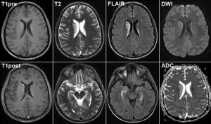
 - CT( computed tomography) or MRI( magnetic resonance imaging) - a method of neuroimaging, in addition to changes in bone structure allows you to determine the severity of intracranial hypertension;
- craniometry - is performed to measure the standard and special parameters of the skull, performed manually and with the help of special computer programs;
- electroencephalography - allows to reveal the degree of functional deviations of the central nervous system;
- ultrasound of the vessels of the head and neck vessels - on the basis of violations of the outflow of blood from the cranial cavity helps to identify and indirectly assess increased intracranial pressure;
- angiography - study of cerebral vessels subjected to contrasting, the method allows to detect abnormal vascular anastomoses, specifies the level at which the outflow of blood occurs;
- pneumoencephalography - visualization of ventricles and subarachnoid spaces( cerebrospinal fluid), is applicable as a method of differential diagnosis;
- Ultrasound of internal organs, ECG, Echo-CG - are performed as additional methods for the detection of concomitant diseases and malformations.
Modern surgery
In case of premature closure of seams, surgical treatment is mainly used, the main purpose of which is to increase the volume of the skull and create favorable conditions for the normal development of brain structures.
 There are a huge number of surgical techniques depending on the type of craniostenosis - craniotomy linear, patchwork bilateral, circular, resection of the cranial vault and others.
The technique of the operation is to perform dissection of soft tissues and bone plastic. During the intervention, skull defects are corrected and intracranial hypertension is eliminated.
It is believed that the operation is appropriate until the age of three, when the most intensive formation of brain structures in the child. Completed in time, it allows you to prevent the compression of the brain structures and almost completely levels out the previous symptoms of the disease, including a gross cosmetic defect.
Operative treatment is not indicated in case of:
- of compensated craniostenosis;
- irreversible loss of vision and mental retardation without signs of intracranial hypertension;
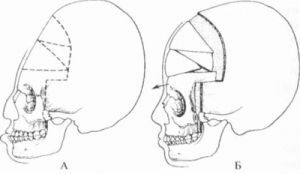
The skull before and after the operation for the correction of craniostenosis
- severe concomitant developmental anomalies( eg, decompensated cardiac malformations).
A relative( temporary) contraindication is an acute disease, either chronic in the acute stage, purulent infections of the skin, recurrent inflammation of the upper respiratory tract.
In the postoperative period, the course of physiotherapeutic procedures and symptomatic drug therapy is shown, as well as long-term follow-up.
Prognosis and prevention
Early diagnosis and timely surgery can prevent the development of serious consequences. In this case, the prognosis for life and normal development of the child is favorable.
Loss of vision, mental retardation, convulsive syndrome, persistent headache and gross deformity of the skull complicate the course of the pathology and lead to disability.
For the purposes of prevention, it is shown:
- conducting medical genetic counseling of couples from at-risk groups;
- restriction of the effects on the body of pregnant unfavorable external factors;
- refusal from smoking and other bad habits;
- prevention and timely treatment of diseases of the future mother and newborn.
And most importantly, if a child develops symptoms of concern, parents should urgently consult a specialist. Timeliness of treatment will avoid the terrible consequences of craniostenosis and will give the child the opportunity for a normal life.