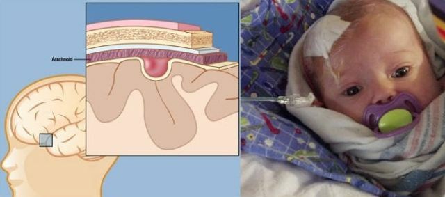
 Porencephaly is a congenital anomaly, in the presence of which the brain surface connects to one of the lateral ventricles( cavities in which there is a spinal fluid) and is covered with cone-shaped depressions( deep into the brain).
Most often occurs in the middle cerebral artery.
Porencephaly is divided into two types: false and true.
False appearance of anomaly
False - has cystic structures in the brain tissues that do not connect to its ventricles, and do not reach its surface, and can communicate with each other.
Basically, these cystic formations are in white matter and can be located in either one or both of its hemispheres. The pattern of the cerebral cortex is preserved in this case, and the walls of the false cyst formations include scar tissue.
True parencephalus
In this case, cystic formations are diverticula of the ventricles of the brain directed toward its surface. Very rarely the cysts are in a separate position with respect to the brain and do not communicate with its ventricles.
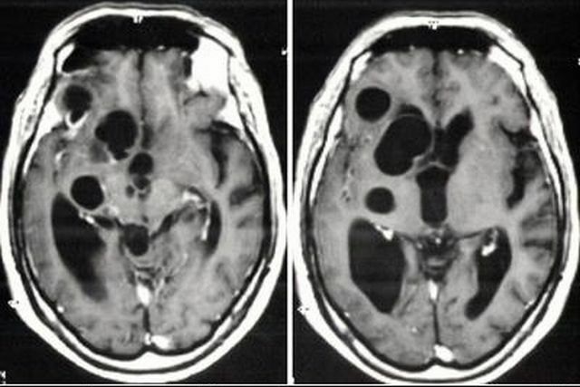
Dimensions of the cystic cavities are very diverse and occupy most of the hemisphere. From the inside, the cavity has a smooth structure and consists mainly of an ependymal-like shell.
In the case when the cyst is isolated and does not have a common passage with the ventricles of the brain - its filling consists of a yellow colored protein, if reported, its content consists of a cerebrospinal fluid.
Most often in the walls of the cystic cavity are traced tissues that have defects in the structure of the cortex of the brain. In some cases, in 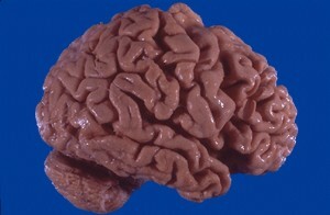 , they detect hemoglobinogenic pigmentation, as a result of hemorrhages, as well as minor anoxic necrosis.
In the area of connecting cysts with the ventricles of the brain, the lateral ventricles are often dilated and have an irregular shape.
The pattern of the cerebral cortex at the site of cystic education is practically absent( this region of the cerebral hemisphere covers a thin layer consisting of a soft shell of the brain and a medullary substance) or immersed radially in the direction of the cavity.
In histories of the cerebral cortex closest to the cyst, histological changes are revealed, characterized by a disturbance in the orientation of the cell layers, as well as the rarefaction of ganglion cells. There may be focal microses in the soft shell of the brain. This type of disturbance is a consequence of true panencephaly.
There are various degrees of abnormal development, but the most pronounced is the "bubble brain"( two large bubbles filled with cystic fluid), characterized by a pathological process involving hemispheres, basal nuclei, as well as the brain stem. It is worth noting that with this type of pathology cystic changes of the cerebellum are extremely rare.
Etiology of violation of
This disease can be caused by a brain injury, an early micromusis of the brain tissue, or a prenatal developmental disorder of the nervous system.
Such defects in the development of the brain as cystic cavities are most often an intrauterine pathology, and not only are a consequence of a violation of the uterine circulation, but also a defect in the process of morphogenesis.
Sometimes, cystic formations can appear at any age and are the result of brain trauma, hemorrhage, infectious and inflammatory processes, various types of tumors of destructive effect on white and gray matter of the brain.
Pathomorphology of anomaly
In the process of filling cavities with cystic fluid, the brain substance shifts, which often causes secondary damage to the brain tissue. There is also a disruption in the orientation of the cell layer.
With this disease, the total brain mass decreases by 20-25%, insufficient development of the frontal lobes and cerebral hemispheres of the  of the brain is revealed, nerve cells are not differentiated and practically absent on the surface of its cortex.
There is insufficient development of the vascular system, as well as disturbed the maturation process of the sheath of nerve fibers.
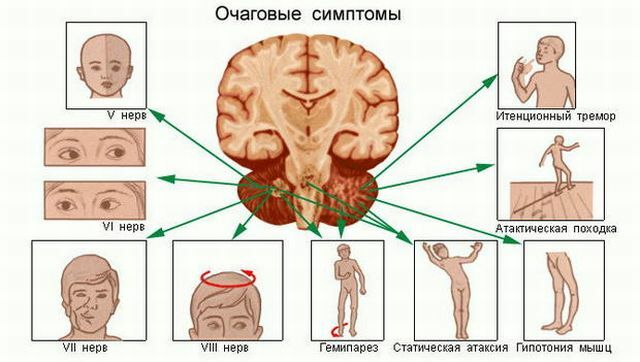
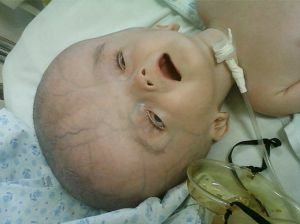
Younger children have seizures of epilepsy, asymmetry of the skull is observed, in which the affected side is somewhat bulging in the temporal region, a significant lag in mental development, disorders of nerve cells in the cranial region, as is paralysis of the limbs.
Dysplasia of the corpus callosum, microcephaly, as well as other defects in the development of organs is often detected.
Causes of the pathological development of
Like most of the defects of prenatal development, the risk of the development of the porencephaly arises from the influence of harmful factors during pregnancy( smoking, alcohol, various types of infection, peculiarities of the structure of the internal organs of the mother), which lead to disruption of the development of the cerebral vascular system.
The same risk factor can be previous abortions, premature pregnancies, the age of the mother(  is the most prosperous age from 17 to 30 years old).
Postponed infectious diseases in the period of gestation can lead to a violation of utero-placental blood circulation, which can lead to intrauterine fetal asphyxia.
In the case of immunological incompatibility, the mother's body produces antibodies that, by breaking the placenta, penetrate the nervous system of the fetus, exposing it to toxic effects.
Diagnosis and treatment options for
Until recently, it was almost impossible to detect the presence of pornecephalus and to distinguish it from other developmental anomalies, and it was detected only during autopsy.
However, to date, there are various research methods that can confirm or deny the presence of this anomaly.
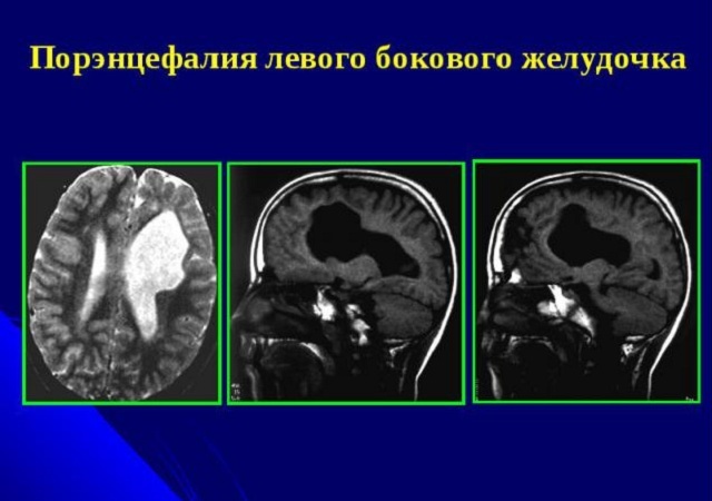
The diagnosis is based on various neurological examinations: ultrasound, CT, MRI of the brain. In some cases, this disease causes hydrocephalus.
Treatment depends on the degree of damage to the brain tissue, as well as on the location of the cysts, their volume and quantity.
Often, patients only show minor symptoms from the nervous system, which is accompanied by normal mental development. In this case, supportive therapy is prescribed, with the help of medicines, physiotherapy.
 In the case of worsening of the patient's condition, accompanied by the development of secondary hydrocephalus and the increase in epilepsy attacks, surgical intervention is performed.
In the process of surgical treatment, the dissection of the porencephalic cysts is performed and the brain tissue damaged by the vascular plexuses located in the depth of the cavity is excised.
In case of ineffective surgical treatment, the ventriculoperitoneal shunt is used to pump the cystic contents of the ventricles of the brain. However, with a large volume of affected areas of the brain, the outcome is unfavorable.
In congenital cerebral porphyria the prognosis is unfavorable, if the cavities( cysts) are large enough, the children die  during the first months of life.
In the case of single cysts of small size, patients experience a delay in mental development, as well as a high likelihood of epileptic seizures and paresis of the extremities.
In the case of false panencephaly( when cyst formation occurs as a result of an infection or head injury), the child develops completely normally before the onset of the underlying disease.