Microsporia - a fungal disease that affects the skin and hair, and in extremely rare cases, and the nail plate. The name of this fungal disease comes from the name of its causative agent - the fungus genus mikrosporum. The disease is also known as "ringworm", due to the peculiarities of its manifestation.
Once on the skin, the fungus is introduced into it and begins to multiply. When the location of hair follicles near the spores germinate, leading to shock of hair. Quite quickly spreading across the surface of the hair, the fungus destroys the cuticle scales which accumulate between disputes. Thus, the fungus surrounds the hair, forming a cover, and dense in the bulb.
Microsporia is the most common fungal infection, not counting the fungus feet. The disease is found everywhere. Microsporia is highly contagious, often children suffer. Adults also suffer from rare - mostly young women. The rarity of the disease microsporia adults, especially with lesions of the scalp, and usually occurs independent recovery at early adolescence due to the presence in the hair adults organic acids, retarding the growth of fungus.
The main source of the disease - a cat (usually kittens), at least the dog. Microsporia Infection occurs through direct contact with the patient or animal subjects infected or wool scales. Once in the soil, the fungus remains viable for only 1-3 months. Thus, the soil is a transmission factor and serves as its natural source.
symptoms microsporia
Manifestations microsporia animals are characterized by patches of hair loss on the face, the outer surfaces of the ears and on the front, sometimes the rear, legs. Often apparently healthy cats can be carriers of the fungus.
Seasonal fluctuations of disease associated with litters of cats, as well as more frequent contact of children with animals in summer. Rise in incidence microsporia begins at the end of the summer, peaking in October and November, the reduction to a minimum occurs in March and April.
The incubation period for zoonotic microsporia is 5-7 days. Character displays microsporia due to location of the lesions and the depth of penetration of the pathogen.
Allocate mikrosporiya smooth skin and mikrosporiya scalp.
Microsporia smooth skin
In place of the introduction of the fungus appears swollen, towering red spot with clear boundaries. Gradually the spot increases in diameter. It formed along the edge of the continuous towering roller submitted by small nodules, bubbles and crusts. In the central part of the spot resolution of inflammation occurs, whereby it acquires a pale pink color, with defurfuration surface. Thus, the hearth is of the form ring.
The number of foci with Microsporum smooth skin is generally small (1-3). Their diameter ranged from 0.5 to 3 cm. The most common lesions are located on the face, neck, forearms and shoulders. Subjective sensations are no worries or moderate itching.
In infants and young children, as well as in young women is frequently a marked inflammation and minimal peeling.
Individuals who are prone to allergic reactions (in particular, in patients atopic dermatitis), The fungus is often masked by the manifestations of the basic process is not always diagnosed in a timely manner. The use of the local hormonal therapy only reinforces the spread of fungal infections.
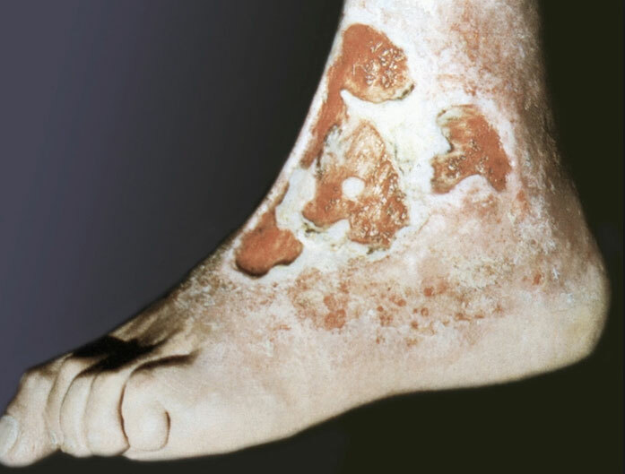
The rare species microsporia should include skin lesions palms, soles, and nail plate. For nail infections isolated lesion characterized by a nail plate, usually its outer edge. Initially formed a dim spot, acquiring over time a white coloring. The nail in whitening becomes more soft and fragile, and can later collapse.

Microsporia scalp
The defeat of the hairy part of the head microsporia occurs mainly in children aged 5-12 years. It is believed that this form uncommon in adults due to the presence in their hair, organic acids, slowing the growth of the fungus. This fact indirectly confirms the independent recovery of children during puberty, when there is a change in the composition of sebum. Interestingly, the mikrosporiya scalp practically does not occur in children with red hair.
Foci microsporia scalp are located mainly on the crown, in the parietal and temporal areas. 1-2 generally present major focus value from 2 to 5 cm, with round or oval outline and distinct boundaries. Along the edge of large pockets may be screenings - small foci diameter of 0.5-1.5 cm. At the beginning of the disease at the site of infection husking portion is formed. In the early days of the fungus is located just at the mouth of the hair follicle. Upon closer inspection, you will notice a whitish ring-flake-like hair surrounding cuff. 6-7 day mikrosporiya extend to the hair, which become brittle, break off above the level of the surrounding skin at 4-6 mm and trimmed to look like (hence the name "ringworm lichen"). The remaining stumps look dull and covered with shaped case grayish-white, which is a fungal spores. If hemp "pat", they deviate in one direction and, in contrast to healthy hair does not recover its original position. The skin in the lesion, as a rule, slightly reddened, swollen and its surface is covered with a grayish-white with small scales.

Photo: site of the Department of dermatology Tomsk Military Medical Institute
suppurative form
When suppurative form microsporia against the backdrop of significant inflammation, formed soft nodes bluish-red color, the surface of which is covered with pustules. When pressed through the holes of pus. Forming suppurative form microsporia promote inappropriate (usually local) treatment, and serious comorbidities later visit to a doctor.
Diagnostics
Diagnosis microsporia carried dermatologists.
To confirm the diagnosis microsporia used fluorescent microscopy and culture studies.
Fluorescent research: the method is based on the detection of a bright green glow of the hair affected by fungi of the genus mikrosporum, on examination under Wood's lamp. The reason for this phenomenon has not yet been established. Fluorescent study should be carried out in a darkened room. Lesions previously purified from crusts, ointments, etc. In a study of fresh glow foci may not be available, due to the lack of hair defeat. In such situations, the hair should be removed from the proposed site of introduction of the fungus, and can be found in the glow of the root portion. At the death of the fungus glow in the hair is preserved.
The luminescence method is used to:
- determining the pathogen;
- determining the infected hair;
- evaluating the results of therapy;
- control of persons in contact with the patient;
- certain infections or carrier animals
Microscopic examination: to confirm the fungal origin of the disease microscopic examination subjected flakes of foci in lesions of the skin smooth and with involvement of the scalp - fragments hair. The scales of the centers on the smooth skin found coiled strands of mycelium. Microscopic study of the affected hair reveals numerous small spores on its surface.
Cultural studies: conducting cultural diagnostics with positive results and fluorescent microscopic research is needed to identify the fungus pathogen. The method allows to determine the genus and species of the pathogen, and consequently to carry out adequate treatment and prevention of disease. Material (scales, hair) is placed on the nutrient medium. Growth of colonies mikrosporum (main exciter microsporia) marked on the 3rd day after sowing.
microsporia treatment
Treatment microsporia smooth skin
When treating microsporia smooth skin without hair lesions used externally antifungals. On the morning of the lesions cause 2-5% tincture of iodine, and in the evening smeared with antifungal ointment. Using traditional 10-20% sulfuric, 10% sulfuric 3% salicylic or 10% sulfuric tar ointment. Twice modern ointment applied on the day:
- clotrimazole,
- ciclopirox,
- isoconazole
- bifonazole et al.
Well-proven drug terbinafine (Lamisil), available as a 1% cream and spray.
In marked inflammation reasonable to administer a combination of drugs having further hormones. By such means include ointments mikozolon and travokort.
If a bacterial infection is useful triderm cream. With deep forms microsporia shown preparations containing dimexide. In particular, in such situations it is widely used hinozola 10% solution (hinozol and salicylic acid at 10.0, 72.0 dimexide, distilled water of 8.0). The solution should be applied 2 times a day until the disappearance of fungi.
With the defeat vellus, and even longer hair must be a systemic antifungal therapy microsporia.
Treatment microsporia scalp
In the treatment of this form of the disease is still the drug of choice is griseofulvin - antibiotic produced by molds. Griseofulvin is discharged in the form of tablets of 125 mg. The drug is taken daily in 3-4 divided doses during meals with a teaspoon of vegetable oil, which is necessary to increase the solubility of griseofulvin and increasing the duration of its validity. Children under the age of 3 designate griseofulvin preferably in suspension, 8.3 ml of which correspond to 1 tablet (125 mg) of the drug. Continuous therapy is carried out prior to the first analysis of a negative result on fungi, whereupon griseofulvin within 2 weeks of receiving the same dose every other day, and then another 2 weeks 2 times week. The total course of treatment is 1.5-2 months.
The treatment must weekly shave and wash your hair 2 times a week. To simultaneously rub to focus any antifungal ointment. In parallel with the reception of an antifungal agent can be carried out manually hair removal preliminary overlapped on the lesion 5% grizeofulvinovogo patch.
Of the side effects of griseofulvin should be noted headache, allergic rashes, uncomfortable feeling in the pancreatic area. Because of the toxic effects on the liver griseofulvin is contraindicated in children who have had hepatitis, or suffering from liver disease. The drug does not designate also renal diseases, gastric ulcer and duodenal ulcer, Neuritis, blood diseases, photodermatosis.
In recent years, an alternative is griseofulvin, terbinafine (Lamisil). In the treatment of Microsporum scalp terbinafine applied in the form of tablets produced at doses of 125 and 250 mg. When treating children terbinafine dose is determined depending on the body weight. Terbinafine take 1 time a day. Good tolerability. Patients may be concerned about a feeling of fullness in the stomach, small abdominal pain. Dieting, aimed at arresting flatulence, Saves patients from the discomfort.
prevention
Prevention microsporia is early detection, isolation and treatment of patients microsporia. periodic medical examinations should be carried out in children's institutions. Diagnosed patients microsporia child must be isolated from other children and refer for medical treatment in a specialized hospital. Things that belong to the patient, to be disinfected.
Be sure to relatives and are examined in contact with the sick person. Particular attention should be given to pets, as they are often the source of infection. Microsporia sick animals or destroyed, or they carried out a full anti-fungal medication.