1 Which method of examination should I choose?
CT of the abdominal cavity is based on a specialized X-ray device. The rays are absorbed by the human body during the study, depending on the density of certain tissues. On the monitor, slices are visible layer by layer. The computer recycles the received data and displays the image on the screen. This kind of research is good for the bone system of the body.
MRI of the abdominal cavity( since the research is based on the application of an electromagnetic field), you can do as many as you want, it's harmless. CT scan - only once every six months, but better a year. Because it is not desirable to expose your body to X-rays.
We recommend that you read the
- What you can not eat before the ultrasound of the abdominal cavity
- What is urine diastase and how to take the analysis?
- When is the GFD of the stomach prescribed and is there a contraindication?
- An effective remedy for gastritis and stomach ulcer
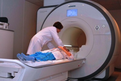
But the advantage of CT is that it is done fairly quickly, only for 8-10 seconds, and the MRI needs up to 20 minutes. Therefore, patients suffering from claustrophobia, such a study is not suitable. If children are manipulated, they will not be able to withstand such a large amount of time in a stationary state. Sometimes diagnostics by means of a tomography to kids spend under anesthesia.
A sufficient number of techniques and apparatuses have been invented in the world to diagnose, with greater or lesser precision, a number of diseases of the abdominal cavity or other organs and systems. In addition to CT or MRI, ultrasound is very popular. But what is more reliable, MRI or ultrasound? In which cases is it better to apply these or other methods?
Ultrasound examination helps to detect quite serious abnormalities in the functioning of the abdominal cavity and retroperitoneal space. And if the disease has already begun to progress, ultrasound can show it best. Neoplasms, tumors are clearly visible during examination on the apparatus. Differences and internal bleeding. When the diagnosis is already known, to confirm it, use ultrasound. If it is necessary to quickly examine the organs of the abdominal cavity with an unknown diagnosis, the doctor directs to ultrasound. The procedure allows you to view organs locally, when you do not need to diagnose several of them at once. This examination in many situations does not require long preparation.
-
 IMPORTANT TO KNOW! Gastritis? Ulcer? To have a stomach ulcer not turned into cancer, drink a glass. ..Read the article & gt; & gt;
Assign ultrasound in the following cases:
- When to look organs of the gynecological sphere.
- To diagnose the condition of the stomach and the gastrointestinal tract as a whole.
- To trace the work of the kidneys, the bladder.
- When it is necessary to know whether everything is in order with the heart.
To determine for yourself what is better - MRI, CT or ultrasound - the simple logic and the current state of the patient will help. If it is necessary to examine a whole complex of organs, it is better to make a tomography scan, the skeleton is CT, but when you need to examine locally the stomach or uterus, ultrasound is suitable. MRI is considered more accurate, because the decoding is carried out automatically by the computer system itself. Conducting ultrasound, the doctor can make mistakes.
-
 Gastroenterologist. IMPORTANT: "I beg you, if you began to worry about abdominal pain, heartburn, nausea, do not do gas in any way. .."Read more & gt; & gt;
Information about the diseases will be most reliable if the patient passes both ultrasound and resonant diagnostics. In this case, the doctor performing the MRI, it is necessary to bring the results of all previous analyzes.
2 Who should do the research?
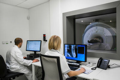
What does MRI of the abdominal cavity show? This question is asked by many patients. MRI examines the organs of the digestive tract, lymphatic system, vessels. The technique allows you to refine their localization, size, shape, find pathology and understand its characteristics, to find out whether there is an anomaly connection with neighboring systems and organs. So tomography allows doctors in advance of the development of a dangerous disease to suspect an undesirable process, identify it and eliminate it.
Who should do the research? If soft tissues and organs suffer, the tomography of the abdominal cavity is irreplaceable. It reveals tumors:
- In the fat layer.
- In the abdominal cavity.
- In the pelvic organs.
Magnetic radiated techniques are applicable for the inspection of the following organs:
- liver;
- pancreatic;
- spleen;
- lymph nodes;
- soft tissue of the digestive tract;
- intestines.
Study of other abdominal organs that is part of the MRI diagnostic system:
- adrenal glands;
- of the kidney.
Resonance technique is indispensable in the detection of cysts, cystic tumors, the definition of their stage. A great help to such diagnostics is provided to the surgeon before the operation. You can take MRT on your own initiative, and afterwards, if your suspicions about the diseases have been confirmed, go to the doctor with the results. The doctor will refer to the procedure when the patient:
ADVICE FROM THE MAIN GASTROENTEROLOGIST
Korotov SV: "I can recommend only one remedy for the rapid treatment of Ulcer and Gastritis, which is now recommended by the Ministry of Health. .." Read testimonials & gt; & gt;
- Injuries in the abdominal cavity.
- Suspicions for enlarged spleen, liver.
- Jaundice.
- Inflammation of the pancreas.
- Ascites( fluid in the abdomen).
- Inflammation of internal organs.
- Liver pathology( cirrhosis, dystrophy).
- Thrombosis.
- Lipoma.
- Suspicions on the cyst.
- Tumor( benign or malignant).
Before surgery, the tomography of the abdominal cavity helps to determine the presence of:
- anomalies in the development of certain systems;
- foreign bodies;
- circulatory disorders;
- local inflammatory process;
- dystrophy of organs.
But there is one caveat for those wishing to carry out the procedure without a doctor's recommendation. MRI does not diagnose stones in the kidneys, ureters and gallbladder. Deposits of calcium salts( without a drop of liquid in the structure) a high-tech apparatus will not see simply because its purpose is to treat soft tissues.
3 Contraindications
Patients with metal implants in the body, devices of the electronic type to maintain the heart, do not go to the MRI.Cardiosystems or hearing aids will become unusable when exposed to a magnetic field.
People with claustrophobia will not be able to pass a survey, this is possible only with an open-type device. Nervous pathologies, in which the patient can not be immobilized even for a short time, will not allow to endure the research process. We mean the weight of a man. If he exceeds 130 kg, doctors will not be able to carry out manipulation. You can not do MRI for pregnant and lactating women, patients with kidney failure.

MRI of the abdominal cavity and other organs is done on high-quality tomographs. Previously, for the procedure, the patient held his breath for several seconds at intervals. Now you do not need to do this. The system itself is guided by the breathing of the subject and is adjusted to it. But the immobility will have to be maintained until the end of the diagnosis. You'll need to lie down until half an hour. If during the manipulation you accidentally make even a slight swaying, the result will be unreliable. For the comfort of the examinee, it is possible to fix his hands and feet so that he does not overexert himself, controlling himself.
4 Recommendations for procedure
Preparation for MRI of the abdominal cavity requires a serious approach. For a couple of days before the manipulation should not eat fresh fruits and vegetables. Contraindicated: rye bread and Borodino, carbonated drinks, soybeans, beans, beans, peas, products based on sour milk.
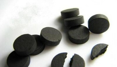
If the patient suffers from constipation, swelling, it is necessary to take laxatives, activated carbon. Immediately on the day when you are going to the diagnostician's office, you can not drink tea or coffee. Women are advised not to use cosmetics for face, hair.
Before you prepare for an MRI, you need to be patient. There are allowed 10 hours before the procedure and only after it. The liquid should be discontinued for 5 hours and take a spasmolytic pill. Do not tolerate, if before MRT you want to go to the toilet. Be sure to release the bladder.
Why preparation for MRI of the abdominal cavity should be performed on an empty stomach? Doctors explain this simply: even a little food in the stomach is a strong impediment to image quality.
If you are researching the pancreas, spleen, liver, then doctors recommend sticking to a carbohydrate diet. But if you need an emergency MRI procedure, it is done without preparation.
Survey does not carry any threat. The capsule, although it is closed, but has space inside, is illuminated, there is an opportunity to negotiate with a doctor who conducts the study.
In some cases, before imaging, a coloring harmless substance is injected into the patient's vein. So it is easy to know the speed of blood spreading through the body, to trace the circulatory processes. Once it is distributed over the vessels and tissues, the organs and systems can be seen more clearly. This is especially important for diagnosing different types of tumors, determining their structure, shape, and size. The fluid that is injected into the veins is called "contrast."
To prepare for MRI with contrast, it is necessary:
- A few days before the procedure, adhere to a more strict diet than is shown with conventional MRI.
- Do not use beans, apples, milk.
- To do the manipulation is recommended on an empty stomach( the last meal on the eve of the examination should be no less than 12 hours).
- Tablets antispasmodic is required before the procedure.
MRI with contrast has contraindications. Pregnancy is the first of them. It is equally important to know the doctor if there are implants in your body.
5 Structure, size, form
Hemangiomas, cysts, metastases, adenomas, cancer and all other types of liver tumors in diameter more than 1 cm can be determined by MRI.No less details can be examined and the gallbladder, its ducts, biliary tract. The procedure reveals anomalies of the gallbladder, its lesions and pathologies, such as cholangitis.
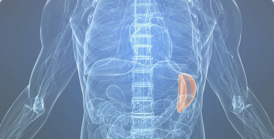
The role of MRI examination in the ailments of the pancreas is great. It is this study that allows you to find out exactly what the organ suffers from. Tumor processes are especially well defined. No less clear are the effects of pancreatitis, both acute and chronic. A diet is required before the examination of the pancreas by means of an MRI.
The spleen is no less important organ that can be examined with the help of tomography. The functions of this body are many. Diseases occur as a result of the effects of neighboring organs. Through the examination, look at the structure, size and shape of the spleen. Indications:
- Increase in size.
- Cysts.
- Localization in a location uncharacteristic for this organ.
- Changes in the structure.
- Inflammatory process.
- Developmental flaws.
MRI is also useful in the examination of the kidneys and adrenal glands. Not only tumors are detected, but also various injuries of these organs. MRI of kidneys was shown and those patients who do not tolerate specialized preparations containing iodine. They are used for urography( one of the methods of studying the kidneys).
If ultrasound is suspected of pathology and neoplasm, using MRI can confirm or exclude them.
Anomalies in the development of the excretory system and propensity to hereditary pathologies are the causes of the direction of MRI.
- 1 Which survey methodology should I choose?
- 2 Who should do the research?
- 3 Contraindications
- 4 Recommendations for procedure
- 5 Structure, size, form
To obtain reliable data on the state of the digestive system, use the MRI of the abdominal cavity. This type of survey makes it possible to see sections in several planes. The pictures are clear. This survey is currently the most accurate and informative.
Tomography of the abdominal cavity is done in a number of cases. If a person has unpleasant sensations in the abdomen, swelling, pain. The doctor appoints a wide range of examinations. MRI of the abdominal cavity and retroperitoneal space is often included in this list. The doctor may be interested in the result of CT( computer tomography) - this procedure is identical to the X-ray, but the CT of the abdominal cavity organs is done with greater radiation and the result is treated differently. MRI is performed using a magnetic field. The impulses that are sent make the atoms of hydrogen move in the body. Due to this effect with the help of equipment, the doctor receives a three-dimensional image on the monitor.
Do you have gastritis?
GALINA SAVINA: "How easy is it to cure gastritis at home for 1 month. A proven method - write down a recipe. ..!"Read more & gt; & gt;


