In dental practice, a tooth cyst is quite common. If the size of the lesion is small, the patient will not disturb the pain syndrome. There is a cyst inside the gum, as a consequence of untreated dental diseases and is characterized by suppuration directly near the root. It affects not only the main dentition, but also wisdom teeth. Since this education is very common, it is necessary to have an idea of what factors the cyst develops and how to treat it. It is also important to know what the cyst looks like on an X-ray.
Cyst of tooth on X-ray
Content of material
- 1 How to recognize the cyst on its own?
- 2 What does the neoplasm look like in the image?
- 3 Types of cystic neoplasms
- 3.1 Video - Cyst of the tooth
- 4 How is the image taken?
- 5 What are the contraindications?
- 5.1 Video - Removal treatment of a tooth cyst
How to recognize a cyst by yourself?
It is almost impossible to diagnose a tumor at an early stage. This is explained by the fact that at the first time the actual cyst does not show any alarming symptoms. The patient begins to feel the first uncomfortable sensations when the neoplasm reaches considerable dimensions, but before it does not bother.
The process of cyst development is very long, so for a long time a person can not guess that the periosteal tissue is affected by neoplasm. If you are attentive to your health, you can identify one of the first symptoms of a cyst. These include:
- on the gums near the aching tooth there is a slight swelling, which gradually increases in size;
- in case of localization of the cyst near the maxillary sinuses, headaches occur, which is extremely difficult to eliminate even with the help of drugs;
- fistula formation, which acts as a tunnel, warning of the onset of an inflammatory process from the outside;
- can often increase body temperature.
What is the tooth cyst
? This symptomatology is superficial, therefore it is recommended to visit the dentist as often as possible and do not ignore routine dental check.
What does the neoplasm look like on the picture?
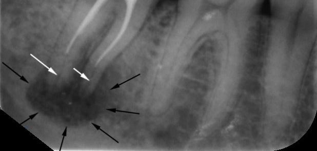
Radiography is one of the best methods for diagnosing cystic neoplasms on dental roots. The picture shows a cyst in the form of a darkened area that has a round or elongated shape with clearly outlined contours. In other words, the mapping of the cyst is defined as a homogeneous formation with even edges located in the bone structure.
On the example of the picture, which is indicated below, the cyst affects the basal area in the lower jaw. The formation of a cyst in this case arose as a result of an inadequately sealed tooth canal, which had previously been poorly cleaned. White arrows indicate the place where the filling material did not pass. Black pointers define clear boundaries of the cyst.
Cyst of the tooth
If we talk about the size of the cyst, then definitely they can be, both large and minor. A small cystic lesion can affect the root of only one tooth, large localized on the root system of two or more teeth. Most often, such formations affect the upper jaw. In childhood, cysts are mostly dangerous for the lower jaw.
Warning! If the picture gave fuzzy information about the lesion, then it is necessary to conduct a repeated X-ray examination, only from a different angle. A CT scan may also be required.
Types of cystic neoplasms
Depending on where the parodental cyst is located, several species are distinguished.
| Cyst type | Short description |
|---|---|
| Radical | This kind of dental formations is also called basal. Provokes an ailment that has entered the periodontal tissue, infection, which leads to severe suppuration. The source of infection can be periodontitis, pulpitis, which was improperly treated. Also, sepsis provokes the occurrence of the |
| cyst Residual | May occur after tooth extraction if an infection has occurred in the tissue |
| Follicular | These are cysts that occur in children. X-ray images are very similar to the radical, that is, the dark focus of the infection with sufficiently clear contours. On children's teeth, it is seen how the root of the root tooth is immersed in the inflammatory focus |
| Paradental | The location of the lesion is determined from the lateral part of the teeth |
Note! With the follicular cyst, pressure on adjacent root rudiments occurs, which leads to difficulty in normal cutting.
Video - Tooth Cyst
How is the picture taken?
If a dentist suspects a pathological lesion, the patient is referred for radiology. There are two types of snapshot - intraoral and panoramic. The latter is a display of the oral cavity with a full image of all teeth. Thus, the doctor makes a conclusion about the integrity of the dental structure, bone tissue and, in general, about the anatomical features.
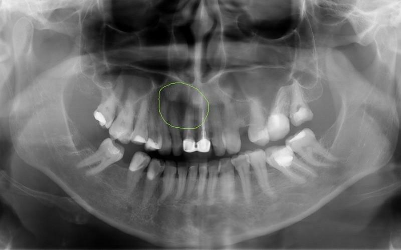
Attention! Below is an example of a snapshot( panoramic view), which clearly shows the status of all available teeth. Here you can see a large number of cysts in the bottom row.
In order for the treatment to be effective, it is necessary to find out the size and shape of the lesion, and for this an aiming image is taken. It involves taking a picture of only one tooth. Based on the results of the sighting, the tactics of treatment are determined.
In the following case, a snapshot of the sighting type is presented, which confirms the presence of a radical cyst in the upper jaw that is located between the first and second teeth.
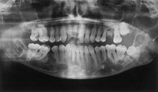
Panoramic tooth shot
What are the contraindications?
The first thing to be said is that there are no absolute contraindications for the X-ray study. On the contrary, there are relative:
- . If this research is done for children or pregnant women, then only modern digital equipment should be used, which minimizes investment. In addition, it should only be carried out if there is a direct threat to life.
- When feeding a baby, there are several important points. If you are scheduled to take a picture, then after the study, you must necessarily express the milk. The child can be fed only three hours after the X-ray exposure.
All other cases do not provide any obstacles for the performance of an X-ray. In this case, the specialist should always paste into the patient's card all x-ray loads, in order to further have an idea of the dosage of irradiation. The lead doctor then monitors that the patient does not receive a high total dose, which can harm the health.
You can find out how the cyst is treated from the video.