With the ultrasonic diagnostic brain and neck defined pathology in their receptacles narrowing of the vessel and its length, revealed the presence of thrombi, plaque, examines the state of the arteries detected defects.
The method laid vascular scanning principle that allows you to see their work from the inside. Contraindications to study there, you can perform on a regular basis, without fear of harmful effects on the body.
The content of the article:
- 1 What is an ultrasound of the brain vessels
- 2 Indications for diagnosis
- 3 Are there any contraindications?
- 4 Advantages and disadvantages
- 5 The ultrasound MRI better?
- 6 Types of ultrasound head and neck vessels
- 7 Preparation for ultrasound
- 8 How do vascular ultrasound?
- 9 That shows Doppler ultrasound of vessels of head and neck?
- 10 Decoding results
- 11 Addresses of Moscow and the cost of the study clinics
- 12 Addresses of clinics in St. Petersburg and study price
- 13 Video of the ultrasound of the brain vessels and neck
What is an ultrasound of the brain vessels
US cerebrovascular and neck is a diagnostic procedure, with which the measured blood flow efficacy in arteries, veins head. During the procedure the patient does not feel discomfort, the results can be copied to a portable device, or print the image.
Now there are other diagnostic options, such as MRI, but ultrasound is an affordable and versatile way.
Ultrasound performed just vessels of the head makes it possible to identify:
- disorders of the brain in the bloodstream;
- oxygen enters the insufficient;
- signs of stroke;
- vascular disorders;
- effectiveness of prescribed drugs.
vascular pathology study to determine:
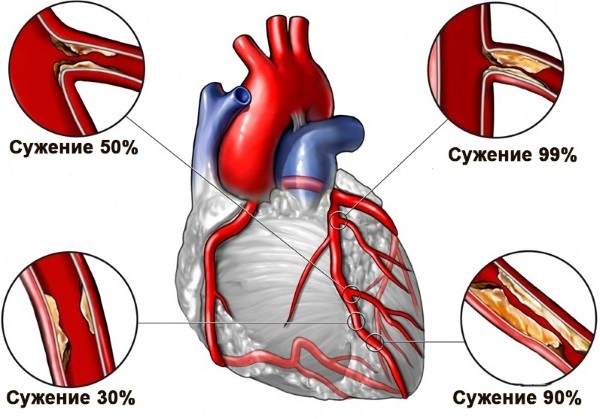
- atherosclerotic disorders in the arteries - determined quantity of plaque, the level of vasoconstriction - high rate when tested connective tissues of the neck;
- violation of blood flow;
- deformation of arterial walls;
- destruction of vessels and arteries;
- possible vasoconstriction;
- degree of compression of tissues;
- deviation obtained at birth;
- Pathology of blood supply to the brain.
Indications for diagnosis
Conducting diagnostic vascular system of the brain is performed after ultrasound of the arteries and veins of the neck, because the causes of the disease are looking for in this area. The fact that the vascular system of the head takes the foundation of the great arteries, which include carotid and vertebral veins.
there are clear indications for ultrasound:
- osteochondrosis of the cervical spine;
- arrhythmia;
- VVD;
- high rate of cholesterol;
- pressure;
- atherosclerosis;
- fatigue;
- excess weight;
- elective surgery;
- aneurysm.
Ultrasonography of the brain vessels and neck must be held in case of symptoms:
- recurring headache;
- tinnitus;
- loss of consciousness;
- sudden loss of attention, hearing;
- impaired coordination of movements;
- insomnia.

Some people are advised to regularly perform the study, regardless of the appearance of certain symptoms:
- age greater than 45 years;
- diseases of the musculoskeletal system;
- bad habits, including smoking;
- stroke;
- injury and a concussion;
- high blood pressure;
- diabetes.
Are there any contraindications?
Any method of research vessels is not harmful to health, so it can perform again. It is not possible to diagnose if the test area is closed fat mass or bone tissue.
Difficulties may arise in the following cases:
- pathologies of the heart;
- circulatory disorder;
- abrasions, cuts on the skin;
- the inability of the patient to take a horizontal position due to the disease;
- plight of the patient.
Advantages and disadvantages
Ultrasonography of the brain vessels and neck distinguish the security, availability, absence of contraindications. Ultrasound shows even infants.
The advantages of this method of research are:
- no pain at the time of;
- three-dimensional image field of study;
- re-examining, if necessary, without any harm to health;
- accurate result;
- reasonable price;
- the ability to analyze the soft tissue.

The downside of ultrasound is the inability to visualize some areas because of the projection overlap.
Among the disadvantages of isolated:
- low resolution in comparison with MRI;
- excess body weight may hinder the diagnosis;
- visualizing complexity bone.
The ultrasound MRI better?
There are basic differences between the two methods of research:
- Different sensitivity of the equipment - with the help of ultrasound can be performed monitoring the course of the disease.
- US defines large tumor, whereas the scanner identifies tumors at an early stage, as characterized by high accuracy.
- US more affordable way to study.
Comparative analysis of ultrasound and MRI:
- Both methods of investigations are performed for 20 minutes, is painless, has no side effects, safe.
- Ultrasound can be done during pregnancy, while MRI is eliminated in the first trimester.
- MRI determines the cause of the disease, ultrasound allows to establish disease, without revealing the reason.
- Excellent imaging in two methods.
Ultrasonography of the brain vessels and neck is used when you need to quickly diagnose the disease. Identify effective vascular permeability, nature of the blood flow. The analysis result is presented as an image 3 waves: initial, middle and final - thereon physician defines the disease.
In the absence of pathologies graph will be symmetrical with the same distances.
If you need a detailed study, the use of MRI - diagnostics of brain diseases by obtaining 3D-images in real-time, it provides an accurate determination of deviations, reveals multiple sclerosis and hidden violations. With the help of MRI can determine the probability of developing the disease.

US regulations:
- the wall thickness of the head and neck arteries should not exceed 1,1 mm;
- free blood within the vessels;
- the absence of eddy blood flow;
- diameter arteries - 2 mm;
- no reduction in the blood flow velocity in blood vessels;
- the absence of vasoconstriction.
After the decryption, a neurologist diagnoses and prescribe treatment.
Types of ultrasound head and neck vessels
Ultrasound is performed on a special unit consisting of a console with an electronic display screen and the signal converter required for scanning. The sensor represented as a device connected to the scanner cord. Based on the type of research, the depth passing vessels, use different sensors.
Typically use linear transducers, they are effective in the analysis of closely spaced vessels. The resulting pattern of the test vessel corresponds to a sound signal that is input from arterial pulsing in the different phases of heart contractions. The patient and the doctor will hear this sound.
Modern equipment is equipped with a recording function to capture the most important stages of research and printing images. All the methods of analysis are carried out on the principle of action of ultrasonic waves, using them identifies static movement in the body.
There are three types of procedures:
- Conducting research vessel in the two-dimensional systemIt shows the structure of blood vessels. In two ways: ultrasound of large arteries and blood vessels of the neck. For the procedure, the transducer is placed in the large arteries of the location area. If the veins are in non-standard locations, the procedure is not carried out. The disadvantages of this method is the inability to detect the blood flow velocity. Benefits - identifying disease at an early stage.
-
Duplex scanning - used when it is already known for the diagnosis, identify the causes of pathologies that allows you to see a picture of the blood supply in the arteries.

- Triplex-scan - it allows you to get the compressed data on the flow rate of the blood. An advantage of the method is an accurate reflection of vascular permeability. Disadvantage - does not allow to determine the blood flow parameters, additional research is required.
Preparation for ultrasound
Special training is not provided. Prior to the appointment procedure, the patient should seek advice from a therapist, to determine whether the need to abandon the use of cardiac drugs. Ultrasonography of the brain vessels and neck requires the same day exclude from the strong tea, energy drinks, coffee and beverages containing ginger.
About 4 hours before the scheduled time should give up eating. The fact is that in the fed body increases blood flow, which may adversely affect the diagnosis. If ultrasound is assigned a child, it is necessary to feed one hour.
An hour before the diagnosis should stop smoking. Before the procedure is necessary to release the head of the chain to secure the sensor, you must remove the long hair in a ponytail.
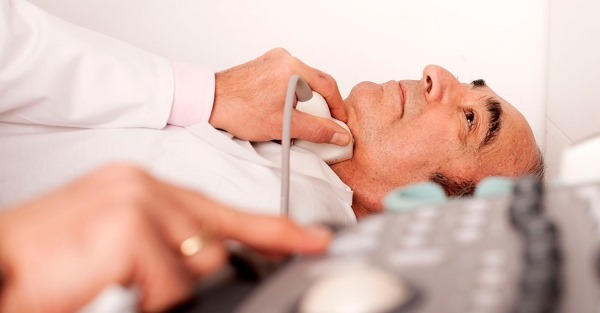
How do vascular ultrasound?
The patient must lie down, turn his head to the ultrasound machine. The first examines the carotid arteries. The doctor turns away a patient's head to give access to his neck. Using the sensors are diagnosed lower region of the carotid artery.
Subsequent research carried out along the neck to detect operation of the vessel, and determining the patency of its region where it splits into a plurality of arteries. With special mode enable physician diagnoses artery and outgoing vein, which allows to determine vascular pathology.
If defects are identified, assigned another study, it can determine the level of vascular lesions and the further course of the disease.
Further research carried out of the vertebral arteries, directing the converter to the neck. During the procedure the sensor passes through the hairy part so used the gel for easy sliding. Analyzes the blood flow rate in the veins, allowing you to identify the emerging pathology.

Overlay sensor temporal zone allows to analyze the condition of vessels, to determine their thickness and permeability. Analysis occipital zone is used to determine the pathology of venous and vertebral arteries.
When the doctor will recommend procedures:
- get up;
- not breathe;
- Quick blinking;
- breathe deeply.
Another recommendation of the doctor is a vessel clamping finger - determines the nature of the circulation of blood flow. Ultrasound can easily tolerate even the children, the procedure is not accompanied by pain.
In rare cases there may be a manifestation of thirst or increased heart rate. During the study, it is possible to hear the sounds of pulsating, changing of the pitch and appear in time with the heartbeat.
That shows Doppler ultrasound of vessels of head and neck?
Ultrasound reveals the deformed veins, congenital abnormalities, blood flow velocity and indicators that assess the nutrition of tissues. Vascular ultrasound head determines the structure of veins, branching blood flow velocity in the brain. On analysis provides information about the existing barriers: plaques, thrombi - allows you to organize data and to find pathology, inflammation, aneurysm.
Using the data obtained during vasospasm neck revealed their functionality, latent potential for normal blood supply.
Neurologist on the basis of these studies, defines the pathology, its progression, depending on the patient's symptoms. The received information is systematized and make recommendations about the course of the disease, methods of treatment and the possible consequences.

Used to decrypt the data:
- Blood flow velocity;
- heart rate;
- vascular thickness.
The results of the hidden data on vascular function, there are deviations. If instability is detected veins, the seal wall occurs - stenosis, the exponent of less than 15% - is ascertained the presence of atherosclerosis. Ultrasound helps to identify plaques, determine whether they are the cause of a lack of oxygen tissues - this information will prevent the development of stroke.
Increased wall thickness can talk about inflammation of the veins. Identifying unusual vasculature indicative of venous anomalies.
The main signs of deviations:
- atherosclerotic lesions - in the area of the carotid artery plaques are fixed, which later will bring to the narrowing and blockage of veins. The initial phase of vascular obstruction characterizes a thickening of the vessel to 1.5 mm and above this figure - the presence of plaque.
- The destruction of the arteries - atherosclerotic changes arise from the sudden pressure drop.
- venous disease - the defeat of the arteries of a circular nature, affects the entire wall of the vessel, which does not allow to separate the individual component parts of the study.
- structural changes - appear in patients with diabetes is characterized by impaired metabolism.
- Disintegration arterial walls - occurs after injury, characterized by peeling the top wall portion and to hit the blood, and then formed thrombi.
- The lack of circulation of venous blood in the brain - appears as a result of reducing the diameter of the vein, the high velocity blood flow.
- arterial thrombosis - a high level of vascular obstruction in the course of developing the disease pathology of arteries.

Ultrasound of the head and neck defines dangerous deviations and prevents the development of disease.
Decoding results
US physician or radiologist presents information obtained during the procedure. Then the doctor makes the data in the card and makes a conclusion. In severe cases, it requires additional research to confirm the disease, taking into account a certain period of time. This method of investigation is required after the operation, the passage of medical therapy.
Standard ultrasound determines the result:
- diameter vessels and its freedom from plaque;
- condition of the walls of veins;
- the presence of destruction;
- value venous constriction;
- blood flow velocity of motion;
- diameter arteries;
- view blood flow;
- the overall condition of the veins.
Healthy blood vessels have a good cross, located right in the walls of the layers can be discerned, which is readily determined by pathology and developing atherosclerosis. We analyze the value of paired arteries - have significant differences. The diameter of the spinal arteries directly affects the blood supply to the brain.
The normal rate of 3-4 mm. Upon detection of a value less than 2 mm or more and 5 mm - stated pathology. Ultrasound analysis protocol comprises anatomical characteristics neck artery blood flow movement, the presence of deformations vascular obstruction.
Jugular veins must have an oval shape, in the commission of pressing should be easily compressed, or a statement of possible blood clot.
Proceedings veins - rectilinear, equal size not exceeding three values of the carotid artery, vein spine should not be more than 2.5 mm. Blood flow in the neck should correspond to tact breath, indicator must be in the range of 30 cm / s.
Addresses of Moscow and the cost of the study clinics
US recommended in any clinic, health center, equipped with the equipment. In the clinic using ultrasound to determine the identify vascular pathology symptoms. The best way - to go study in special centers at the highest level to give a competent and decryption purpose of correct treatment.

Diagnostic value depends on the area of the procedure. When choosing a medical center should focus on the best price and quality. Treatment of identified vascular disease surgically produced by large hospitals, it is preferable to stop the choice on such institutions.
Enroll for an ultrasound at any clinic in Moscow:
| Name and address of the clinic | cost of |
| Miracle Doctor - street School, 49 | 1500 rubles. |
| Orange Medical Center Clinics - Novoyasenevsky Avenue, 13, building 2 | 2100 rubles. |
| Evromedklinik - Lilac Boulevard, 32a | 2420 rubles. |
| Cecil Clinic Plus - 1st Tverskaya Lane, 13/5 | 2500 rub. |
| MEDSemya - Krasnodar street, 57a | 1400 rub. |
| Clinic ViTerra Belyaevo - Profsoyuznaya Street, 104 | 3300 rubles. |
| Health Clinic - Clement lane 6 | 2500 rub. |
| Medical Center MEDEO - 1st street Engineering, Building 2/7, Building 1 | 2000 rubles. |
| Children's Center AvroMed - Tolbukhina Street, 13, Block 1 | 2800 rub. |
Addresses of clinics in St. Petersburg and study price
In St. Petersburg, do ultrasound in the following medical centers:
| Name and address of the clinic | cost of |
| Al'termed - Avenue Bolshevikov 7, the housing 2 | 2200 rubles. |
| Medical and Diagnostic Center - Lena Street 19, Building 1 | 2800 rub. |
| LabStori - Baseina Street 45 | 2500 rub. |
| Valmed Clinic - Prospect Moscow, 73, housing 4, the room 27-H | 1850 rubles. |
| Therapeutic and diagnostic medical center TSMRT - street Tipanova 12A | 3750 rubles. |
| Clinic Al'termed - street Oleko-Dundycha, 17, Building 1, letter A | 2200 rubles. |
| Medical Clinic Family Doctor - Petrograd district, street Academica Pavlova, 5E | 2200 rubles. |
| The first Neva Clinic - Street Esenina, 1k1, 1st floor | 1400 rub. |
| International Clinic MEDSI - street Marata, 6 | 3996 rubles. |
Vascular ultrasound detects abnormalities in the functioning of the blood flow in the brain and neck, allow the doctor to develop a treatment method and prevent complications. Research is harmless and does not require preconditioning of the patient and is an effective diagnostic method.
Registration of the article: Lozinski Oleg
Video of the ultrasound of the brain vessels and neck
Indications for ultrasound of the head and neck, and the procedure to: