
Many have heard about the neck of the femur in a human, but few know where it is. But everyone is convinced that a turning point in this part is dangerous, especially for older people.
The content of the article:
- 1 What is the femoral neck, which is a person?
- 2 At what age is the greatest number of fractures?
-
3 Types of hip fractures
- 3.1 By the presence of a wound
- 3.2 The shift of rubble
- 3.3 Depending on the location
- 3.4 Relatively damage angle
- 3.5 The placement of the fragments
- 3.6 Depending on the nature of the injury
-
4 Symptoms of hip injury
- 4.1 fracture
- 4.2 Dislocation
- 4.3 Crack
- 4.4 Necrosis
- 5 First aid for fractures
- 6 Diagnostics
- 7 Treatment
- 8 conservative
-
9 Surgical intervention
- 9.1 open surgery
- 9.2 Closing operations
- 9.3 osteosynthesis
- 9.4 Endoprosthesis
-
10 The program of rehabilitation after a hip injury
- 10.1 LFK
- 10.2 Massage
- 10.3 Psychotherapy
- 10.4 nutritional care
- 11 The duration of recovery
- 12 The consequences of fractures, and the forecast
- 13 Videos about hip
What is the femoral neck, which is a person?
Hip situated in the area of the lower extremity, hip bounded above and below - knee joint. Form hips like a truncated cone, the base of which is looking up. One of the biggest bones of the human body - the femur - controlled muscle groups responsible for the crook of the leg and hip movement.
Above femur below the inguinal lymph nodes and fold are located neurovascular bundle. Everything is located close to the skin surface. The main blood vessels are the femoral artery and Vienna. In the subcutaneous tissue passes a large venous trunk, Vienna subcutaneous thigh.
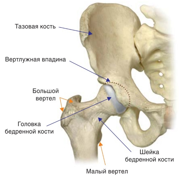
The upper end of the thigh bone is connected to the acetabulum, where you can see the head and neck. Femoral neck at the top of the femur, where the rounded head is connected with the long bones of the body. It is located at an angle to the underlying bone. Due to the fact that it has an angled shape when dropped easily broken.
At what age is the greatest number of fractures?
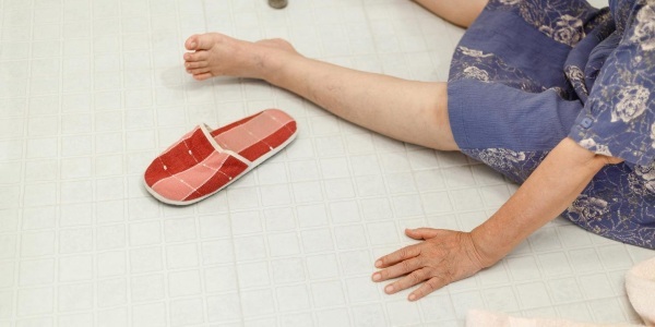
Femoral neck (where a person can see in the picture later in the article) - it is the most delicate and fragile part of the femur. level of injury increases with age. External factors affecting less than the internal, since changes over time, the structure of bone tissue, bone fragility increases.
hip fracture is more common in women, especially after 65 years. This is explained by the fact that took place in the body hormonal changes have reduced bone density. The following risk factors - osteoporosis, which often develops in postmenopausal.
Injury is considered serious, because the integrity of the bone recovery is difficult and slow.
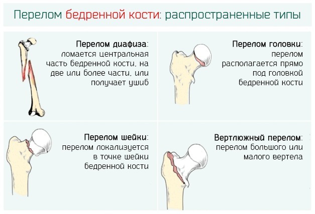
Types of hip fractures
Fractures occur in departments:
- The upper part of the bone around the joint (which includes the neck, head, trochanteric region).
- The middle part of the femur.
- The lower region.
According to the anatomical characteristics of fractures are:
- at the base of the femoral neck;
- extending through the neck;
- at the head of the femur.
By the presence of a wound
The neck of the femur (where the human and recovering from damage described later in the article) can be affected with open fault. This occurs when the soft tissue wound or gunshot wounds. The severity of damage depends on the state of the blood vessels. This may occur extensive muscle breakdown, hip nerves.
If excessive bleeding superimposed bundle introduced novocaine blockade, antibiotics, painkillers.
The shift of rubble
Complete fracture occurs with displacement of fragments or not.
For this purpose, the classification of fractures of Garden depending on the bias:
- 1 degree - incomplete without displacement.
- 2 degree - full without offset.
- 3 degree - completed with partial displacement.
- Grade 4 - completed full displacement.
Depending on the location
If hip damage occurred just below the femoral head, in which case the bone fusion forecast most unfavorable, because the head is poorly supplied with blood. If the fracture line passes through the center of the neck or at the beginning, the recovery prognosis is more favorable.
Relatively damage angle
When it traumatizing important to consider not only where the line is damaged, but its angle. The greater the angle of displacement, so it is likely that the fracture is displaced, and the bone is not srastetsya.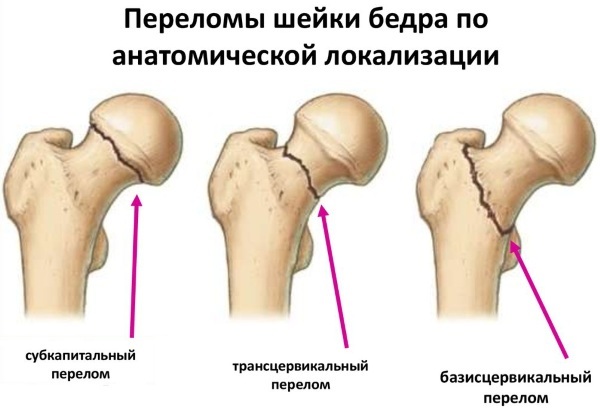
Classification offset angle:
- first power offset corresponds to the angle of inclination of the fracture plane in 30˚ or less;
- second power corresponds to the angle to 50˚;
- third degree - a corner of 50˚.
With a large angle of inclination of the fracture plane of 60˚ or higher, the probability of occurrence of adverse trends to shift the wreckage and the formation of false joints.
The placement of the fragments
Most of the fractures of long bones, which include the thigh, accompanied by the presence of fragments. They are certain difficulties in the treatment, because the fragment is deployed and is often at a considerable distance from the site of injury.
Depending on the nature of the injury
neck fracture can be caused by minor situations:
- jumping from a step;
- a sharp turn of the trunk and legs;
- badly bruised with a subsequent fall.
In this impacted injury can occur in which a fragment fused with the bone body. These cracks are not very explicit, and motor function almost not violated. And also there is a second type of injury - offset at which otkolok moves away from the bone.
Symptoms of hip injury
Femoral neck fracture much pain, and pain is localized mostly in the groin. The horizontal leg can look shorter, it can be twisted. With a slight turn impacted the sufferer can go and experience a little pain.
fracture
When passing the bone fragments of a person can not walk, stand, move your foot. In the event that a fault in the place of hurt blood vessels and follows a lot of blood, a person experiences dizziness, weakness. Swells damaged portion, is formed at this place a large bruise.
Dislocation
Dislocated joints accompanied by severe, sharp pain at the time of its occurrence. 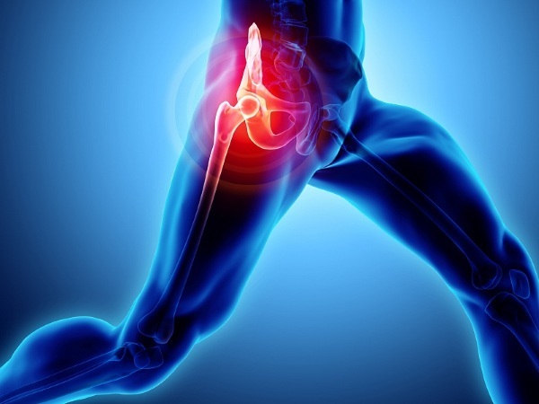 Pain develops incrementally scale increases with the load on the leg. Dislocation is accompanied by joint deformation.
Crack
The fissure is one embodiment of lung injury, the damaged portion does not intersect the plane of the entire cervix, it is limited. In such cases, the conservative treatment is assigned, the gypsum bandage is applied with a load extractor. Rehabilitation of young people going on for 3-5 weeks in the elderly, it takes up to 8 months.
Necrosis
Femoral neck (where a person which arteries and veins surround it, you can see in the photo below in the article) at fracture is often accompanied by damage to the subcutaneous blood vessels.
In this case, the vessels feeding the bone, broken, and the bone fragment that was left in the capsule remains without power. After a while, there is necrosis of the bone site. The ongoing process is called osteonecrosis.
First aid for fractures
When injury to the upper bone segment about joint pain appears not very intense, and the final takes place outside. Occasionally the leg becomes shorter by 3-5 cm, it often occurs in older patients.
First aid:
- Straighten a bent leg should not.
- It is necessary to fix the leg still. This requires a splint or tightly primotat injured limb to a healthy tissue, which will perform the function of the tire.
- The injured person can not sit or lean on the affected limb.
- If a patient wants to move independently, move the required limit, because in the future it will worsen the bone healing.
- In open fractures, complicated by bleeding, shock, first aid is to stop the bleeding. For this purpose, a sterile bandage applied, and analgesics are administered urgently admitted to hospital.
- If the patient's pain after injury worries, edema, hemorrhage, but the mobility of the joint with it is not broken, it is necessary to make the cold and before the termination of pain symptoms to use a cane or crutch.
- In the case of dislocation, which is considered a serious injury, the foot turns out, people can not pick it up, and when moving pain intensifies. It is in an unnatural position, often deployed out either side. Before the arrival of doctors can not touch it and try to fix it. Splinted joint via a tissue, the tissue in the position in which it is located. Avoid any motions, as can be affected by the sciatic nerve.
After applying plaster bandage dries immediately. Therefore it is impossible to close the space blanket fracture. fracture site should be protected from crushing, kinking. Certainly better to lay on the platform, in order to prevent tissue edema. After hardening plaster can immediately start gymnastics, it improves blood circulation, accelerates healing. It is important for elderly patients.
Diagnostics
fracture diagnosis is based on: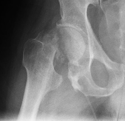
- Complaints of the patient (the inability to rely on the leg, pain in the groin).
- History.
- Clinical picture, when the injured limb is shortened, inside-out, the injured person can not tear from the surface of the heel (heel stuck symptom).
- X-ray data.
Treatment
Assessment of the patient is determined on the basis of:
- physical health;
- intensity of pain;
- survey data;
- vein thrombosis.
After administration of anesthetic drugs in the receiving compartment of the patient is sent to the casualty department, where the treatment of the patient starts on the basis of its state:
- Skeletal traction. The technique is not applicable for patients older than 50 years.
- Analgesia. Preoperative pain relievers drugs are prescribed, taking into account the scale of analgesia performed ambulance service.
- Preoperative period (in the case of appointment of an operational method). This period should not last more than 6-8 hours.
- Anesthesia.
- Surgery.
If there is no need of surgery, conservative treatment is carried out.
conservative
The neck of the femur (where the person trauma, determined by hardware diagnostics) is treated, in some cases, a conservative method. In this case there is an incomplete fracture without displacement and the tear line does not exceed the angle under 30˚.
It includes:
- hip fixation;
- Tensile (for medical reasons);
- reduction therapy using drugs;
- Complex physical therapy.
For relief of pain appointed anesthetic in the hip nerves. The procedure is called nerve blockade.
Drugs used in the treatment of:
| drugs group | Name | Act |
| Anesthesia pain |
ketorol | Admission to 10 mg three times a day for a week |
| Ksefokam | Intravenously, 8 mg, 2 times a day | |
| Emodol | Intramuscularly every 2 hours after the operation, then after 4-6 hours | |
| Ketanov | Intramuscularly, the patients older than 65 years, 10-15 mg, 10 mg tablet up to 4 times a day | |
| ibuprofen | 1200 mg per day, at moderate pain syndrome | |
| fentanyl | Intramuscularly or intravenously, depending on the patient's weight | |
| Lipidolor | Intramuscularly, the volume depends on the weight of the patient | |
| antispasmodics | Serdalud | 2 mg three times a day |
| soothing | Donormil | Up to 2 tablets. per day |
| Motherwort extract Motherwort Forte or | 1 hour before meals for 1 tab. 1-2 times a day | |
| endorntikoagulyanty | Warfarin (if conservative treatment) | |
| soothing ointment | Dolgit Traumeel |
Rubbed 2-4 times a day |
| antiedematous | torasemide | From 20 to 40 mg maximum |
| veroshpiron | From 100 to 200 mg per day | |
| diakarb | Is assigned by a special scheme |
All patients after seeking medical help conduct thromboprophylaxis, which refers to the emergency measures. The risk of venous thrombosis is present from the moment of injury.
Surgical intervention
about hip fracture surgery reduces the time of bed rest, reduce the risk of inflammatory complications. operation type depends on the type of fracture.
Drug assigned anesthesia 24 hours after surgery. For this purpose prescribed opioid analgesics reception circuit which is made according to the severity of pain.
Postoperatively appointed anticoagulants:
- Vitamin K antagonists (warfarin).
- Apixaban.
- Rivaroxaban.
As well as prescribers based on the patient's general status analysis. It can be medicine for the treatment of the gastrointestinal tract, nervous and cardiovascular system.
open surgery
At the turn of the neck in the area of bone just below the tops of open surgery is performed, during which a small incision is made in the thigh. During the operation, special screws are inserted to hold the bone in the correct position.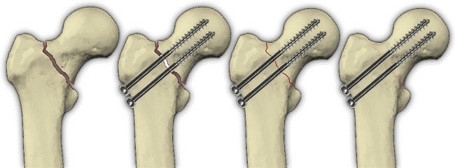
Open operations are conducted at the total hip replacement. If the ends of the broken bone are displaced or damaged, remove the head and neck of the femur and the prosthesis is set to replace them. Partial replacement is recommended for patients who have health problems, cognitive impairment.
Closing operations
Fractures of the femoral shaft treated surgically using intramedullary nails. Thus quadriceps femoris less damaged after surgery retained a small percentage of purulent complications. Closed reduction reduces blood loss, keep the blood supply to the femur.
The operation is performed on a special radiolucent table. Fixation of bone parts in this way is a minimally invasive surgery and the next day after surgery allows the patient to rely on a limb.
osteosynthesis
Surgical comparison of bone fragments by means of screws, fasteners, designed to create a fixed connection. This technique is more effective than conservative treatment, but can postoperative complications such as scarring, osteomyelitis.
Endoprosthesis
joint replacement arthroplasty is recommended in case of:
- bone fracture close to the head;
- the emergence of the risk of avascular necrosis;
- the large number of small fragments;
- strong fragmentation of the bones.
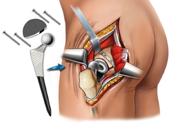
The entire operation takes less than 1-15 hours. 10-12 cm incision, whereupon he sutured cosmetic seam. Endoprosthesis is conducted more frequently in patients older than 70 years.
The program of rehabilitation after a hip injury
The main condition after the operation - observe safety that was not dislocated the operated joint. When lying under the feet should underlay cushion to feet are not meshed, you can sit down, pulling the leg forward. On the first day after the operation the patient planted and raised, put on its feet on a walker - on the second day. For two months, the patient could move with support.
Rehabilitation course is divided into early and late postoperative period. The first begins on the intensive care unit, and continues after the discharge in a rehabilitation center.
The first stage of the recovery period aimed at the prevention of post-operative complications, trophic disorders reduction, wound tissues traumatized during surgery. He continues 1-2 weeks. The second stage begins with 7-14 days after surgery.
It includes:
- initial recovery period, which takes place in conditions of rehabilitation hospital;
- secondary period which may be long.
All periods solve problems recovery support function, mobility, self-service. Upon completion of all stages of recovery the patient is sent to outpatient follow-up care. He offered to take courses of massage, exercise therapy.
rehabilitation program should be regularly and aims to improve the circulation of non-development of necrotic disorders in the buttocks and sacral region.
LFK
The course of physical therapy begins a week after surgery. Sessions may continue up to 1 year, with a gradual increase in the load on the muscles. The main task - to prevent the atrophy of muscle tissue, to restore motor function. ALWAYS exercise on the development of breath, in order to avoid stagnation in the lungs.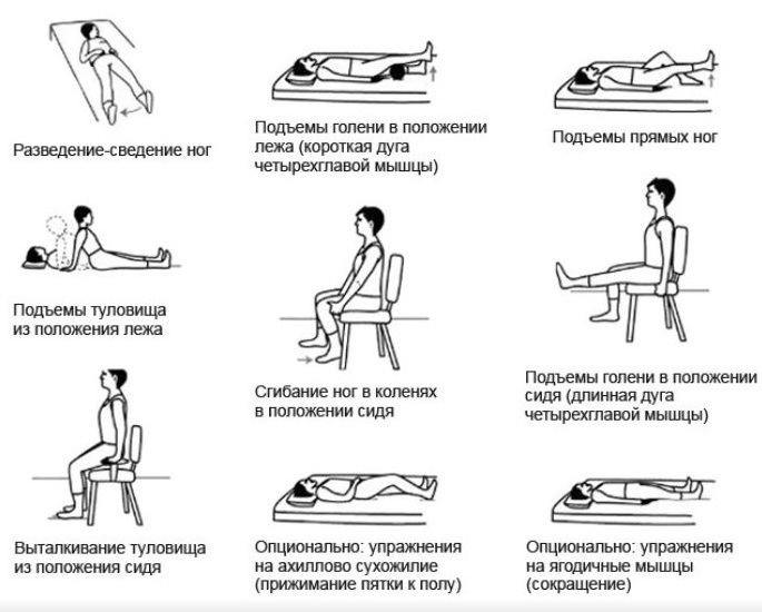
LFK complex comprises 10-13 tonic exercises for the muscles that alternate with breathing exercises.
Massage
Manual and hardware medical massage improves circulation, raises muscle tone, relieves pain. With the massage prevents pressure sores, congestive pneumonia.
Psychotherapy
Sessions conversations with the therapist to the patient based on the suggestion of optimism, lifting depressed mood, anxiety, eliminate power disturbances, appetite disorders.
nutritional care
Diet containing foods with calcium, vitamin D is necessary, but more important is diet, which neutralizes hypodynamic constipation. For this purpose, patients should give products containing vegetable fibers, as well as excessive drinking.
Healthy foods:
- a fish;
- milk, cottage cheese;
- Orange juice;
- nuts (almonds, cashews);
- beans, rice;
- dried fruit (prunes, figs);
- green vegetables, broccoli;
- bran bread;
- wheat bran;
- persimmon.
The duration of recovery
Femoral neck (where the person shown in the photo in the article) is recovered after the fracture after the operation from 2 to 12 months. This occurs provided rapid onset of action to restore the authority of efficiency. Prolonged recovery occurs after conservative treatment, the patient stand on crutches is permitted a few months after extraction.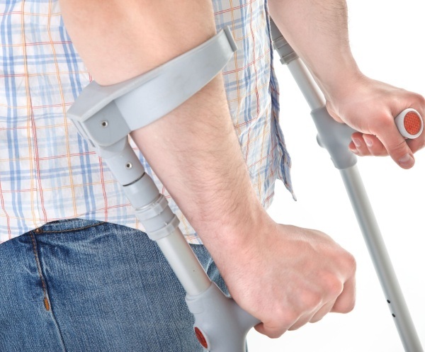
Postoperative recovery process starts on day 2-3. With 5-7 days you can do massage, 10 days later appointed courses physiotherapy, therapeutic exercises.
Independent lifting and walking with crutches is allowed after 1-2 months, and recommended a full load on the joints after 3 months. Timing represent approximate, it depends on the severity of the fracture, body condition of each patient.
To prevent recurrent fractures, doctors recommend taking bisphosphonates as reducing the risk of osteoporosis.
The consequences of fractures, and the forecast
Consequences largely depend on the fracture of the patient's age, state of health, the tactics of treatment and rehabilitation. Often, surgery does not give a full guarantee that the person will be able to move independently. Following surgery and a long recovery period is possible aseptic necrosis of the femoral neck, which requires joint replacement surgery.
Possible complications after the fracture:
- congestive pneumonia associated with physical inactivity in older patients;
- pressure sores in the area of the blades, shanks, buttocks;
- limitation of passive movements in joints;
- deep vein thrombosis in the lower extremities;
- respiratory insufficiency;
- disorder psycho-emotional sphere sick person.
Many years of hip fracture in a person's middle and old age often lead to death. This time there was an opportunity for the continuation of life in patients, if they are in the zone of increased attention and set them proper care.
Author: Anna Belyaeva
Registration of the article: Vladimir the Great
Videos about hip
Anatomy of the hip joint:


