
Diagnosis of diseases of internal organs using MRI is a modern method. It is done to identify pathologies of the brain apparatus, bone and soft tissues. The study helps to effectively diagnose disorders of the spine area.
Record content:
- 1 Why do MRI of the spine
- 2 The principle of operation of the MRI machine
- 3 Spine MRI technology
- 4 Rules of procedure
- 5 Advantages of MRI over other diagnostic methods
- 6 Indications and contraindications for MRI
- 7 Contraindications for the MRI procedure for children
-
8 MRI of various parts of the spine
- 8.1 MRI of the cervical spine
- 8.2 Lumbar spine
- 8.3 MRI of the spine of the chest
- 9 MRI of the spine: price
- 10 Spine MRI video
Why do MRI of the spine
MRI of both the spine and other internal organs allows for a detailed diagnostic study of the inner region of the human body.
The mechanics of the method consists in the effect on the body of electromagnetic waves by means of a special apparatus "MR Tomograph". The main advantage of this technique is that ionized radiation does not affect the body.
Diagnostics is carried out by creating a complex of images that allow the doctor to determine the nature of the disease at an early stage of development and begin its timely treatment.
The tomograph reproduces images that inform about the state of soft and bone tissues. In addition, it is possible to add a contrast agent to the procedure, which makes it possible to more accurately visualize pathological changes.
The principle of operation of the MRI machine
MRI of the spine is done using a tomograph, an apparatus that is a rounded scanning camera. The patient's body placed in the device is susceptible to the influence of electromagnetic waves. These waves interact with hydrogen atoms, set them in motion.
The hydrogen nucleus consists of one proton with its own spin. Being in a powerful magnetic field, the hydrogen nucleus changes its spatial orientation. Thus, the tomograph records the vectoriality and spins of protons, which makes it possible to determine the location of a specific hydrogen atom in tissues.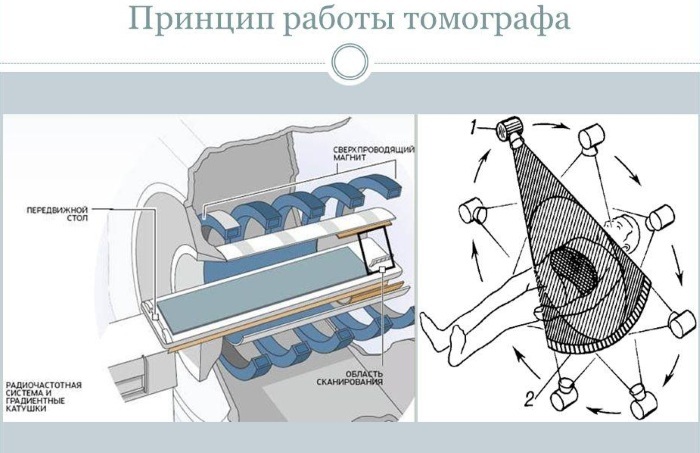
Under the influence of a magnetic field, hydrogen atoms move. Information about the dynamics of hydrogen nuclei is transmitted to the computer. The data processed on a computer gives a clear image of the structures of the spine and its individual regions in longitudinal and cross sections.
Spine MRI technology
The examination is carried out in a clinical setting using integrated equipment. The radiologist is responsible for the procedure and interpretation of the images.
The research is carried out in stages:
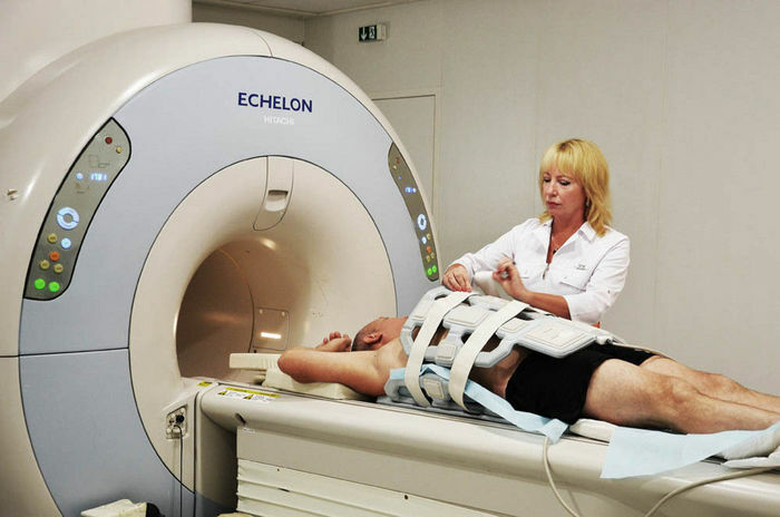
- The patient is placed in a horizontal position on a special table, fixed with straps.
- The patient, lying on the table, moves into a special camera, an MRI machine.
- The patient's body goes through the scanning process.
- The received data is processed on a computer.
- The data is visualized in snapshots.
To suppress the noise from the operation of the device, headphones or earplugs are put on the ears of the subject. The procedure requires complete immobility from the patient. The duration of the research session is at least 30 minutes. The condition of specific areas of the spine is diagnosed taking into account medical advice.
Rules of procedure
The principle of the study with an MRI apparatus is to influence the body of a magnetic field. Consequently, the procedure requires compliance with the necessary conditions. Before being placed in the chamber of the device, there must be no metal objects on the patient's body.
At the preparatory stage of the procedure, the doctor examines the patient's anamnesis and medical history, including information about current and past pathologies. This approach allows the doctor to eliminate errors in the study of MRI.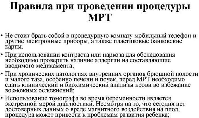
Patients of all ages should familiarize themselves with the MRI procedures before the procedure. In addition to the individual physical characteristics of the organism, the neuropsychic state of the subject is taken into account. Especially in this regard, it is difficult to prepare a child.
Depending on the area of the spine being examined, an enema may be necessary, especially if suspicion of pelvic disease (metastatic tumors to the skeletal tissue, other pathologies). The procedure requires the patient to have no metal and magnetic paraphernalia (keys, jewelry, mobile phones, plastic magnetic cards).
A child placed in an enclosed space of an MRI machine must be at rest for at least 30 minutes, which is very difficult for young patients. The increased activity of children makes the procedure impossible without the use of medicines.
Therefore, the doctor will be able to perform the MRI procedure using sedatives or psycholeptics. These drugs are not dangerous and will allow you to carry out the procedure without problems, and you are guaranteed to get clear pictures.
Advantages of MRI over other diagnostic methods
For patients who feel painful symptoms in the spine and decide to undergo the procedure, the doctor will quickly determine the nature of the pathology using MRI and prescribe adequate treatment.
Compared to other methods, magnetic resonance imaging offers a number of significant advantages, including:
- accurate analysis of the spine, revealing the presence of pathologies in its structure;
- effective diagnosis of intervertebral disc disorders;
- identification of pathologies of the facet joints of the spine;
- determination of the state of the nerve endings of the spinal cord in specific vertebral zones;
- detection of tumor neoplasms;
- identification of postoperative changes;
- detecting the presence of inflammation;
- determination of the presence of pathologies in the spinal membranes and local vascular formations;
- lack of exposure to ionized radiation;
- obtaining high-precision layer-by-layer images of the spine sections;
- accurate visualization of the endings of the nerve processes;
- assistance in carrying out large-scale diagnostics, which makes it possible to compile the most complete picture of the patient's spine condition;
- with the correct use of the tomograph, absolute safety for the human body.
If the treatment of the identified pathology requires surgical intervention, the surgeon will receive a clear picture of the disease based on MRI scans. Magnetic resonance imaging is indicated for the physician to monitor the dynamics of the patient's condition in the postoperative period.
The discussed technique is not shown to everyone, since it includes a number of contraindications. Consequently, the passage of an MRI is prescribed exclusively by the attending physician after a detailed external examination and on the basis of anamnesis.
Indications and contraindications for MRI
MRI examination of the spine is prescribed by a surgeon, traumatologist, orthopedist. The patient can independently decide to undergo the procedure, provide the results of the tomography to the doctor, and receive qualified assistance.
MRI of the spine is done if the following diseases are suspected:
- neuralgic diseases;
- pathological disorders in the spinal cord identified by x-ray;
- pathologies of the intervertebral discs;
- complications after traumatic injuries of the spinal column;
- oncological diseases
- characteristic symptomatology of neoplasms in the cranio-vertebral junction.
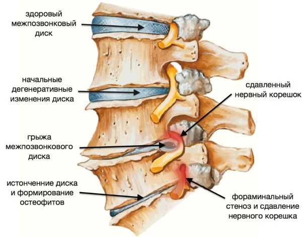
In addition to the pathologies mentioned, MRI helps diagnose background diseases:
- osteochondrosis;
- intervertebral hernias;
- pathological bulging (protrusion) of the intervertebral discs;
- scoliotic disease;
- kyphosis, of various origins;
- displacement of the vertebra relative to the bottom-lying (listez);
- misalignment of the spine;
- spinal vascular disorders;
- tumor formations;
- infectious diseases;
- diseases of the nervous system with damage to the myelin sheath of neurons.
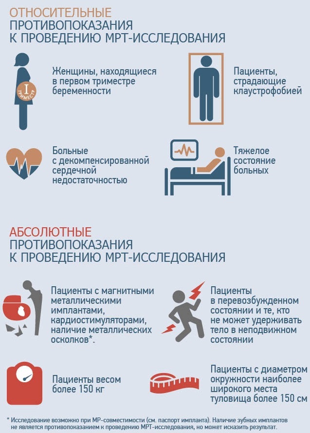
The MRI procedure is contraindicated in the following cases:
- The body contains metal objects. The magnetic field can damage metal implants present in the body.
- Women during breastfeeding. Breastfeeding of an infant is prohibited for 2 days after passing an MRI scan.
- The presence of allergies. Possible manifestations of allergic reactions in patients arise from the components that make up the contrast fluid.
- Claustrophobia. Contraindication is due to the process of passing the procedure in a closed chamber. Consequently, patients with claustrophobia may experience an attack of panic in a confined space.
- Nervous disorders.
- Restlessness of the patient. Mainly for children, since the procedure takes more than 30 minutes. resting state.
- Overweight. The technique is not allowed for patients with a body weight of more than 130-220 kg, depending on the type of apparatus.
- Pregnant. The impact of MRI on the body of pregnant women is poorly understood. Therefore, the procedure is not recommended for patients who are carrying a fetus.
MRI is contraindicated for patients with diseases of the spine who have tattoos on their bodies made with paints with metal inclusions. If these factors are present, the procedure can harm the patient.
Contraindications for the MRI procedure for children
According to experts, conducting an MRI scan is safe even for newborns. Nevertheless, there are a number of contraindications that make the procedure unacceptable to children.
For children, MRI is contraindicated in the following cases:
- In the postoperative period.
- If there are auxiliary devices in the body that ensure vital activity (hemodialysis, hearing aid, ventilator, pacemaker, defibrillator).
- The child is allergic to the basic components of the contrast agent composition.
The procedure is effectively used in pediatrics. The safety of the study makes it possible to carry out magnetic resonance imaging in children, of all ages, including newborn infants.
Given the fragility of bone structures in children, the study is carried out not only to identify pathologies of the spine, but also for prophylactic purposes. Yet doctors do not recommend MRI for children under 5-7 years oldexcept in emergencies.
MRI of various parts of the spine
MRI of the spine is done for effective differential diagnosis using modern magnetic resonance imaging equipment.
The following diagnostic measures are carried out using the tomograph:
- Virtual endoscopy. Allows you to get 3D images.
- MR diffusion. Aimed at tracking hemodynamics, and allowing to determine the degree of ischemia of the cerebral apparatus.
- Diffusion-weighted MRI. Examination of the body, at the cellular level, contributing to the diagnosis of oncological pathologies.
- MRI brain perfusion. Analyzes the blood circulation in the brain by injecting a contrast agent based on gadolinium. The technique is aimed at identifying pathological disorders of the brain structures.
- Proton magnetic resonance spectroscopy. This young method of magnetic resonance imaging is aimed at determining the biochemical composition of fluids and tissue structures.
-
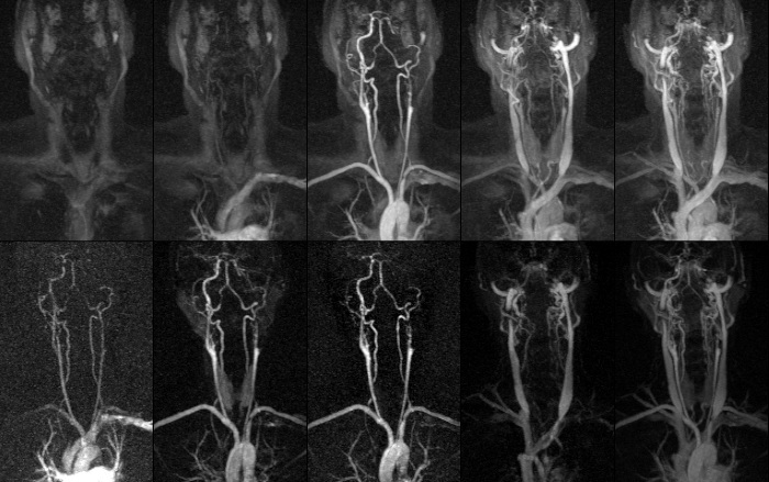
MR angiography. Analyzes the vessels in the section, determining the anatomical and functional features of the blood vessels.
A complete MRI scan allows you to visualize the condition of the spine everywhere. Depending on the symptomatology, the spines of various areas of the body are examined.
MRI of the cervical spine
An important study of the cervical spine is the assessment of the functionality of neural endings and the general picture of the state of the cervical vertebrae.
Examination of the cervical spine is mandatory for the following symptoms:
- no causal pain in the upper extremities and neck area;
- tinnitus, loss of auditory function;
- decrease in visual functions;
- dizziness;
- pronounced arrhythmia;
- muscle spasms;
- numbness of the hands.
The study will help prevent pathologies of the following nature in time:
- heart attack, spinal cord and brain;
- stroke;
- intervertebral hernia;
- perforation of the intervertebral discs;
- intra-vertebral cavity stenosis, other pathologies.
Problems in the vascular system of the cervical spine causes a violation of the blood supply to the brain. Consequently, brain activity is impaired. Timely diagnosis will reveal the disease at an early stage of development.
Lumbar spine
The most common causes of lumbar abnormalities are:
- excessive physical activity;
- inactivity;
- traumatic injury;
- lifting heavy objects.
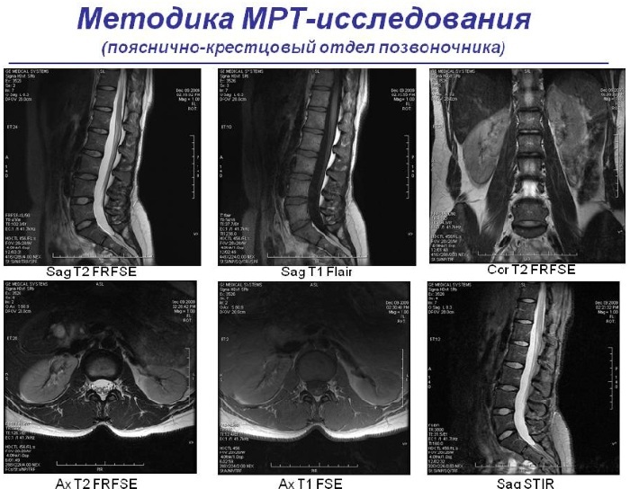
MRI of the lumbar spine. How they do and how it looks
In addition to problems with the spine, pain in the lower back can occur with uterine pathologies, diseases of the small pelvis.
The main indications for an MRI procedure are due to the following symptoms:
- pulling pain in the sacrum and lumbar region;
- increased pain in the lower back during the implementation of rotational motor skills of the trunk;
- loss of sensitivity in the toes and feet;
- spasmodic phenomena in the spinal and gluteal muscles;
- painful injuries, fractures, bruises.
Timely diagnostics will help localize pathological disorders of the lumbar spine at the developmental stage. MRI diagnoses tumor formations, hernias, diseases of nerve receptors, assesses the clinical picture after injuries and fractures. Consequently, the procedure will help prevent newly emerged pathologies in time.
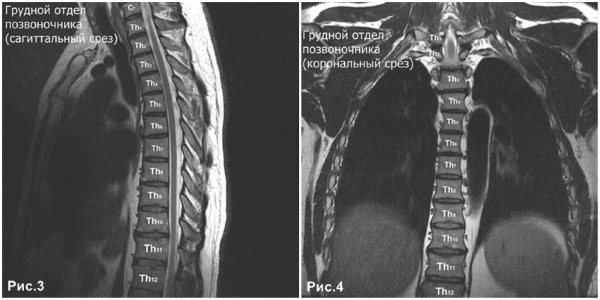
MRI of the spine of the chest
The structure of the body of the thoracic spine is formed by 12 vertebrae.
Symptoms for an MRI indication of this area include:
- pain, intrathoracic region;
- interscapular pain;
- stiffness of limb motility;
- hypesthesia of the upper and lower extremities.
MRI of the spine is done to identify diseases with any of the symptoms mentioned.
Such diseases can be:
- infectious and fungal pathologies;
- inflammatory processes;
- diseases of a degenerative dystrophic nature;
- osteomyelitis;
- bone tuberculosis;
- bulging and protrusion of intervertebral tissues;
- abnormal formations of the thoracic spine area;
- areas of the spine damaged by injuries;
- arthritic deformities;
- scoliotic disorders;
- oncology with metastasis in the thoracic region.
An MRI examination visualizes the exact location of pathological abnormalities in the thoracic spine. Further, the doctor determines the degree of pathology, develops an adequate treatment strategy. Dysfunctions of the thoracic spine entail disturbances in the functioning of the digestive system (stomach, intestines).
MRI of the spine: price
The cost of diagnostics using MRI is quite high, but the effectiveness, safety and efficiency of the procedure pays for the money spent. The average price for an MRI session is 4000-25000 rubles. It depends on the location and scope of the study.
| MRI of various parts of the spine: prices | ||
| Spine areas | Price, rub.) | |
| Moscow | SPB | |
| Whole spine (3 sections) | from 4500 | from 6600 |
| Cervical | from 2000 | from 2200 |
| Thoracic department | from 3500 | from 2200 |
| Sacroiliac region | from 2200 | from 2200 |
| Lumbar | from 2000 | from 2600 |
| Coccyx | from -2350 | from -2800 |
The highest cost for a comprehensive study. The average price for a complete MRI scan is 20,000 rubles.
Spinal pathologies are considered the most severe diseases in the musculoskeletal system. Pathological disorders entail curvature of the spine and other deformities with concomitant local pain, painful syndrome of the brain region, visceral organs.
The examination of the spine, soft tissues, nerve endings and blood vessels using MRI is done for the timely detection of many diseases. This effective diagnostic method will help the doctor identify the disease in time at the initial stage of its development, increasing the chances of curing the disorder without surgery.
Spine MRI video
Spine MRI: