
MRI contrast involves the introduction of special substances that increase the contrast of the image. This makes it easier to visualize abnormal formations and speeds up the final diagnosis.
For contrasting, paradigmatic preparations are used that do not have properties for accumulation and negative impact on the body. The procedure is considered safe, although it has some contraindications.
Record content:
- 1 Indications for research
- 2 Contraindications
- 3 Views
-
4 Preparation and implementation
- 4.1 General preparation rules
- 4.2 Preparing for an abdominal MRI
- 4.3 Preparing for an MRI of the brain
- 4.4 Carrying out the procedure
- 5 Decoding the results
- 6 When to see a doctor
- 7 Possible complications
- 8 Contrast MRI video
Indications for research
Contrast with MRI is an advanced diagnostic method using not only an MRI scanner, but also a special contrast agent that is injected into the area of interest in various ways. The contrast agent improves image quality and enhances the visualization of tissues and organs.
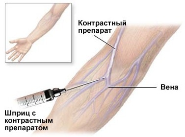
It is administered intravenously, after which the drug is distributed through the vessels, and then accumulates in the focus of the pathological process. The contrast agent concentrated in a certain area stands out brightly, which gives the doctor the opportunity to determine the activity of the pathological process and its localization.
A study is prescribed within the framework of diagnostics for suspected various diseases of the internal organs and the musculoskeletal system.
Any doctor who is faced with the need for a deeper diagnosis can issue a referral for examination:
- oncologist;
- mammologist;
- surgeon;
- gynecologist;
- neurologist.
The indications for the appointment of an MRI are the following symptoms:
- frequent headaches;

- dizziness;
- fainting;
- decreased sensitivity of the limbs or facial nerves;
- convulsions;
- getting a craniocerebral injury;
- deterioration in general health indicators for no apparent reason;
- disruptions of the menstrual cycle;
- sharp fluctuations in weight;
- severe pain of a constant nature in the local area.
Tracking an already identified pathological focus is also considered an indication for conducting a study. Contrast-enhanced MRI is also prescribed to assess the patient's condition after surgery.
Contrast-enhanced MRI reveals the following pathological processes:
| Brain research |
|
| Abdominal organs |
|
| Chest organs |
|
| Pelvic organs |
|
| Various parts of the spine |
|
| Joints |
|
Contraindications
Despite the safety of the procedure, contrast-enhanced MRI has contraindications. Often, they are associated with exposure to a contrast agent.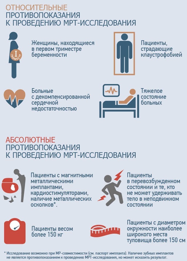
Contraindications to MRI can be conditionally divided into 2 groups:
- Absolute - contraindications, the presence of which makes the procedure impossible on an ongoing basis. These include:
- the presence of a prosthesis or electromagnetic implant;
- the presence of a pacemaker or artificial valve in the heart;
- renal failure;
- severe bronchial asthma;
- individual intolerance to the injected drug.
- Relative - temporary contraindications that open the prospect for the procedure in the future. Such contraindications are:
- pregnancy;
- lactation period;
- the patient's predisposition to panic attacks;
- patient weight exceeding 110 kg;
- general serious condition.
Views
MRI contrast is a procedure that can be performed using different types of contrast media.
According to the method of administration, contrast substances are conventionally divided into groups:
-
Intravascular. The drug is injected in full into a vein.
- Drip. The contrast agent is administered intravenously using a dropper. The scanning procedure and drug delivery are carried out simultaneously.
- Oral. The introduction of the substance is carried out orally and is used to scan the organs of the gastrointestinal tract.
If we talk about the types of tomographs that are used for MRI with contrast, there are:
- Open type apparatus. Does not have a closed scanning chamber. The patient is placed in a special room equipped with magnetic field sources. A low-power magnetic field is used for examination, which affects the quality of the images obtained.
- Closed apparatus. It is a closed wide tube into which a patient is placed, placed on an examination table. The diameter of the tomograph chamber is 60 cm, and the length of the tunnel reaches 2 m. The advantages of the device include a high power of the magnetic field and obtaining a high-quality image.
Preparation and implementation
The nature of the preparation for the procedure depends on which anatomical area of the body will be diagnosed. Specific preparation for MRI with contrast enhancement is required only in the case of examining the abdominal organs.
General preparation rules
Contrast with MRI is a research method that requires adherence to certain preparatory steps:
- First, it is necessary to exclude the likelihood of allergic reactions to the injected contrast agent - gadolinium, omniscan or primovist. Before conducting an MRI, a test is carried out, the essence of which is to inject a small amount of the drug subcutaneously. If after the procedure there is no hives or signs of anaphylactic shock, we can talk about the absence of allergies.
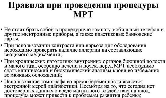
- Before conducting the study, the doctor clarifies the patient's information about the presence of chronic diseases, previous operations, possible installed implants, taking medications.
- 4 hours before the procedure, you should refuse to eat, and 2 hours before the procedure. This will help to avoid the manifestation of nausea or exclude the likelihood of vomiting during the study.
- Before you go to the examination table, you need to remove all metal objects from yourself, and also make sure that there is no mobile phone nearby.
- The medical worker should be warned about the presence of tattoos made using metallic paint, which can provoke a burn or pain.
Preparing for an abdominal MRI
Examination of the abdominal cavity deserves special attention in preparation. MRI with contrast agent is performed on an empty stomach.
The patient needs to endure a 12-hour fast and adhere to some rules:
- follow a diet with a low carbohydrate content for 3 days;
- 4 days before the examination, exclude foods that provoke increased gas production from the diet;
- for 3-4 hours, fluid intake should be limited;
- on the day of the procedure, it is necessary to cleanse the intestines with an enema;

- if necessary, on the eve of the study, drugs are taken that inhibit peristalsis and intestinal motility.
Preparing for an MRI of the brain
The preparatory stage for the MRI of the brain with contrast enhancement involves the implementation of the general rules of preparation.
It also follows:
- remove dentures, if any, since they contain ferrimagnetic metal alloys;
- remove all metal objects from the head and face area: piercings, earrings, jewelry;
- women need to remove cosmetic products from their skin that contain chemicals that can distort the test results.
Carrying out the procedure
The contrast-enhanced MRI procedure consists of a number of stages. The contrast agent can be injected both immediately before the examination and during the scanning process. For contrasting, paramagnets are used that do not react with biologically important substances of the human body and are excreted naturally through the kidneys.
A single injection of the drug is performed before the patient is sent to the tomograph capsule. The puncture is carried out into a vein located in the area of the elbow bend. The dose of the substance is calculated based on the patient's body weight. For 1 kg of weight, there is 0.2 ml of the drug.
Contrasting may also be needed after the study. This is due to the need for a more detailed study of pathological areas.
When examining images obtained by MRI without contrast, problems with visualization of problem areas may arise. Then the doctor decides on the introduction of a contrast agent. After 2-3 minutes after the administration of the drug, the patient is returned to the tomograph capsule for further examination.
The drug can also be administered as a bolus using a special injector or infusion pump. Such an introduction involves a slow drip of the substance. Before the procedure, a peripheral venous catheter connected to the injector is installed in the patient, and the machine injects the drug according to the established scheme.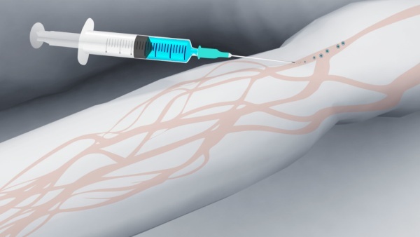
Conducting an MRI with contrast enhancement consists of the following stages:
- The patient is asked to remove clothing and dispose of all metal objects. A special disposable cape is issued for a change of clothes.
- The patient assumes a horizontal position on a retractable examination table. Then he is sent to the tunnel of the apparatus.
- During the procedure, the patient should be in a stationary position.
- The duration of the study depends on which organ or system can be diagnosed. Examination of the brain takes about 40 minutes, the diagnosis of the spine or joints takes about 1 hour. Intravenous administration of contrast increases the duration of the procedure by 15 minutes. With a drip of contrast agent, when it is required to make a series of images in dynamics with time intervals, the study can take about 2 hours.
There are times when anesthesia is required for an MRI scan.
This is required in order to achieve complete immobility of patients, including:
- Small children;
- people who are claustrophobic or mentally ill;
- people experiencing severe pain syndrome.
Depending on the clinical case, the following types of anesthesia are used:
- sedative - the use of medications injected into the vascular bed or muscle tissue;
- inhalation - the drug is administered through the respiratory tract.
When anesthesia is administered, special dispensers for intravenous administration or light face masks for inhalation are used.
Decoding the results
Interpreting the results of MRI requires a high level of knowledge of the topography of human organs, pathological physiology and anatomy. Therefore, the radiologist or the attending physician who sent the patient to undergo the study is engaged in the study of the images.
If, after receiving the images with the results of MRI, it is not possible to contact a specialist, you can decode the image yourself. When conducting MRI tomography, organs are examined in 3 projections - sagittal, coronal and transverse.
Different tissues are displayed in different shades in the image, so it is better to find images of the same anatomical areas in normal conditions on the Internet and compare them with individual results.
Also, changing the projection and using an additional light source will help to deal with the pictures. When reviewing the results, pay attention to all asymmetric areas. In addition to the images, the patient receives a written opinion from a specialist in radiation diagnostics. The description will indicate the presence or absence of pathological areas.
When examining images on your own, it is recommended to pay attention to:
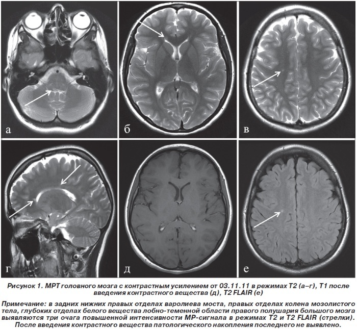
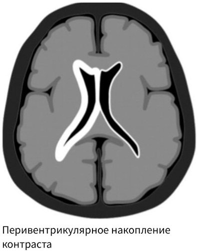
- On the localization of the pathological focus. Its location will be evidenced by the changed areas. Normal tissue can be recognized by a grayish tint of varying intensity. The presence of pathology will be indicated by areas that will significantly differ in color from the general picture of the visualized area.
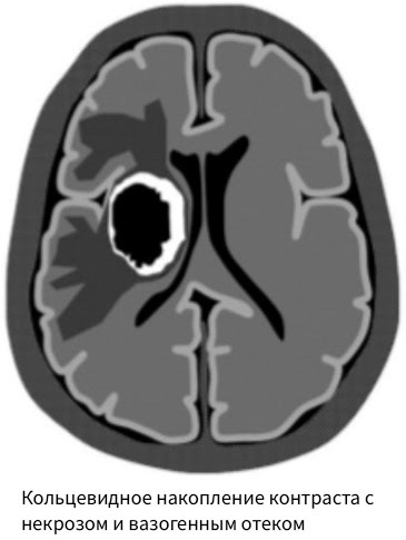
- On the size and shape of the hearth. The presence of a benign tumor will be indicated by foci of the correct shape, with clearly contrasting and even edges. The malignancy of the process may be evidenced by multiple irregular neoplasias, which indicate metastasis.
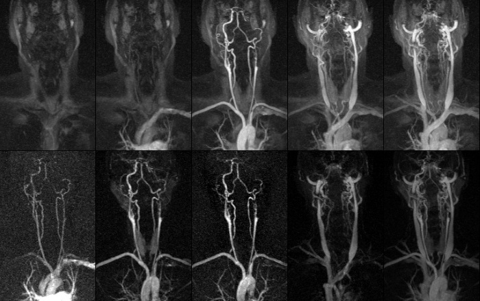
Contrast-enhanced MRI, used for vascular diagnostics, detects various vascular pathologies. The development of atherosclerosis will be indicated by narrowed vessels that are filled with a contrast agent unevenly and not completely. Also, light plaques will be visible in the walls of the vessels.
It should be borne in mind that you can decipher the results yourself when a large, pronounced pathology is visible in the picture. In other cases, reading the results is possible only when contacting a doctor.
When to see a doctor
Doctor's help after MRI with contrast enhancement is required when pathological foci are found in the images. The specialization of the doctor will depend on which anatomical area has been examined. If the brain was diagnosed, you should visit a neurologist.
In the case when the need for MRI was associated with traumatic brain injury, you need to contact a surgeon or neurologist. If the pituitary gland has been diagnosed, an endocrinologist's consultation will be required.
When examining the abdominal organs, you should consult a gastroenterologist. The gynecologist is engaged in the study of the results of MRI of the pelvic organs.
Possible complications
Contrasting with MRI is a medical procedure that can lead to adverse consequences in the form of unpleasant symptoms.
These include:
- labored breathing;
- dizziness;
- hives;

- burning in the eyes;
- the appearance of a feeling of heat;
- general anxiety;
- nausea.
These symptoms can appear at the time of the study or immediately after it. They are temporary and self-destruct within 24 hours after MRI with contrast.
Contrast in MRI is a highly informative procedure that can significantly improve the image quality during the study. The contrast agent tends to accumulate in pathological foci and tumors. This helps to distinguish one pathology from another, to judge the activity of the inflammatory process, or to evaluate the effectiveness of the treatment received.
Contrast MRI video
What to prepare for and what are they afraid of from MRI with contrast: