
Excretory urography (EU) is a diagnostic test for assessing the condition of the kidneysas well as urinary tract (MP). The procedure implies the appropriate preparation of the patient, indications and contraindications.
Despite the fact that research of this kind is less popular today than ultrasound, there is still a mass pathologies that can be diagnosed much faster and more successfully precisely due to excretory urography.
Record content:
-
1 General understanding of the procedure
- 1.1 Genitourinary system
- 1.2 Intravenous contrast urography of the kidneys
- 1.3 Plain urography
- 1.4 CT urography
- 1.5 MRI urography
- 1.6 Retrograde urography
- 2 Purposes and indications
- 3 Restrictions on performing urography
- 4 Pros and cons of the technique
- 5 Preparatory stage. How to prepare for a patient?
- 6 How is intravenous urography performed?
- 7 How is infusion urography performed?
- 8 Contrast agents for urography: routes of administration and doses
- 9 Possible adverse reactions to the administration of a contrast agent
- 10 Term of receipt and decoding of the urogram
- 11 Features of carrying out excretory urography for children
- 12 Research cost
- 13 Video about urography
General understanding of the procedure
This type of study belongs to the category of X-ray measures based on visualization of internal organs, which cannot be seen during standard X-ray.
The method is based on the fact that renal tissues are able to excrete (that is, excrete) contrast agents previously injected into the patient's body. As a result, the contrast agent begins to illuminate and provides the clearest picture of the state of the MP organs.
Thanks to this procedure, it becomes possible at an early stage to identify any changes in the renal structures and urinary tract. In this case, as many pictures can be taken as required for the formulation of a particular diagnosis.
The procedure is prescribed by a urologist most often for violations of the outflow of urine. But there are a number of other symptoms that require diagnostics in this way. In order to better understand how the procedure proceeds, it is worth considering the basic concepts and features of its implementation.
Genitourinary system
MP consists of several vital organs: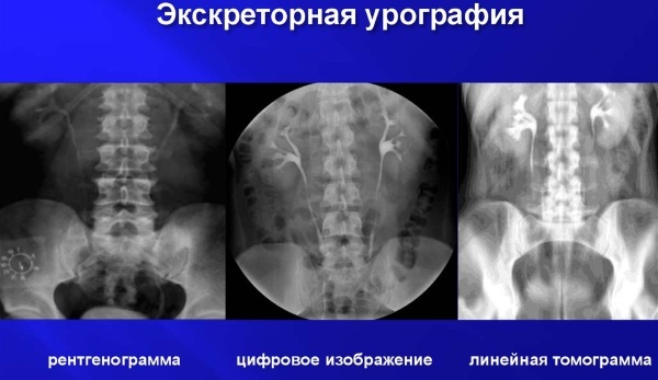
| Organ of the genitourinary system | Peculiarities |
| Kidney | Located in the lumbar region. They accumulate urine, which passes further into the ureters. |
| Ureters | Pass urine and send it to the bladder. |
| Bladder | It stores urine in itself until it is excreted through the urethra. |
| Urethra | The last link in the movement of urine. Through the urethra, urine is excreted from the human body. |
In the process of passing through all these organs, urine is filtered. If the kidneys fully perform the excretory function, then the contrast introduced earlier into the patient's body will be released at a 5% concentration. Coloring of the urine will also occur, and the doctor will see a clear outline of the bladder in the picture.
Intravenous contrast urography of the kidneys
Excretory urography is performed by intravenous administration of a contrast agent, which begins to spread through the patient's body with blood. In 10-15 minutes after the administration of the drug, images are taken to assess the condition of the organs.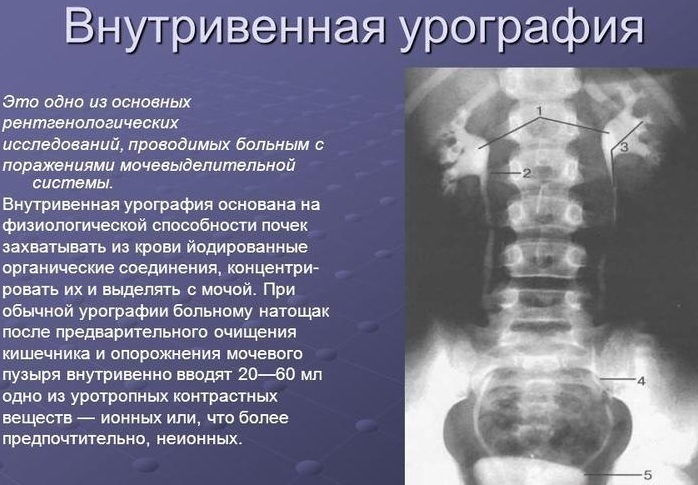
If any of the zones are colored unevenly or not at all, this may indicate different pathologies or anomalies. However, before performing such a procedure, the urologist usually recommends an overview urography.
Plain urography
This procedure also applies to X-ray examination of the MP. It is performed before EH, since sometimes the doctor only needs a general X-ray to make a final conclusion about the patient's condition.
Despite the fact that an overview diagnosis may seem uninformative, this method is more than enough to identify hematomas, kidney stones, MP anomalies. The study covers almost the entire urinary system, as well as the spine.
CT urography
In the standard EI procedure, an X-ray is taken to obtain an image, and for a CT urography, the image is created by a CT scanner. This is a combined and more modern diagnostic method that allows you to obtain a layer-by-layer image of the organs under study.
CT urography is a more informative method and is usually done when kidney stones are suspected. or adjacent organs, chronic infectious pathologies and diseases in an exacerbated stage.
MRI urography
This is another type of power plant, during which a tomograph is used, only in this case it is magnetic, and not a computer one. A combined examination of this type allows obtaining high-resolution images, which greatly facilitates the diagnostic process.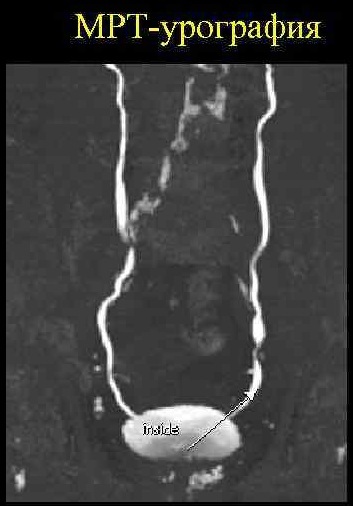
Directly in the process of MRI urography, the doctor can observe the state of organs in real time, looking at the computer monitor. A study of this type allows you to determine the presence of tumors of a malignant and benign type, edema, abscesses and other conditions that are dangerous to human health.
Retrograde urography
Excretory urography (patient preparation is one of the important stages of the study) can be performed by a method in which the contrast is injected into the human body directly through the urethra itself, that is, the introduction of the substance is carried out ascending by. In this case, the coloring composition moves in the reverse order.
Purposes and indications
First of all, EI is done to determine if there are blood impurities in the patient's urine. Thanks to the study of this type, it is possible to roughly say how exactly the red blood cells could get into the urine.
Also, studies of this type are carried out in case of complaints of pain in the MP area (for example, if the patient says that he has pain in his lower back and groin) or in case of impaired diuresis. EH can be triggered by swelling and so-called gratuitous hypertension.
Excretory urography is prescribed for:
- Urinary tract infections. As a rule, such pathologies are characterized by frequent relapses. An examination of this type makes it possible to exclude the likelihood of a recurrence of the development of the infection.
- Identification of zones in the tissues that are characterized by a deformed structure.
- Changes in the size or shape of organs. For example, the diagnosis of ES is prescribed for dystrophy or hypertrophy.
- The appearance of symptoms that indirectly indicate the possible presence of stones in the kidneys and other organs of the MP.
- Suspicion of ureteral obstruction (i.e. blockage).
- Suspected complications after a previous operation.
- Diagnostics of genetic type anomalies.
- Traumatic injuries in order to identify additional injuries.
Also, activities of this type are performed if the patient suffers from pyelonephritis, cysts, chronic glomerulonephritis or enuresis. It should be noted that EI is prescribed in the event that other diagnostic measures turned out to be uninformative or did not provide the required amount of information.
Restrictions on performing urography
Excretory urography (preparation of the patient is carried out regardless of the type of EI) differs in a number of contraindications, therefore it is not always used as a diagnostic study.
For example, studies of this type are strictly prohibited if the patient has an allergic reaction to the contrast agent used. In this case, there is a huge risk of complications and dangerous side reactions. EI is also not performed if the patient has only a mild form of drug or iodine intolerance.
Excretory urography is not indicated for patients with too high creatinine levels. In this case, the risk of complications after diagnostic measures is too high.
Also contraindications include:
- high urine density (a urine test is taken before the examination);
- thyrotoxicosis (change in hormone levels);
- active tuberculosis;
- shock or collapse in the patient;
- pheochromocytoma (a tumor in the hormonally active stage);
- diabetes mellitus, if treatment is carried out using glucophage;
- renal failure in the acute or chronic stage;
- glomerulonephritis (kidney disease) in the acute stage;
- liver failure and any severe damage to this organ;
EH may be prescribed for patients with hypertension, but this type of study is not done where possible.
Pros and cons of the technique
Excretory urography requires not only proper preparation of the patient, but a preliminary examination in order to identify pathologies that may interfere with the procedure. In the medical field, ES is considered informative research.
In addition, it is worth highlighting the following advantages of such a diagnosis:
- The doctor receives the most accurate and detailed data on the state of the patient's urinary system. In this case, there is no need to carry out catheterization or use endoscopic devices.
- The procedure does not have any age restrictions, therefore it is one of the few diagnostic methods applicable to children. However, regardless of the age category of the patient, EI is performed only if ultrasound is not informative.
- If a person has suffered from an injury, then an ultrasound study demonstrates a smaller amount of necessary information, while EI allows a detailed assessment of the injury, to determine its nature. For example, excretory urography makes it easier to diagnose rupture of the ureter or bladder, kidney injury, and other serious injuries. It is also important that EI allows the most accurate determination of the localization of damage.
- EI belongs to the category of non-invasive research methods. This means that during the procedure, the patient does not experience any painful sensations. Some people only complain of minor discomfort.
- After diagnosis, only a small percentage of patients experience complications directly caused by EI.
- During the procedure, the patient is not put into a state of anesthesia. The only exceptions are babies and very young children.
There are no serious drawbacks to this procedure. It can only be noted that a contrast agent is used during the study, however, this type of activity is always performed using contrast. Also, the disadvantages include insufficient contrast of the renal pelvis. Against this background, additional diagnostic measures have to be performed.
Preparatory stage. How to prepare for a patient?
Excretory urography (patient preparation allows for the most accurate results) demonstrates high accuracy.
To minimize possible complications and not provoke false readings, you need to adhere to the following rules:
- You need to change your diet 3-4 days before doing EI. It is important to exclude any foods that can provoke the fermentation process in the body. It is necessary to give up the use of legumes, fresh vegetables, fruits, sweets. Also, you can not use alcohol and dairy products.

- If the patient has a tendency to flatulence, then the doctor may advise a few days before the diagnosis to start taking activated charcoal in a certain dosage.
- 1 day before the event, you need to reduce the amount of fluid you drink. During this time, you can drink water or weak tea. Coffee, juices and other drinks must be discarded. It is not recommended to drink any liquid 8 hours before the examination.
- In case of constipation or difficulties in the process of emptying the intestines, an enema is required (the amount of liquid should be small). You can give 1-3 mild enemas on the evening of the previous day or in the morning on the day of EH. If it is not possible to use an enema, then you can take a mild laxative diluted in a small amount of water.
- In case of severe nervous tension against the background of the upcoming procedure, a mild sedative can be taken a few days before the diagnosis. The specific drug must be agreed with the attending physician.
- If the examination is performed on a child under 1 year old, then in the morning on the day of the procedure, feeding should not be carried out. You can give your child a small amount of weak tea.
If the patient is taking medication on an ongoing basis, then it is imperative to notify the doctor about this. It is important to list all drugs. Some of them may not work with the contrast agent or provoke an allergic reaction. In some situations, the doctor may indicate the intake of medications.
How is intravenous urography performed?
Diagnostic measures of this type are carried out in an office specially equipped for EI.
The procedure for intravenous urography itself is as follows:
- First, the patient is informed in detail about how the study will proceed. After that, he must sign an official document confirming that he agrees to this procedure.
- After that, you need to remove jewelry, glasses, any metal objects. If there are any removable dentures on the body, then they must be removed as well. After that, the patient needs to change into disposable clothes, which are provided by the medical staff.
- If a person experiences too much nervous excitement, then he is given a sedative or anesthetic.
- At the next stage, the patient should take a horizontal position on a special table. If, for one reason or another, he cannot lie down, then the diagnosis is carried out in a standing position.
- An overview of the kidneys is taken and only after that the doctor allows the administration of contrast.
- Next, the vein located at the elbow bend is treated and the slow injection of contrast medium is started. This may take 2-3 minutes.
- The first X-ray is taken 5 minutes after the injection. If the patient suffers from a decrease in renal function, then the primary scan is performed later, after 10-15 minutes.
- Additional images are taken for the next 40-60 minutes. Their number is determined by the doctor, however, as a rule, 3-5 images are sufficient for making a diagnosis.
After completing the examination, the patient can go home.
How is infusion urography performed?
This procedure is carried out by analogy with the previous one. The main difference is that the contrast is not injected with an inkjet, but with a drop method. In this case, the amount of the drug differs from the volume of the injected fluid in the case of intravenous urography.
With the infusion method, the volume of contrast is higher, but this is not more dangerous or painful for the patient. In addition, in the process of supplying contrast, the doctor can stop the procedure at any time. In some situations, patients complain of a feeling of heat, mild nausea.
Contrast agents for urography: routes of administration and doses
In the ES process, non-ionic iodine-containing preparations are used. There are quite a few of them, so the doctor selects a remedy depending on the characteristics of the patient's condition. Most often they use Urografin, Omniopak, Urokon.

With intravenous urography, a catheter or dropper is used (much less often). The amount of contrast is calculated based on the patient's weight - 0.5 ml of the drug per 1 kg. Therefore, the volume of the substance can be about 30-50 ml.
With the diffusion method of drug administration, 1 ml of contrast is required per 1 kg of human weight. This will provide a clearer picture of the condition of the renal system. The contrast agent is mixed with 120 ml of saline or glucose solution.
Possible adverse reactions to the administration of a contrast agent
As a rule, the ES procedure itself is not capable of provoking a deterioration in the patient's condition. But in some situations, a side effect is observed after the administration of a contrast agent. For example, the patient may feel fever, nausea, dizziness, or a metallic taste.
Since a person is constantly under the supervision of a doctor, it is possible to quickly stop such conditions. If a person has an allergic reaction, the procedure is immediately interrupted. But, for example, mild dizziness and a metallic taste in the mouth are considered normal reactions to contrast administration.
If the patient has signs of hives, the skin begins to itch and itch, then this is a sign of a more severe allergic reaction. In such a situation, there is a risk of developing anaphylactic shock.
In such a situation, the diagnosis is interrupted. The same happens in the case of an acute allergic reaction (shortness of breath, swelling). In this case, the doctor takes measures to relieve the allergy attack.
After the procedure, some patients complain of pain in the place where the contrast was injected. This is also considered normal. If, due to inexperience, the medical professional introduced the contrast past the vein, then this can provoke severe edema or tissue infiltration.
Term of receipt and decoding of the urogram
Excretory urography (preparation of the patient can be supplemented with additional measures that are determined by the doctor) allows you to obtain comprehensive data during the examination.
Therefore, almost immediately the doctor can voice his initial opinion. But, an accurate diagnosis is made only after a more detailed study of all the images obtained. Your doctor may need to consult another specialist.
As a rule, ready-made images are given to the patient or sent to him by mail in electronic form. It is difficult to figure out the image on your own. This is done by professional diagnosticians, so the patient himself will not be able to make a diagnosis.
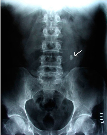
Independently, from the images, you can only determine how correctly the kidneys are located in relation to each other (for example, in some pathologies, one of them drops slightly). The bladder can be recognized by the shape of an ellipse. It is visible as a shadow.
Features of carrying out excretory urography for children
If the patient is a child, then it is worth noting several nuances of the ES:
- A few days before the diagnosis, the doctor prescribes antihistamines to the patient. This is necessary in order to exclude even the minimal likelihood of an allergic reaction during the study using contrast.
- Since young children are more active during diagnostic activities, the doctor takes into account the fact that the information content of EI will be lower.
- Children need a lot of psychological preparation for this type of examination. If the child is already able to understand speech, the parents must explain to him the need for the procedure and that in the process of diagnosis he must remain calm and immobile.
- For children, doctors make more accurate calculations of the amount of contrast agent. Also, the medical staff must be prepared for any situation.
Research cost
Despite the fact that urography is a simple procedure, its cost can range from 4 to 9 thousand rubles. rub. It all depends on the specific region. In large cities, a referral to EI can be obtained at a polyclinic.
In this case, the cost of the procedure may be covered by the health insurance policy. But in this situation, a longer waiting time for the diagnosis may be required. If you need to do the research quickly, you can go to a private medical center. But, it must be borne in mind that the cost of the procedure in this case will be higher.
The procedure for excretory urography is one of the informative diagnostic methods. If the patient has properly prepared for the examination, does not get nervous and does not move during the examination, then the diagnosis will be the most accurate. However, in some situations after EHM, it is worth listening to the opinion of other experts. Especially when it comes to a rather difficult diagnosis.
Video about urography
Excretory urography:
