Content
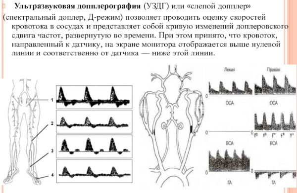
- Features of USDG vessels
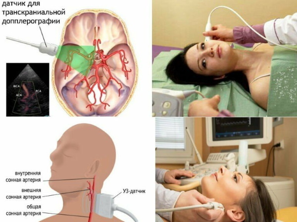
- Transcranial
- Extracranial
- Indications
- Contraindications
- Possible complications
- Training
- Procedure step by step
- Advantages and disadvantages
- Decoding the results
- Diseases that can be detected through research
- Video about Doppler ultrasound (Doppler) of the vessels of the head and neck
Doppler ultrasound (also called Doppler ultrasound) of the vessels of the head and neck Is a method for diagnosing blood flow, its condition and speed. The price of the procedure depends on the location, region, equipment. In clinics on an insurance policy, UZDG is done free of charge, in private clinics, the cost varies depending on the area and scope of the study - from 2,000 rubles.
Features of USDG vessels
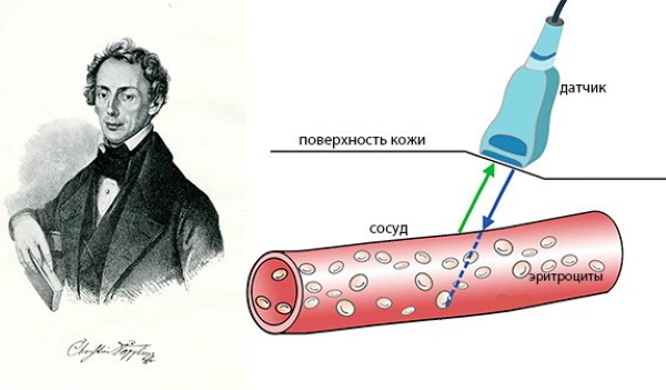
Doppler ultrasound is based on the ability of ultrasonic waves, reaching moving red blood cells, to push off from them. The data is captured by the sensor and redirected to the computer. There, instant processing of information takes place, and they are displayed on the monitor screen in the form of an image. It shows all the veins and arteries, blood flow, you can determine its speed.
Doppler ultrasonography allows you to assess the state of the circulatory system - the thickness of the walls, see cholesterol plaques, blockage of the channels. The examination helps the doctor to understand the reasons why there is a violation of blood circulation in the great vessels.
Doppler ultrasonography is of several types:
- MAG - scanning of the main large vessels;
- BCS - study of vascular anomalies;
- BCA - study of the carotid, subclavian, vertebral arteries.
Doppler ultrasonography of the vessels of the head and neck (the price depends on the scanning method) can be performed in three modes:
- IN - black and white version, which allows to assess the thickness of blood vessels, the presence of neoplasms, tumors and the condition of the valves;
- D (spectral) is a graph of changes in blood flow in real time;
- colored - tissue dopplerography, showing the relief, all bulges or depressions, the blood flow rate.
For scanning, one of two methods is used - transcranial or extracranial. Choose the one that is more appropriate for the field of study.
Transcranial
The probe is applied to the cranial bones with the smallest thickness. This helps to assess the condition of the arteries and veins of the brain. The examination is carried out through the temporal holes - anterior and middle. They are located in front of and above the conch of the ear.
This method helps to examine the main arteries of the brain, the veins of Rosenthal, Galen. The scan helps evaluate the rectus sinus and posterior artery. To check the ocular and the state of the internal carotid - the sensor is moved to the closed eyelid.
Extracranial
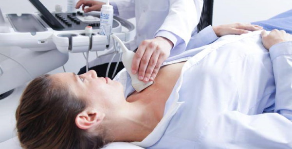
The method is used to diagnose large vessels that are located in the neck. These include the subclavian, carotid and vertebral arteries, jugular veins. The examination starts from the lower part of the carotid artery - on the right side. Then the sensor is moved up to the lower jaw. This helps to find the place where the vein is divided into external and internal, to assess the correct location of the vessel, its depth.
During the examination, atherosclerotic plaques can be detected. Also, Doppler ultrasound can be pulse-wave, when changes in blood flow are visible only in a certain vascular area. With a constant wave, the blood flow velocity can be estimated in the range of 5-50 m / s. Scanning can be three-dimensional when a volumetric image is obtained, duplex when B-mode and Doppler effect are combined. This method shows the veins, allows you to study their condition.
Triplex scanning - when color and duplex methods are combined.
It is considered the most effective, as it allows you to study the speed of blood movement, its direction. The image clearly shows the tortuosity and patency of the veins. Duplex and triplex methods are additional examinations that are assigned after a standard scan.
Indications
Doppler ultrasonography (Doppler) of the vessels of the head and neck (the price for urgency may be higher than with a routine examination) is done after the patient turns to headaches, sudden dizziness, spasms.
Other indications for diagnostics:
- arrhythmia;
- osteochondrosis of the neck;
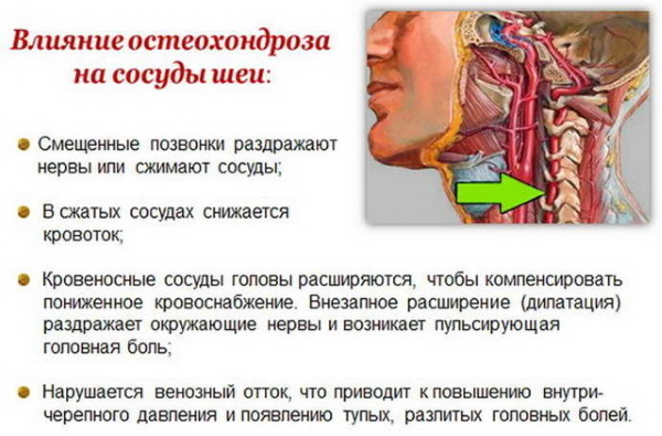
Doppler ultrasonography (Doppler) of the vessels of the head and neck is done for osteochondrosis of the neck. - diseases of the central nervous system;
- thrombosis of large vessels;
- elderly age;
- trauma to the brain, head, or neck;
- chronic hypertension;
- a sharp decrease in vision;
- diabetes;
- intoxication of the body with chemicals, paints, varnishes;
- obesity;
- lack of pulsation in the vessels of the hands;
- neoplasms (or suspicion of them) of the brain;
- compression of the arteries;
- identification of the location of cholesterol plaques;
- vegetative dystonia;
- vertigo;
- mental disorders;
- scoliosis;
- excessive fatigue;
- ischemic attack;
- excessive alcohol abuse, smoking;
- vascular diseases (aneurysms, atherosclerosis, or stenosis);
- noises, ringing in the ears, head;
- throbbing in the neck;
- blood pressure surges;
- bad habits that increase the risk of vascular diseases - smoking, frequent drinking);
- violation of coordination of movements;
- obesity (overweight is often accompanied by vascular disorders);
- sleep disturbance (in particular insomnia);
- any neurological symptoms;
- high blood cholesterol;
- memory impairment;
- fainting;
- swollen veins in the neck;
- tremor of arms, legs;
- speech problems;
- fluctuations in hormonal levels for an unknown reason;
- genetic predisposition to vascular disease;
- taking hormonal drugs;
- pregnancy;
- dystrophic processes;
- malfunctions in the functioning of the cervical intervertebral discs;
- pre- and postnatal period;
- a history of heart attack or stroke.

Doppler ultrasonography is mandatory before cardiac surgery. For children, examination is prescribed for developmental delay or scoliosis. Doppler ultrasonography should be performed annually as a prophylaxis. Men need to be examined after 40 years old, women - over 45 years old.
Especially if a person works in hazardous industries or the activity is associated with heavy physical exertion. Also, the risk of vascular disorders increases if they were in the history of relatives.
Contraindications
Doppler ultrasound has no absolute contraindications. But open wounds or damage to the skin where the sensor slips can become a limitation. Then the examination is postponed until the violations are eliminated. If you urgently need to assess the state of blood vessels and blood flow, alternative research methods may be prescribed.
Doppler ultrasonography of the vessels of the head and neck (the price of a routine examination is from 1,500 rubles) can be difficult if the area being examined is under the hypodermis or bone. Also, slow blood flow or arrhythmia that changes it can become an obstacle to the examination. In this case, a different transducer location or re-examination may be required.
Possible complications
There can be no complications after Doppler ultrasound, since the scanning procedure does not damage the tissue. Irritation or allergic reactions sometimes occur if scans are performed with contrast.
Another option is when a catheter is inserted into the jugular vein. But this is an additional operation, to which the complication directly belongs.
Training
No special preparation for Doppler ultrasonography is required. But before the examination, a consultation with the attending physician is necessary. You may need to temporarily withdraw antispasmodics, certain vascular drugs, or other medications. They can affect the tone of veins, arteries, capillaries, and as a result, the test result will turn out to be inaccurate and you will have to carry out the procedure again.
Also, before ultrasound Doppler ultrasound, do not drink alcohol, smoke, drink coffee or strong tea. Before the examination, the patient removes the jewelry from the neck so that they do not interfere with the sliding of the sensor. Usually, the diagnosis is carried out in the supine position.
The examination can be carried out with the introduction of contrast agents. This helps to obtain more accurate results. Iodine-based preparations are used as a contrast. They rarely cause allergic reactions. But before the Doppler ultrasound, you need to do a test. He will show if there is a negative reaction to the contrast agent.
Procedure step by step
Doppler ultrasonography can be performed while sitting or lying down. A special gel is applied to the neck. It removes the air cushion between the skin and the device, which ensures a snug fit of the sensor and a clear image. Then the device is driven over the gel, pressing lightly on it. If necessary, the doctor asks you to turn your head, hold your breath, or take frequent breaths in and out. If the vessels of the head are scanned, then the gel may not be applied.
The duration of the procedure takes from 20 minutes to an hour. The survey can be carried out in two ways. Its choice depends on which arteries and veins need to be examined. First, the procedure is performed in black and white. Then the device is switched to color mode. It allows you to explore areas where blood flow is disturbed, to identify cholesterol plaques, blood clots, abnormal abnormalities in the structure of the vascular walls.
Then the vertebral arteries are scanned, the main diagnostic parameters are measured - diastolic and systolic blood flow velocities and their ratio. Doppler ultrasonography of the vessels of the head and neck (the price depends on the need for additional studies) may require additional tests. For this, vascular drugs, hyperventilation, pressure on the vessels with fingers can be used.
In urgent cases, doppler ultrasonography is done. The reflected signals are then converted into sound. Such an examination is often prescribed for seriously ill people, bedridden patients (for this, portable devices are used). The full examination takes no more than 45 minutes, the minimum - 15 minutes.
Advantages and disadvantages
The advantages of Doppler ultrasound include the lack of special training, safety and painlessness of the study. The procedure does not cause discomfort, is easily tolerated by children, and can be performed as many times as necessary without time constraints. There is no harmful radiation during scanning. Doppler ultrasonography can be performed for everyone, regardless of age, during pregnancy and lactation.
The examination is affordable, the result is handed out immediately. Doppler ultrasonography of the vessels of the head and neck (the price depends on the type of examination) has several disadvantages. Scanning does not show the cause of vascular stenosis, it is impossible to determine the type of tissue of veins and arteries. Doppler ultrasound is less informative than alternative diagnostic methods - MRI or CT.
Decoding the results
When decoding the results, the obtained indicators are compared with the norm (they are individual for different types of vessels). During the examination, the thickness of the walls, their diameter, the resistive and pulsation index are assessed. Check the phase and nature of the blood flow, the symmetry of the arteries. Measure the minimum and maximum speed, their ratio. Then the data is written into the card. Based on the information received, taking into account laboratory tests and symptoms, the doctor makes a diagnosis.
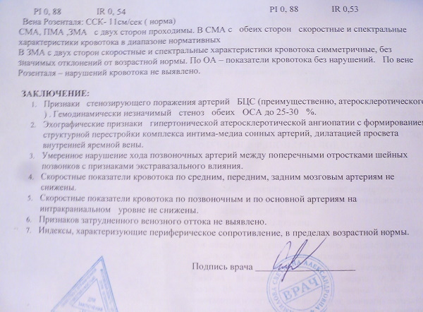
Normal:
- the lumen of the vessels should be free, without neoplasms and plaques;
- wall thickness - no more than 0.9 mm, for some, a value of up to 1.1 mm is permissible;
- blood flow velocity in the veins of the spine up to the 6th articulation - up to 0.3 m / s;
- there must be no malformation (pathological vascular network);
- there should be no turbulent blood flow in places without vascular branching;
- veins and arteries should not experience pressure, if any, additional research is carried out;
- vertebral vessels should be of the same diameter - no more than 2 mm.
To make a diagnosis, only one Doppler sonography is not enough, other studies and analyzes are needed. For example, angiography is additionally prescribed to assess small veins or those located in hard-to-reach places.
Diseases that can be detected through research
What deviations from the norm can reveal:
- Temporal arteritis. In this case, there is a uniform diffuse thickening of the vascular walls, low echogenicity. If arteritis is in an advanced stage, then atherosclerotic plaques are additionally visible.
- Vertebral artery hypoplasia. In this case, the diameter of the arteries decreases to 2 mm or less. This is accompanied by a violation of the entry into the canal of the transverse branches of the cervical vertebrae.
- Thrombosis. In this case, the arterial lumen overlaps the blood clot. The monitor clearly shows inflammation of the walls with a hypoechoic (homogeneous) internal formation. Blood clots have clear boundaries. When they are in this place for a long time, artery atrophy appears.
-
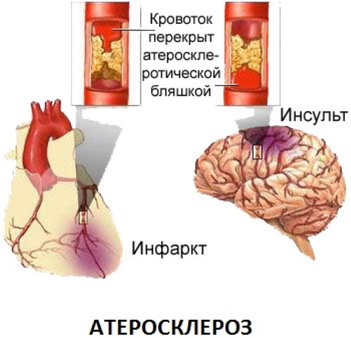
Stenosing atherosclerosis. If present, plaques are detected. Their research helps to conclude whether they are the source of the embolism. At the initial stages of atherosclerosis, while there are no plaques yet, a thickening of the intima-media is visible on the monitor.
- Vascular malformations. An abnormal vascular network is visible, which can be of different sizes. The veins extending from the pathological site are hypertrophied, with signs of calcification and lipous infiltrates. As a result of malformation, there is often a violation of cerebral circulation and the syndrome of "steal".
- Non-stenosing atherosclerosis. At the same time, violations in the structure of large arteries, uneven echogenicity, narrowing of the lumens of 20 percent or thickening of the walls are revealed.
- Vasculitis. With the help of Doppler ultrasound, you can determine the stage and distribution of the disease. More often, vasculitis is characterized by diffuse changes in the vascular walls, disruption of layers, signs of inflammation and changes in echogenicity.
- Congenital pathologies and deformities. With congenital malformations, excessive tortuosity is observed (in the form of the English letter S). Bends may be visible when the bend angle is sharp or reaches up to 90 degrees. This disrupts hemodynamics. Loops also belong to malformations. They are circular configurations. They appear due to the pathology of angiogenesis during the formation of the fetus, but then they do not affect the quality of life of the child. Less common is hypoplasia of vertebral vessels, when their diameter decreases less than normal values.
- Takayasu's disease (autoimmune aortoarteritis). Doppler ultrasonography shows the absence of pulsation in the arteries. There is a violation of blood circulation. Reveal complete or partial occlusion of the branches of the aorta. With an illness, the blood supply to the brain decreases, signs of inflammation are visible.
- Metabolic angiopathies. In this case, the lesion covers all vessels, regardless of size. At the same time, there is a decrease in their tone and blood supply, narrowing (spasms). A violation of the circulation of connective fluid tissue is diagnosed. If the cause is an infectious lesion, phlebitis or vasculitis, then mainly destruction and narrowing of one vessel occurs, and rarely several. In this case, the localization of the violation is clearly visible.
Also, during the examination, neoplasms can be detected that disrupt blood flow by squeezing the vessels. With the help of Doppler ultrasound, the incorrect communication of the arteries and veins is clearly visible. Persistent hypertension will be indicated by thickening of the walls of the arteries and loss of their elasticity. In an aneurysm, they stratify and the blood flow changes direction.
Doppler ultrasonography of the vessels of the head and neck can be performed in polyclinics or private clinics. The price depends on this, the quality of the equipment and the region. The average cost of diagnostics is 3000-5000 rubles. If the scan needs to be done urgently, then the procedure can be done in any private clinic (most of them have modern equipment).
Video about Doppler ultrasound (Doppler) of the vessels of the head and neck
What is ultrasound (Doppler) blood vessels: