Content
- The essence of electroneuromyography
- Aims and objectives of the diagnostic method
- Indications
- Contraindications
- Types and stages of EMG of the lower extremities
- Needle
- Stimulation
- Preparation for research
- Procedure and duration of the procedure
- Decoding the results
- Lower extremity EMG cost
- Video about electromyography
EMG (electroneuromyography) lower limbs - This is a hardware diagnostic method, which is carried out in relation to patients with signs of a decrease in the functional activity of muscle tissues and peripheral nerves.
Causes of the pathological condition of the legs, impaired mobility, the occurrence of severe weakness during walking, running or exercising can be set using this method survey. Electroneuromyography is divided into superficial (stimulating) and acicular.
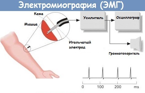
The essence of electroneuromyography
EMG is one of the methods of neurophysiological diagnostics, which allows detecting pathological changes in the functioning of the peripheral nerves and muscle tissues of the lower extremities. The essence of this research method is to timely track the complications and negative consequences of existing diseases of the body, which are manifested by a weakening of the tone of the muscles of the legs, a violation of the conduction of neural impulses along the nerve fibers.
Electroneuromyography is used once to make a diagnosis, or it is performed regularly to monitor the dynamics of treatment and prevent exacerbation of diseases. For example, people with muscular dystrophy or diabetic neuropathy undergo EMG in order to track pathological changes in the structure of the muscles and nerves of the legs. This makes it possible to timely begin treatment aimed at eliminating the pathological process.
Aims and objectives of the diagnostic method
EMG of the lower extremities is a kind of hardware diagnostic method, the use of which allows you to realize the following goals and objectives of a comprehensive examination of a patient:
- confirm or deny the presence in a person of this or that disease, the development of which is accompanied by damage to the peripheral nerves and muscles of the lower extremities (for example, diabetic neuropathy, muscular dystrophy);
- establish the extent of the spread of the pathological process when the diagnosis has already been made;
- adjust the course of drug and physiotherapy treatment;
- track the dynamics of the restoration of the previous functions of the nerves of the peripheral system and the muscles of the legs in patients whose therapy has been completed;
- to prevent the development of complications and negative consequences associated with the progression of acute diseases of the musculoskeletal system;
- to provide timely prevention of mono- and polyneuropathy, muscular dystrophy of the lower extremities.
 Examination of patients using EMG allows solving only part of the diagnostic problems. Therefore, this method of studying the tone of muscle tissues and the level of conductivity of peripheral nerves lower extremities should be used in combination with other methods of apparatus and laboratory diagnostics.
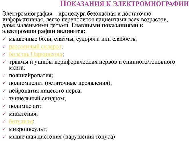
Indications
EMG of the lower extremities is a modern method of examining the functional state of peripheral nerves, which is indicated for the following clinical cases:
- all types of diseases that reduce the activity of neural impulses;
- atrophic process in the muscle tissues of the lower extremities and lumbar spine;
- amyotrophic lateral sclerosis;
- radiculopathy of non-vertebral and vertebrogenic type;
- myelopathy;
- complications of diabetes mellitus associated with impaired peripheral nerve conduction;
- severe weakness in the muscles of the lower extremities of unknown etiology (in this case, all actions doctors are aimed at establishing the causes of a decrease in the functional activity of muscle tissues legs);
- recovery period of the musculoskeletal system after severe injury or surgery, as a result which the patient was bedridden for a long period of time, deprived of the possibility of independent movement on own legs;
- postradiation polyneuropathy of the peripheral nerves of the lower extremities;
- suspicions of the presence of a tumor neoplasm in the structure of bones, soft or muscle tissues of the legs, which disrupts the normal functioning of the lower limb, causes periodic attacks of acute or aching pain;
- the consequences of extensive intoxication of the body with chemicals or biological poisons, which led to partial or complete paralysis of the leg muscles;
- infection of the body with a bacterial or viral infection, the pathogens of which cause destruction peripheral nerves directly involved in the innervation of the muscles of the lower limbs;
- recovery after previous traumatic brain injuries and surgical interventions on the tissues of the brain, which are responsible for the functional mobility of the legs;
- muscular dystrophies of hereditary genesis, when dysfunctional disorders of the peripheral nerves of the legs progress to throughout the entire independent life of a person (in most cases, such pathologies are the result of genetic mutations).
Electromyography is necessary for the timely identification of complications and negative consequences, caused by an underlying disease of the internal organs, the peripheral and central nervous system of the patient. In most cases, EMG is used as an auxiliary research method in combination with other types of hardware diagnostics.
Contraindications
EMG of the lower extremities is a diagnostic procedure that is characterized by the presence of a minimum number of restrictions on its use.
Electromyography is contraindicated for use as the main or auxiliary examination method in the presence of the following circumstances:
- the patient's lower limb is plastered, or a fixing splint, splint, tissue bandage is applied to its surface;
- in the soft tissues of the leg, acute or chronic inflammation of an infectious nature of origin occurs (in this case the patient must undergo a course of antibiotic therapy, and only then return to the issue of diagnostics using EMG);
- the skin surface of the lower limb is damaged by trophic ulcers, abrasions and other injuries;
- there is an individual intolerance to electric current impulses, which is manifested by local allergic reactions.
The doctor performing the electromyography procedure conducts a preliminary examination of the general condition of the patient's lower extremities. In the absence of visual contraindications to the use of EMG, the doctor examines the patient's medical record for the possible presence of chronic diseases that can cause complications.
For example, pathologies of the cardiovascular system, in which a person is forced to constantly wear a pacemaker. The EMG procedure involves the impact of electric current pulses on certain parts of the patient's body, and this can lead to disruption of the functions of this apparatus of the heart muscle.
In addition to the above contraindications, electromyography has the following restrictions on its use as a primary or auxiliary diagnostic tool:
- muscles and peripheral nerves located deep in the tissues of the lower extremities may be inaccessible for carrying them objective examination (this fact is taken into account by the attending physician, who diagnoses a patient with pathologies of the lower limbs);
- it is impossible to assess the functional state of too thin nerve fibers of the autonomic system of the body;
- the presence of a severe form of obesity (the results of the study can be distorted by poor conduction of electrical impulses through a thick layer of adipose tissue);
- severe edema of the lower extremities, regardless of their nature of origin.
The electromyography method also has limitations on its use in relation to patients with signs of impaired trophism of the tissues of the lower extremities. Before the start of the examination, each patient undergoes a preliminary medical examination.
Types and stages of EMG of the lower extremities
EMG of the lower extremities is a modern method for diagnosing the level of innervation of the muscles, which is classified into 2 main types. This is needle and stimulation electromyography. Each technique for examining the functional state of the peripheral nerves of the legs has its own purpose, and also allows you to implement specific diagnostic tasks.
Needle
Needle electromyography is an invasive diagnostic method that involves inserting the electrode into the soft tissues of the lower extremity rather than superficially. This method of examining the functional state of the peripheral nerves of the legs is more painful and requires high qualifications on the part of the doctor performing the study.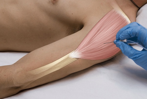
During needle electromyography, a sterile disposable electrode is used, which is disposed of immediately after the completion of the diagnostic process. At the moment of implantation of the electrode into the tissue of the lower extremity, the patient feels a slight pain from the injection, the intensity of which is comparable to the consequences of an intramuscular injection.
The advantage of using needle electromyography is that this technique makes it possible conduct the most objective assessment of the level of electrical conductivity of the muscle tissues of the lower limbs.
This diagnostic method is considered the most informative for examining patients with signs of the following pathologies:
- peripheral motor neuron diseases;
- muscle atrophy of the spinal type;
- dystrophic changes in the muscles of the legs;
- polymyositis;
- different types of myopathy.
Needle electromyography of the lower extremities can be performed at the moment when the muscle tissues of the legs are in a state of complete rest, as well as at the moment of their physical exertion. Based on the results of the diagnosis, a detailed medical report is drawn up.
Stimulation
Stimulation electromyography is a non-invasive method of apparatus examination, which involves the use of surface-type skin electrodes. Weak impulses of electric current are applied to the patient's skin surface, which irritate the receptors of the peripheral nerves.
At the moment, the doctor performing the diagnosis is fixing the physiological reaction of the nerve endings in response to an external stimulus. At the time of stimulation electromyography, most patients complain of mild discomfort in the form of tingling of the skin and muscles in the area of the installed electrodes.
Twitching and involuntary contraction of the leg muscles is not excluded. This is a normal reaction of the peripheral nervous system in response to electrical impulses. The method of stimulation electromyography is considered one of the most informative methods for determining chronic diseases of the peripheral nerves.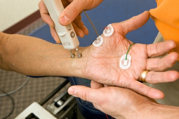
For example, during a slowly progressive poly- or mononeuropathy that developed against the background of diabetes mellitus, tumor neoplasms in the structure of the spinal column, the consequences of craniocerebral injuries of varying degrees severity. Stimulating electromyography takes less time to complete.
Preparation for research
Before conducting a diagnostic examination using the electromyography method, it is not required to perform a large number of organizational steps.
It is enough to observe the following recommendations:
- stop taking drugs Proserin, Kalimin and other drugs from the pharmacological group of anticholinesterase drugs 24 hours before the appointed date of the examination;
- on the day of the examination, you must take a shower, put on clean clothes, and also observe the general hygiene rules, since this type of diagnosis involves direct contact of the doctor with the skin surface patient;
- do not apply moisturizers, lotions and other types of cosmetics to the skin of the feet;
- on the day of electromyography, depilation of the skin of the lower extremities should not be performed, as this can lead to the formation of scratches, local irritation, the manifestation of which will be aggravated by the influence of electrical impulses current;
- prevent hypothermia of the muscle tissues of the legs, dress in accordance with the weather conditions.
On the day of electromyography, patients are allowed to eat any food, drink coffee, tea, water, juices and other drinks. It is recommended to arrive in advance at the medical facility, where the functional state of the nerves and muscle tissues of the legs will be diagnosed.
This is necessary in order to slightly decrease the muscle tone of the lower extremities. Each patient must have a referral from the attending physician, as well as accompanying documentation, which indicates the purpose of the examination and the diagnosis. It is advisable to have a clean diaper, towel or sheet with you.
Before starting electromyography, the patient should warn the diagnostician about the possible presence of the following health problems:
- Propensity for periodic syncope.
- Epileptic seizures, regardless of the frequency of their manifestation.
- The presence of an installed pacemaker.
- Infection with HIV, hepatitis virus, syphilis and other especially dangerous infectious diseases with a chronic course.
- Damage to the skin with dermatological diseases that cause edema, itching, irritation, the formation of trophic ulcers.
- Taking medications from the pharmacological group of anticoagulants, as well as other medications that help thin the blood.
- Previous fractures of the lower extremities.
This information must be communicated to a specialist in order for him to calculate all the risks and potential complications that may arise during the diagnostic process, as well as after it completion. Women who are pregnant must also notify the doctor about this fact. In this case, the period of development of the fetus does not matter.
Procedure and duration of the procedure
The table below details the step-by-step process for performing lower limb electromyography.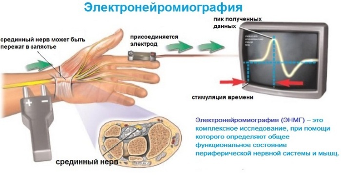
| The order of diagnostic manipulations | Description of the survey process |
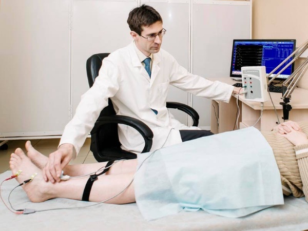
| Step 1. Prepare the body for diagnostics. | The patient takes off his clothes, exposing the skin of the lower extremities. |
| Step 2. Take a comfortable position. | Depending on the type of electromyography, the patient is in an upright position, sits down or lies down on the couch. |
| Step 3. Disinfection of the working area. | The specialist performing electromyography performs antiseptic treatment of the skin of the legs in the electrode fixation zone. An ethyl alcohol solution or other disinfectant is used. |
| Step 4. Fixation of electrodes. | Surface electrodes are attached to the patient's skin surface or a needle type of this equipment is inserted. The fixation of the device is carried out using special velcro, which come with the electromyography device. |
| Step 5. Electrical stimulation. | Electrical impulses are supplied to stimulate the functional activity of the peripheral nerves of the lower extremities. |
| Step 6. Adjustment of the amperage. | The specialist gradually reduces or adds the current strength depending on the projection and depth of the nerve fibers that need hardware diagnostics. |
| Step 7. Changing the position of the needle electrode. | In the case of using the needle electromyography method, a medical worker changes the position of the electrode in the area of the muscle under study 3 to 4 times. |
| Step 8. Completion of the diagnostic procedure. | The patient's skin surface in the area where the electrodes are inserted is re-treated with an antiseptic. The patient gets dressed and leaves the diagnostic room. |
The total duration of the EMG diagnostic procedure depends on a large number of factors. This is the type of underlying disease that is diagnosed in the patient, the current symptoms, the patient's age, the presence or absence of concomitant pathologies of the muscles and soft tissues of the legs. In uncomplicated cases, the average duration of electromyography is about 30 minutes, but always it must be remembered that this diagnostic process may require much more time.
Decoding the results
Deciphering the results of electromyography is carried out by a physiotherapist, rehabilitologist, neuropathologist, and neurologist. The doctor performs a comparative analysis of the information data recorded by the electroneuromyograph with the indicators of the norm. At this point, the patient's age, the type of underlying disease and the severity of pathological symptoms associated with dysfunction of the lower extremities are taken into account.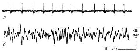
Deciphering the results of electroneuromyography is an individual process, but as reference values, it is taken into account that the change in amplitude fluctuations in the potential of muscle tissues does not exceed a few millivolts with a total duration of 20 to 25 ms.
Lower extremity EMG cost
The average cost of stimulation electroneuromyography of 1 motor nerve on only one lower limb is 2850 rubles. The price of a similar diagnostic method, during which a study of all peripheral nerves of the leg is carried out at once, is in the range of 6750 rubles. The final cost of the examination can be adjusted by the management of the medical center, depending on the complexity of the clinical case.
EMG of the lower extremities is an informative diagnostic method that is prescribed to children, adult men and women with signs of weakening of muscle tissue tone, impaired peripheral nerves. This area of neurophysiological examination of the body involves the use of electroneuromyograph, equipped with Velcro surface electrodes, or needle sensors.
The latter type of EMG is invasive, since the electrode is inserted into the muscle tissue of the examined leg. This type of diagnostic procedure is performed under sterile conditions using disposable consumables. The average examination duration is 30 minutes.
Video about electromyography
Doctor about EMG: