
Content
- Views
- By the principle of education
- By morphological characteristics
- Stages and degrees
- Symptoms and Signs
- Causes
- Diagnostics
- Treatment methods
- Groups of drugs for treatment
- Surgery
- Possible consequences and complications
- Video about skin hemangioma
Hemangioma on the skin Is a benign formation of vascular tissue. Outwardly, it looks like red, crimson or cyanotic raised spots (shown in the photo). They are more often formed in infants, but they can also appear in adults. Hemangioma is diagnosed by ultrasound, treatment is conservative or surgical.
Views
Hemangioma on the skin can be congenital, when it is formed during intrauterine development, or acquired (if it appears in a baby - an infant). The photo shows that in structure they are single, multiple. According to the structure, hemangiomas are venous, arterial, capillary.
By the principle of education
- Local, growing from one point. They have smooth contours, small sizes.
- Segmental. They differ in uneven edges, grow large. Often they develop against the background of a combination of several diseases at once (for example, in the chest and pelvis, heart defects, aorta).
By morphological characteristics
According to morphological characteristics, it is divided into types:
-
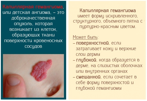
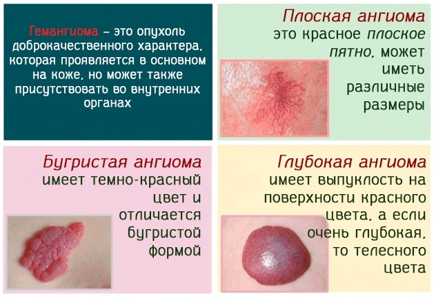
Capillary (simple or strawberry nevus) forms on the surface of the skin. Soft education up to 8 cm in diameter, with clear edges, red or purple with blue in color. When pressed, it fades, but when the pressure disappears, the shade is restored. It forms mainly on the face, head, back and chest. Most often it is formed immediately after the birth of a child, and by 5-12 years old it disappears, leaving scars, coarsening of the skin, tissue atrophy.
- Cavernous (cavernous) formed under the skin due to the abnormal structure of the vascular system, subcutaneous tissue. This is a nodular, bumpy spot with a soft elastic consistency, uneven contours. Inside the hemangioma there are cavities filled with blood. If you press on the tumor, a pit is formed, but it quickly disappears. In the first months, the hemangioma grows rapidly, then the process stops. In this case, there are no symptoms. The prognosis is favorable, but it is necessary to periodically check the condition of the internal organs, limbs, and the nervous system.
- Combined, when all layers of the skin are involved in the process. Combines all the features of a capillary cavernous appearance.
- Mixed, when the process involves not only the skin, but also muscle and nerve fibers. This neoplasm has a complex structure. It captures nerve, vascular, connective and lymphoid tissues. Their appearance, shade and consistency - depending on the structures involved in the pathological process.
- Arterial is formed from birth. It is characterized by multiple bright pink spots with even outlines. In size, hemangiomas can be either small or large. More often, the tumor appears on the face, mucous membranes, behind the neck.
- Venous. It is a soft dark blue or purple papule that forms on the ears, lips, and face in older people. Venous hemangiomas can be single or multiple, but they do not cause much concern.
- Arachnid (star-shaped). This is a small red papule with a network of capillaries diverging from it. These are single hemangiomas, up to 1.5 cm in diameter. Most often appears on the face, hands and forearms.
- Keratinizing (angiokeratoma). A neoplasm forms on the penis or scrotum. Appears as numerous dark red papules.
- Arcal arteriovenous. These hemangiomas appear after 30 years. They look like red or bluish spots with a diameter of 0.5-1 cm, but there are also much larger ones. Hemangiomas appear on the face, if multiple are located in groups. The walls of the vessels are thickened, similar to arterial walls, but without an inner elastic membrane.
- Target-like hemosiderotic. Appears after trauma to an existing hemangioma, with the further development of a blood clot. It is characterized by a rapidly growing major brown or purple papule. It is surrounded by a paler ring that gradually dissolves. But the main papule disappears slowly. A tumor can form anywhere.
- Microvenular. This type of hemangioma rarely occurs, sometimes during pregnancy, after prolonged use of hormonal drugs. The tumor is 0.5-2 cm in diameter and grows slowly. It is localized on the hands, more often on the forearms, but can form on the legs, body, face.
- Sinusoidal. It is an acquired vascular tumor that mainly occurs in women. A solitary nodule forms under the skin.
- Senile. They form in the form of bright red or purple hemispheres or multiple dotted spots. Senile hemangiomas form on the face, lips and body. The first signs appear after 30 years, then their size and number increase. Sometimes spots come and go suddenly.
 In 95% of cases, simple forms of neoplasms are detected. Symptoms appear externally, other signs of discomfort and itching are rare.
Stages and degrees
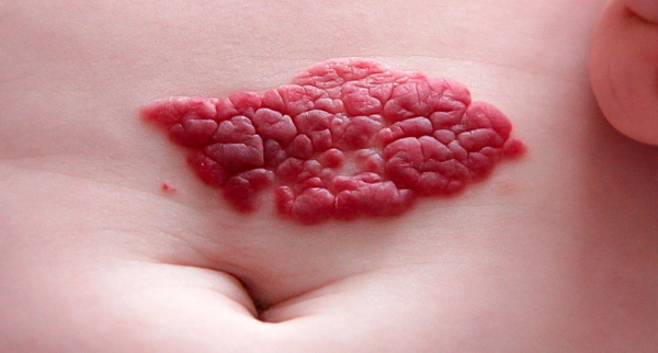
Hemangioma on the skin (the photo shows how large the tumor can grow) usually appears in a child in the first days of life. In 80 percent of cases, a neoplasm occurs only in one place, and in girls it is 7 times more often than in boys. Numerous skin lesions are extremely rare, but in this case they spread throughout the body.
Infant hemangiomas often go away on their own; as they grow older, no treatment is required. A neoplasm can affect fatty tissue, bones and internal organs of a person. The most dangerous are hemangiomas that form on the internal and genital organs. Injury to them can cause bleeding and infection of damaged tissue.
In children, hemangiomas may appear in the first days after birth, and the peak of growth occurs at 2-3 months. Then they can continue to increase up to 6-9 months. Then the neoplasms stabilize (plateau effect) and the reverse development of the tumor begins, which can last for years.
In adults, the neoplasm develops quickly - in 2-3 weeks. It goes through several stages - growth, stabilization and spontaneous regression. Complications are possible in all phases. Hemangioma can behave unpredictably. Sometimes it begins to grow rapidly, affecting healthy tissue.
Symptoms and Signs
Hemangioma on the skin (the photo shows that the tumor can be in the form of a small speck or cover a large area of the skin) is usually a small convex spot with a diameter of 2-3 cm. Most often it appears on the chest, neck or face, less often on other parts of the body, especially around the ears.
It can be difficult to detect a hemangioma at an early stage. First, a small, pale pink, irregular spot appears, which gradually begins to turn red. This plaque is easy to catch with your fingernail. If there are many spots, then gradually they begin to unite into one whole.
A large tumor appears in the center, from which small capillaries diverge. Formed completely visible on the skin due to its bright red or crimson color, rises above the surface. A characteristic symptom of a hemangioma is a temperature difference. When pressed, it will be warmer than healthy areas of the skin.
Causes
The exact cause of the formation of hemangioma has not yet been established. But in babies, it can appear due to a strong proliferation of connective tissue, as a result of which a lump and swelling forms.
Also possible reasons are:
- Genetic predisposition. If a hemangioma has been diagnosed in parents or close relatives, then there is a chance that it will appear in children as well.
- Negative environmental impact. This category includes bad ecology, hazardous production, radiation.
- Influence of ultraviolet radiation. Prolonged exposure to the sun or its too aggressive effect provokes the development of various types of tumors.
- Diseases of internal organs, vascular system.
- Late or multiple pregnancy.
- Low birth weight.
- Sedentary lifestyle.
- Violation of the formation of the vascular system of the embryo.
- Fetal hypoxia.
- If there was a threat of miscarriage or placenta previa.
- Respiratory viral acute infections. Especially those carried over in the first trimester of pregnancy.
- High estrogen levels.
- Inflammation or abruption of the placenta.
- Preeclampsia.
- Infectious diseases that the pregnant woman suffered during gestation.
There is a possibility that malnutrition can provoke the development of hemangioma. Especially if the menu contains a lot of fried and fatty foods. Also at risk are people with bad habits - smoking or drinking alcohol often or in excessive quantities.
Diagnostics
Most often, hemangioma is found in infants. It can be seen with your own eyes or identified by palpation. When pressed against the formation, it will be warmer in temperature than the surrounding tissue. They also consult a dermatologist. They donate blood for analysis - they do general and biochemistry. Cost - from 1900 rubles.
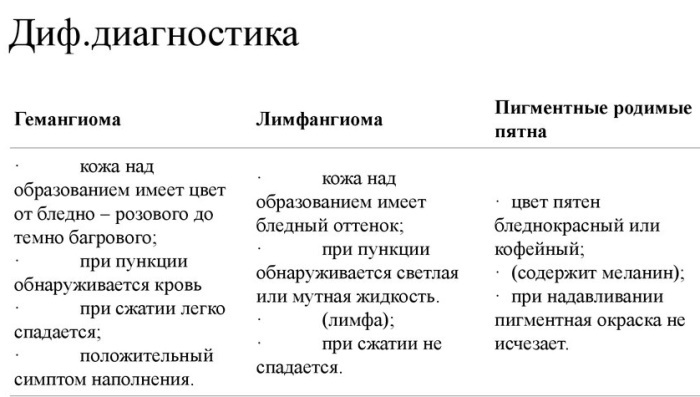
Hemangioma is found on Doppler ultrasound, contrast angiography, CT, or MRI. Depending on the location of the neoplasm, it may be necessary to consult an ophthalmologist, urologist, gynecologist or other doctors.
If the examination is carried out in a polyclinic, then if you have an insurance policy, it will be free of charge. When they turn to a paid medical center, ultrasound will be cheaper - from 500 rubles, CT or MRI are much more expensive - from 3000 rubles. Prices may vary depending on the region.
Treatment methods
Hemangioma on the skin (a photo at different periods helps to determine how fast the tumor is growing and to prescribe the most effective treatment) does not always require surgical intervention. In children, it can go away on its own until the age of five.
Groups of drugs for treatment
If treatment is required, then conservative methods are first tried. For this, medications are prescribed.
| Groups of drugs | Brief description and treatment |
| Beta-blockers | They are among the most effective drugs. They have a low likelihood of side effects. They constrict blood vessels, stimulate apoptosis. The drugs are administered in the form of injections or use a solution for lotions. For systemic therapy, apply:
|
| Hormonal | They also prevent active tumor growth. The drugs can be taken in tablets as a systemic therapy. But hormonal agents are selected individually and carefully in each case. They are rarely used, as they are highly addictive and have a large number of side effects. On average, the course of treatment is 4-12 weeks. They are sometimes taken throughout the year. Glucocorticoids can be injected into the neoplasm. Most often used "Triamcinolone". The drug is administered intramuscularly, at 40 mg, with an interval of a month. If necessary, the dosage can be doubled, the interval between injections is 14 days. The drug is also available in the form of an ointment for external use. It is applied to hemangiomas 1-3 times a day. The course of treatment is 5-10 days, but not more than a month. |
| Cytostatics | They stop the growth of tumors, but at the same time there is a risk of developing a cancerous neoplasm - angiosarcoma. Most often, "Vinblastine" is used, which induces apoptosis. It is injected once a week through a catheter. The dosage is determined individually - depending on age. |
| Interferons | Suppress the growth of hemangiomas, promote necrosis of neoplasm tissues. Interferons are prescribed only if hormone therapy is ineffective. They need to be taken in long courses, the effect will be visible only after 4 months of treatment. The dosage is prescribed individually, depending on the age of the patient and the size of the tumor.
|
If the hemangioma does not bother and does not grow, the risk of embarrassment is absent or minimal, then expectant tactics are most often chosen.
Surgery
Hemangioma on the skin (a photo of different types of neoplasms shows that sometimes they are removed only with the help of an operation) is often treated surgically. The indications for this are the rapid growth of the tumor, the risk of embarrassment if it interferes with the normal functioning of organs.
Hemangioma can be removed in several ways:
- Electrocoagulation. The tumor is exposed to an electric current. As a result, the damaged tissue is gradually destroyed. The operation has disadvantages - the appearance of scars, burns.
- Cryodestruction. The tumor is exposed to very low temperatures. The neoplasm freezes, then dies off and disappears completely. Liquid nitrogen is used for the procedure. But this operation has its drawbacks - often scars and scars remain on the skin. Cryodestruction is difficult to use for large tumors; it is more effective for small tumors.
-
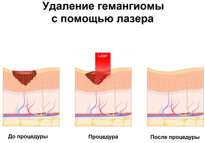
Removal of neoplasms with a laser. It is processed with a neodymium laser. Its luminous flux is absorbed only by the affected tissues, and healthy ones are not affected. As a result, the vessels heat up and merge. It takes time to remove the hemangioma, but the procedure goes without side effects.
- Sclerotherapy. A special adhesive solution is injected into the tumor vessels. The method is used if other methods of treatment have not yielded results or can lead to complications and undesirable consequences. For example, when a hemangioma is formed on the eyelid. The disadvantage of the operation is that the injected solution can cause an allergic reaction.
- Treatment with a demato-oncological magnifying glass. The hemangioma is treated with high-frequency radiation. As a result, the affected cells disappear. Again, the tumor in this place is no longer formed, scars and scars will not remain.
Of all these methods, laser hemangioma removal is considered the most effective. This method is not only suitable for adults, it is also safe for children. The duration of the procedure is no longer than 20 minutes. If the hemangioma is small, then it can be removed in one procedure. After that, the skin recovers quickly.
Possible consequences and complications
Hemangioma usually does not threaten human health and life. This is a benign tumor. But she is easily injured, as a result of which severe bleeding and infection can occur. As a result, there is a risk that the infection will spread to healthy tissues and organs. This will provoke the development of other serious diseases.
Possible complication when the tumor is on:
- internal organs - mechanical squeezing, disruption of their work;
- bones - their gradual destruction;
- eyes - visual impairment;
- mucous throat - breathing disorder;
- nerve endings - numbness of the limbs, loss of their sensitivity, disruption of the gastrointestinal tract or bladder;
- ears - hearing loss;
- lip - deformation of the teeth, mouth, bleeding gums;
- nose - deformation of the septum, shortness of breath.
When the tumor begins to grow, it can provoke a deterioration in blood clotting, thrombosis, tissue necrosis. If a purulent process joins, then this sometimes becomes the cause of sepsis. Large hemangiomas of the spine provoke the appearance of pain, compression fractures. The most dangerous complication is when the neoplasm transforms into a cancerous tumor.
Hemangioma on the skin usually heals quickly and without consequences. Despite the fact that the tumor may look frightening in the photo, it most often does not cause serious harm to health and complications are extremely rare.
Video about skin hemangioma
Hemangioma of the skin:


