Content
- Olfactory (nervus olfactorius)
- Nerve characteristic
- Functions and properties
- Anatomy and structure
- Diseases and lesions
- Visual (nervus opticus)
- Characteristic
- Functions and properties
- Anatomy and structure
- Diseases and lesions
- Oculomotor (nervus oculomotorius)
- Characteristic
- Functions and properties
- Anatomy and structure
- Diseases and lesions
- Block (nervus trochlearis)
- Characteristic
- Functions and properties
- Anatomy and structure
- Diseases and lesions
- Trigeminal (nervus trigeminus)
- Characteristic
- Functions and properties
- Anatomy and structure
- Diseases and lesions
- Abduction (nervusabducens)
- Characteristic
- Functions and properties
- Anatomy and structure
- Diseases and lesions
- Facial (nervusfacialis)
- Characteristic
- Functions and properties
- Anatomy and structure
- Diseases and lesions
- Vestibule-cochlear (nervusvestibulocochlearis)
- Characteristic
- Functions and properties
- Anatomy and structure
- Diseases and lesions
- Glossopharyngeal (nervusglossopharyngeus)
- Characteristic
- Functions and properties
- Anatomy and structure
- Diseases and lesions
- Wandering (nervusvagus)
- Characteristic
- Functions and properties
- Anatomy and structure
- Diseases and lesions
- Additional (nervusaccessorius)
- Characteristic
- Functions and properties
- Anatomy and structure
- Diseases and lesions
- Sublingual (nervus hypoglossus)
- Characteristic
- Functions and properties
- Anatomy and structure
- Diseases and lesions
- Video about nerves on the face
On the face of a person, many nerves are located that are responsible for the movements of the facial muscles, eyes, tongue and pharynx. They all originate from the brain and exit from the cranium. If communication is broken in the work of even 1 of these nerves, a person loses the ability to use facial expressions, speak, smell and see. Nerves on the face help identify chewing, swallowing and rolling the eyes.
Olfactory (nervus olfactorius)
All facial nerves are paired and may not work simultaneously, performing different functions.
Nerve characteristic
The olfactory nerve goes to the muscles of the olfactory organs from the frontal part of the brain.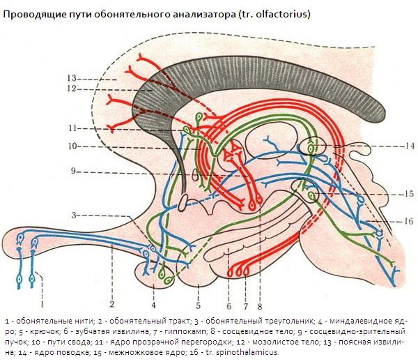
The olfactory nerve carries out its work through neurons that are scattered over the surface of the nasal mucosa. These neurosensory cells receive and transmit information through the neural channel to the parahippocampal gyrus, as to the center of the associative analysis of the olfactory apparatus.
Functions and properties
The olfactory nerve is responsible for smelling, assimilating and detecting odors. The sensitivity of smelling depends on the quality of development of the olfactory nerve. With a fine nervous organization, a person has the ability to recognize the lightest shades of aroma.
Anatomy and structure
The olfactory nerve begins with bipolar neurons, which often cover the mucous membrane of the human nose. The dendrites of these neurons end on the surface of the mucous membrane and intersect, forming the olfactory apparatus. Axions, in the form of thin filaments, connect the olfactory apparatus with the second neurons, which look like small bulbs. The second circle of neurons passes through the channels into the bodies of third neurons at the transparent septum. Neurons of the third circle transmit information along the nerve fibers of the root territory on the cerebral cortex of both hemispheres.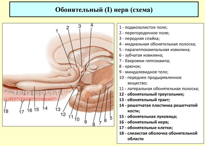
Through the subcortex, information from the nerve endings reaches the region of the parahippocampal gyrus. In this zone, all the information received from the olfactory zone is processed, sending a signal to increase appetite, the appearance of a reflex of salivation or nausea. The transmission of nerve impulses within the system is carried out along the fibers of the medial bundles of the forebrain and cerebral stripes of the optic tubercle.
Diseases and lesions
The main diseases of the olfactory nerve are anosmia and hyposmia. With anosmia, the sense of smell is completely absent, with hyposmia, the sense of smell is sharply reduced. The main causes of diseases are infectious diseases of the nasal mucosa, underdevelopment of the olfactory tract, head trauma and neoplasms. In rare cases, people develop parosmia, or a perverse idea of smell, which develops with schizophrenia, a severe stage of anemia.
Visual (nervus opticus)
Nerves on the face of a person, the location of which is associated with a wide network of blood vessels, include the optic nerve. It has a complex multistage structure with transitions.
Characteristic
The visual canal passes through the entire brain and there is a connection of the paired perception system. This nerve consists of the smallest nerve tissues that penetrate the eyeball and connect to form a single nerve channel. It collects basic information inside the eyeball and transmits it through channels to the brain for processing. At the entrance to the skull, the optic nerves of both eyes unite to exchange information. Then the optic canal is disconnected and connected to the brain through the channels.
Functions and properties
As a result of its complex structure, the optic nerve provides processing of information from the retina of the eye, i.e. a person's ability to see. In this case, the information received from both eyes is analyzed and combined into a single picture with central and peripheral vision. All perception is associated with the vestibular apparatus, speech and sensations.
Anatomy and structure
The optic nerve is very complex in structure and includes more than 1.5 million. nerve fibers. A complex system of nerve tissues is mirrored in the opposite part of the face.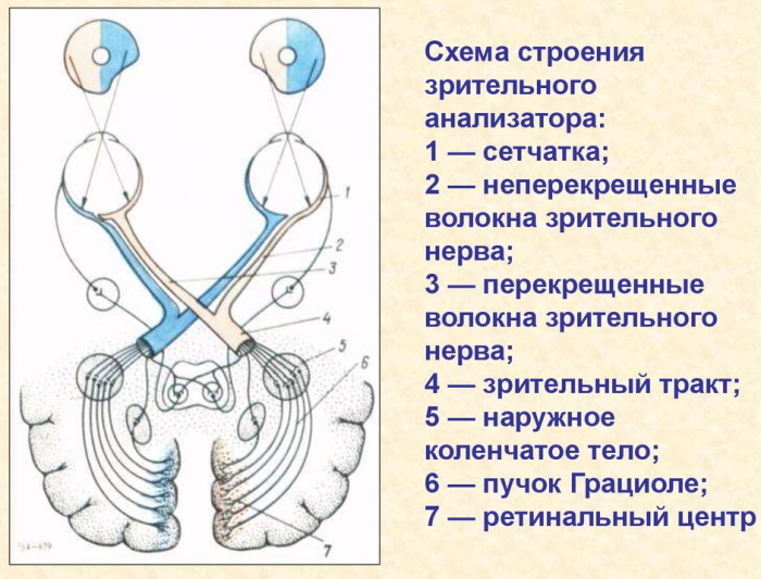
The optic nerve begins from the nerve cells of the retina. The endings from these cells are connected in a disc in the middle of the eyeball. Then, from the disc, thin nerve fibers pass through the sclera and exit in the form of meningeal structures, connecting to the nerve trunk. In this case, all nerve fibers are completely isolated from each other by an intermediate layer of myelin.
The optic nerve enters the optic canal through the tendon ring. Through the orbit, this nerve enters the skull to the surface of the sphenoid bone. At this point, the optic nerves partially intersect and diverge further, each to its own side of the optic canal. The optic canal runs over the stem of the brain and connects to the subcrustal visual center, where it ends. All nerve fibers at the end are joined together in the optic capsule, which extends to the back of the brain.
Diseases and lesions
Nerves on the face of a person, the location of which affects neighboring tissues, are prone to neuroses and injuries.
The main diseases of the optic nerve include:
- decreased vision (farsightedness, myopia);
- amaurosis (rupture of the optic nerve, in which blindness occurs);
- amblyopathy (decreased vision due to increased optic nerve tone);
- scotoma (loss of part of the picture as a result of partial damage to nerve fibers).
Oculomotor (nervus oculomotorius)
The oculomotor nerve is a paired nerve.
Characteristic
It is referred to as mixed nerves because it includes autonomic and motor tissues. This nerve has nuclei that support the activation of the ciliary region and the pupil sphincter.
Functions and properties
The oculomotor nerve is a 3 pair of cranial nerves and is responsible for the movement of the eyeball, eyelids, constriction and dilation of the pupils. It maintains the mobility of the eyelids.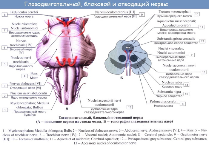 This nerve protects vision in the bright rays of the sun, constricting the pupil, and takes part in the performance of visual function.
Anatomy and structure
The oculomotor nerve originates from the base of the midbrain hills. From the tissues of the medulla, the fibers of the nerve pass along the pedicle and exit through the inter-pectoral pit through the wall of the cavernous sinus. Then the oculomotor nerve passes through the upper part of the palpebral fissure, where it is divided into 2 parts.
The upper part of the oculomotor nerve runs along the surface of the optic nerve and approaches the muscle that lowers the upper eyelid and lifts it. The lower branch is thicker. It passes to the lower oblique muscle of the eye, medial, lower rectus muscles. As well as the ciliary department.
Diseases and lesions
The main diseases of the oculomotor nerve include:
- ptosis or drooping of the eyelid due to paralysis of the oculomotor muscles;
- divergent strabismus due to improper structure of the oculomotor muscles;
- mydriasis, in which the pupil does not respond to light;
- limitation of movement of the eyeballs, which is associated with weakness of the oculomotor muscles;
- Diplopia (split image) occurs due to deviation from the axis of one eye due to muscle weakness.
Block (nervus trochlearis)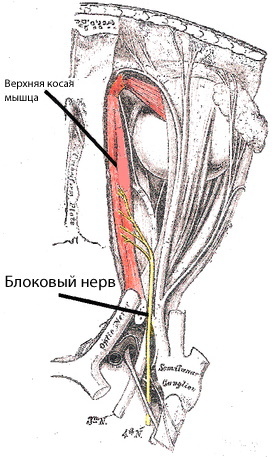
The block nerve is a 4 pair of cranial nerves and is part of the system that performs oculomotor functions.
Characteristic
It is duplicated on the right and left sides of the face. The block nerve provides horizontal and downward movement of the eye, which provides an overview of the peripheral picture.
Functions and properties
The block nerve acts on the superior oblique muscle of the face, which provokes movement of the eyeball in difficult areas of view. This nerve provides movement of the eyes in the form of turning inward, to the tip of the nose and its abduction to the outside of the face, as well as moving downward.
Anatomy and structure
The block nerve originates from the hills of the midbrain. Its nerve fibers bend around the outer side of the gray matter of the brain and exit from the brain stem, where they pass to the side of the brain stem. Coming out of the hole between the temporal part of the hemisphere and the cerebral peduncle, the nerve exits through the palpebral fissure. In the orbital region, the trochlear nerve is located above the tendon ring near the optic nerve and ends in the superior oblique muscle of the eye.
Diseases and lesions
The block nerve is very rarely affected, the main cause of such damage is trauma. In this case, a person develops a complete or partial paralysis of the trochlear nerve, which is expressed in a constant position of the eye at the top, closer to the center. In this case, to obtain a visual image of individual areas, a person has to tilt his head down and to the side.
Trigeminal (nervus trigeminus)
The trinity nerve is a paired and mixed nerve. It belongs to the 5th pair of nerves in the skull. The trinity nerve contains sensory and motor tissues. He participates in the performance of visual function and in the performance of movements of the jaw.
Characteristic
The trinity nerve at the exit from the ternary node is divided into 3 parts. 1 of which goes to the eye, and 2 others to the jaw. The functions of the ternary nerve are mixed. It contains motor and sensory fibers.
Functions and properties
The trinity nerve, due to its complex structure in the oral cavity, provides superficial and deep sensitivity. It provides sensitivity to the eye, jaw, and chewing function of the jaw.
Anatomy and structure
The trinity nerve starts from the median cerebellar peduncle with 2 branches in the form of motor and sensory fibers.
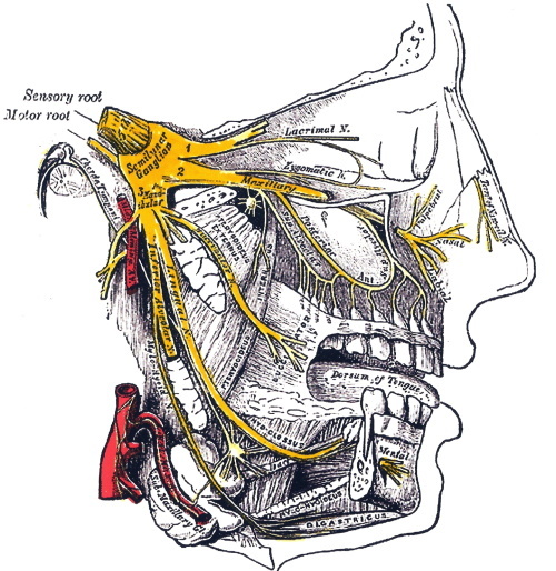
Both parts of the nerve pass into the opening between the layers of the dura mater of the brain and form a triangular cavity at the temporal bone. The lower and maxillary, optic nerves depart from the ternary node. Then the canal goes around the ternary node from the inside and approaches the oval entrance, where it connects to the 3 branch of the ternary nerve.
Diseases and lesions
The trinity nerve has a wide spreading network over the face.
When it is defeated, it occurs:
- persistent or temporary damage to the ternary nerve, which is a loss of sensitivity in the nose, oral cavity.
- neuralgia of the ternary nerve is expressed in the defeat of nerve cells. It causes facial pain;
- Gradenigo syndrome is caused by inflammation and cutting of the ternary nerve, which is accompanied by pain and impairment of the basic functions of the nerve;
- trismus or overstrain of the chewing muscles occurs due to irritation of the nerve fibers, which provokes a violation of the chewing, speaking functions in humans.
Abduction (nervusabducens)
The abducens nerve is paired and belongs to the 6th pair of facial nerves.
Characteristic
It supports eye movement and complements the oculomotor nerve. The abducens nerve is present on both the right and left sides of the face. The abducens nerve is a motor nerve.
Functions and properties
The abducens nerve is the cranial nerve that acts on the lateral rectus muscle. As a result of this effect, the eyeball is diverted outward.
Anatomy and structure
The nerves on the face of a person, the location of which has long been studied, originate from the brain. Likewise, the nerve fibers of the abducens nerve depart from the lower surface of the brain between the medulla oblongata and the pons varoli. This nerve travels through the surface of the brain and exits into the superior opening in the eye socket. The abductor muscle runs over the eye and merges with the lateral rectus muscle.
Diseases and lesions
With congenital and acquired lesions of the abducens nerve, a person develops strabismus, a doubling of the picture in the eyes. With absolute paralysis, the abducens nerve can be blocked and the person's functional ability to move the eye decreases, the visual picture narrows.
Facial (nervusfacialis)
The facial nerve is the paired 7th nerve of the facial nerves.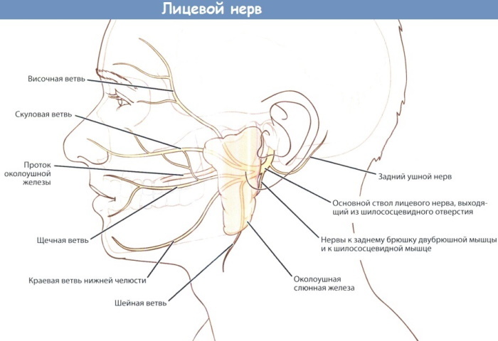 The facial nerve departs from the brain at the point between the pons varoli and the medulla oblongata. It has a complex structure in the front part.
Characteristic
The facial nerve is of mixed importance. It includes both sensory and motor neurons, which are located in both the muscle and soft tissues of the tongue. The facial nerve runs in the right and left sides of the face, which can act separately from each other, therefore, if the facial nerve is damaged on the 1st side, the face asymmetry occurs.
Functions and properties
The facial nerve is responsible for facial expressions, as it affects the facial muscles. It also reaches the lacrimal gland and irritates it. The facial nerve reaches the stapes muscle and is responsible for the sensation of taste in 2/3 of the tongue, and also participates in the work of the tympanic membrane.
Anatomy and structure
The facial nerve nucleus is located next to the abducens nerve nucleus. From the nucleus, the nerve fibers of the facial nerve bypass the lower part of the brain and exit at the medulla oblongata.
The main part of the facial nerve is a motor, but after connecting to the intermediate nerve, it becomes mixed. Connecting with the intermediate nerve, the facial nerve enters the auditory opening and passes into a large canal.
Then, at the temporal bone, the intermediate nerve is divided into:
- a large stony nerve, which, passing through the pterygoid canal, reaches the lacrimal gland and nasal cavity.
- A connecting branch that goes to the petrosal nerve;
- stapes nerve, which goes to the stapes muscle;
- the part connecting with the vagus nerve;
- a drum string that enters the tympanic cavity and connects to the ear node.
Before entering the styloid foramen at the base of the temporal bone, this nerve is divided into the posterior ear nerve, stylohyoid, digastric and lingual branches.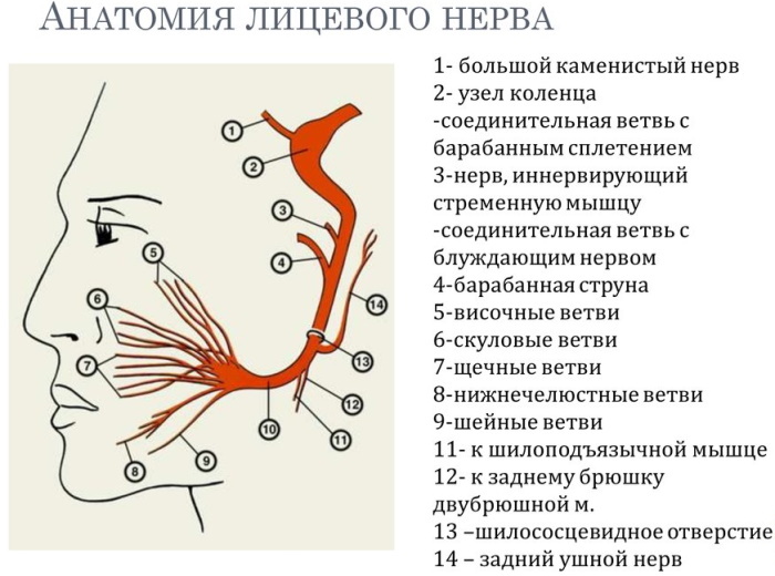
Branches branch off from the parotid joint:
- temporal;
- zygomatic;
- buccal;
- enemy;
- cervical.
All of them are crushed and excreted by the smallest nerve endings in the tissues.
Diseases and lesions
Paralysis of the facial nerve causes an asymmetry of the face, a decrease in taste sensations, an increase in the functions of secretion of lacrimal streams. The facial nerve can be damaged at the beginning of the nerve fibers, along the path of the facial nucleus junction or its attachment to the brain.
Vestibule-cochlear (nervusvestibulocochlearis)
The nerves on the human face, the location of which can be found in the form of a diagram, include the paired vestibular cochlear nerve.
Characteristic
It is responsible for transmitting auditory impulses in humans that come from the inner ear. The vestibular cochlear nerve is a nerve of special sensitivity. It consists of 2 roots of different functionality, 1 of which, in the form of semicircular ducts, performs the functions vestibular apparatus, and the second in the form of a cochlear root, transmits auditory impulses from the cochlear labyrinth.
Functions and properties
The vestibular cochlear apparatus transmits nerve impulses from the middle ear to the brain for analysis. As a result of such work, a person develops the ability to hear. The same nerve is involved in the work of the vestibular apparatus, helps us maintain balance.
Anatomy and structure
Nerve fibers start from the cochlear root and pass into the spiral elements of the hearing aid.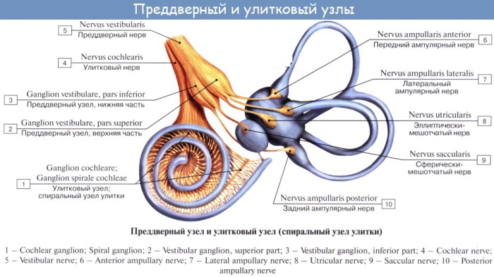 The central processes of the cells of the cochlea form a canal that exits from the pyramid of the temporal bone through the auditory opening into the substance of the brain. The vestibular root departs from the vestibular ganglion and bifurcates into the upper and lower parts. The vestibular cochlear nerve begins in the brain lower than the facial nerve.
Diseases and lesions
In case of a violation in the work of the vestibular cochlear apparatus, a person's hearing drops sharply or hearing acuity decreases. Complete hearing loss may occur in 1 or both ears. When a nerve is damaged in the part of the connection with the vestibular apparatus, a person develops a violation in the work of the orientation system in space. As a result, a person may lose the ability to walk steadily, he may have rapid involuntary eye movements, dizziness.
Glossopharyngeal (nervusglossopharyngeus)
The glossopharyngeal nerve is considered the 9th nerve of the facial nerve system.
Characteristic
It contains sensory, motor and parasympathetic connections, therefore it is a mixed nerve. It interacts with the main reflexes of the mouth and the muscles of the tongue.
Functions and properties
The glossopharyngeal nerve has several main functions, namely:
- makes the stylopharyngeal muscle move, which raises the pharynx;
- carries out a parasympathetic effect on the parotid gland, which helps to carry out secretion;
- provides the sensitivity of the tongue and palate, pharynx;
- provides taste possibilities for a third of the tongue.
Anatomy and structure
On the human face, the glossopharyngeal nerve is located in the mandibular region. Its base departs from the brain, passing through the anterior part of the jugular foramen.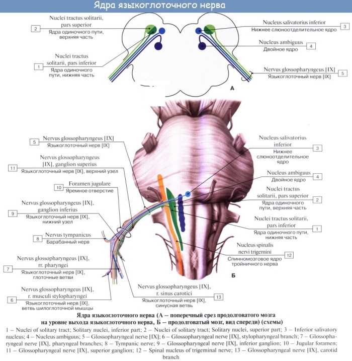 The nerve then thickens and travels down to the carotid artery and goes up and down into the tongue. At the bottom, the glossopharyngeal nerve creates a network of nerve endings that extend deep into the pharynx, stylopharyngeal muscle, tonsils and tongue.
Diseases and lesions
In case of damage or inflammation of the glossopharyngeal nerve in patients, there is a sharp decrease in the sensitivity of the tongue and palate. The loss of taste can be temporary or permanent. Pharyngospasm or muscle spasms may appear, in which the ability to swallow on their own is reduced. Injury to the glossopharyngeal nerve can cause pain around the tongue and throat.
Wandering (nervusvagus)
The vagus nerve refers to the 10th pair of cranial nerves.
Characteristic
This nerve is the longest and runs from the brain to the abdomen. He is responsible for the work of the muscles of the head, neck, abdomen and thoracic region. Due to its complex structure, the vagus nerve is classified as a mixed one, since it includes motor, sensory and autonomic elements.
Functions and properties
The vagus nerve has many functions.
These include:
- ensuring the sensitivity of the oral mucosa of the lower jaw and larynx;
- ensuring the sensitivity of the skin behind the ear and the outer part of the auricle;
- maintaining the work of the tympanic membrane and transmitting impulses from it to the brain;
- receiving information from the hypothalamus, olfactory system, autonomic system and transmitting it to the brain;
- participation in the regulation of blood pressure;
- activation of smooth muscle contractions in the bronchi;
- provokes the occurrence of cough and vomiting;
- activates the muscles of the palate, larynx, pharynx;
- provides innervation of the lungs, stomach, heart;
Anatomy and structure
The vagus nerve begins with 10-15 roots on the lower part of the brain. Exiting through the anterior part of the jugular foramen, it thickens and passes in the neck. Here it connects with the internal jugular vein and the common carotid artery, forming the nerve bundle of the neck. Then the vagus nerve passes through the aorta, bends around the bronchi and approaches the food. At the esophagus, the vagus nerve is divided into 2 parts and covers the esophagus and diaphragm in front and behind. In the abdominal cavity, the vagus trunks penetrate the solar plexus.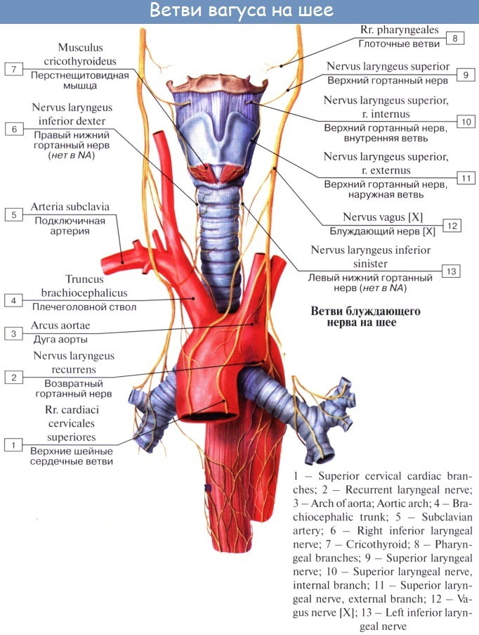
The head section is the shortest. It includes miningeal, ear, connecting branches. The cervical region has a pharyngeal branch, an upper and lower laryngeal branch, a recurrent laryngeal nerve. The thoracic vagus is subdivided into the thoracic, bronchial, pulmonary, and esophageal plexus. The abdominal vagus nerve covers the entire stomach and peritoneum.
Diseases and lesions
With lesions of the vagus nerve, a person may develop:
- neuritis;
- multiple sclerosis;
- paralysis that is fatal;
- dysphagia (swallowing disorder);
- aspiration pneumonia;
- hypoxia.
Additional (nervusaccessorius)
The accessory nerve contains mixed fibers (motor, innervating) and belongs to the 9th pair of muscles.
Characteristic
The accessory nerve is mainly responsible for motor functions in the body.
Functions and properties
The accessory nerve is responsible for the mobility of the sternoclavicular muscle, activates the head turn, shoulder lift. This nerve provokes the pulling of the shoulder girdle back, is responsible for involuntary turns of the head back or to the side.
Anatomy and structure
The accessory nerve consists of 2 parts: spinal and cerebral. The roots of the accessory nerve branch off from the brain and form the cerebral canal. They then thicken and combine to form a spinal root that passes through the foramen magnum in the skull. 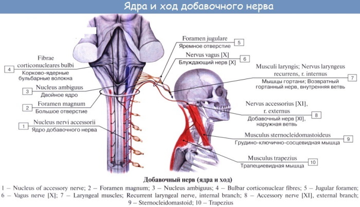 Both of these systems combine into the trunk and exit from the cranial cavity. They are further subdivided into an internal and an external branch. The inner branch goes to the vagus nerve, and the outer branch goes to the sternoclavicular section.
Diseases and lesions
If the accessory nerve is disrupted or damaged, a person develops spastic torticollis, clonic convulsions with twitching of the head, muscle atrophy of one or both shoulders. Less commonly, the patient may experience pain in the back or arm, as well as limiting the movement of the arms.
Sublingual (nervus hypoglossus)
The hypoglossal nerve is a motor nerve and is made up of similar fibers.
Characteristic
It is the 12th pair of cranial muscles. The nerve nucleus is located in the medulla oblongata, and branches from it go into the tongue, maintaining its mobility.
Functions and properties
The hypoglossal nerve provides mobility of the tongue in all directions, participates in the digestion process by maintaining the reflex processes of swallowing, chewing, sucking and licking. It allows us to speak clearly and whistle.
Anatomy and structure
The hypoglossal nerve leaves the medulla oblongata with 10-15 roots, uniting into a single canal. The nerve trunk passes through the hypoglossal nerve canal and then exits next to the jugular vein into the submandibular triangle. 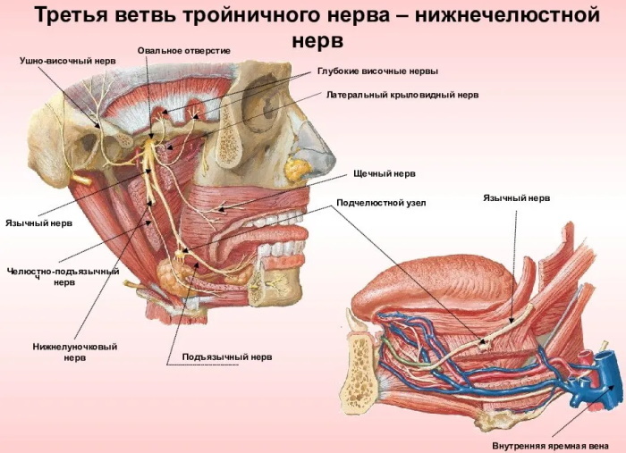 Entering the muscles of the tongue, the hypoglossal nerve is divided into 4 branches, which diverge in a small network of nerve endings.
Diseases and lesions
Nerves on the human face include the hypoglossal nerve, which is located in the mandible and head. With inflammation and lesions of the hypoglossal nerve, a person experiences speech impairment, atrophy of the tongue, inhibition of the reflexes of swallowing, sucking, and chewing food. When the hypoglossal nerve is cut, complete or partial paralysis of the tongue occurs, which is impossible to completely restore.
Video about nerves on the face
Facial nerve. Anatomy. Neurology: