
Shoulder joint performs one of the most important functions in the human body, providing movement in the upper limbs. Disease of the joints ranks third among all pathologies, after diseases of the circulatory and digestive organs. Due to the complex anatomical structure of this joint, the most valuable method for its study is MRI.
Record content:
- 1 What is diagnostics?
- 2 What does the scan show?
- 3 Benefits of Shoulder Diagnosis
- 4 Indications for the procedure
- 5 Basic limitations
- 6 Preparation for research
- 7 Diagnostic steps
- 8 Purpose of examination with contrast media
- 9 Possible complications
- 10 Survey result
- 11 Where to make and what does the cost depend on?
- 12 Shoulder MRI video
What is diagnostics?
MTP of the shoulder joint (the cost of this study is quite high) is one of the most relevant diagnostic methods, which is based on the action of a magnetic field.
The essence of the method is that the hydrogen nuclei in the human body under the influence of a magnetic field capable of absorbing and emitting in response radio waves of a certain frequency, which are recorded by the receiver.
For this purpose, it was hydrogen that was chosen, since it is part of the water molecule. And the fact that all cells in the body are 70% water makes it possible with the help of this study to examine all human tissues. Subsequently, the signals are converted by computer programs into digital images and recreate detailed information about the state and structure of the organ.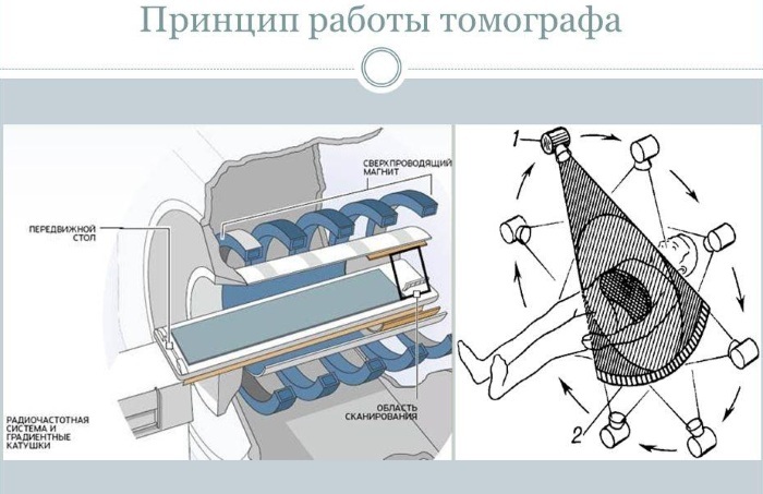
The study can be performed with devices of several types. Closed-type tomographs have an annular shape limited on all sides, into which the table with the patient is immersed. A high magnetic field is created in it, which makes it possible to examine in detail the deep structures of the organ.
Open-type tomographs have more free space, they have a magnetic coil only from above, it creates a comfortable environment for claustrophobic patients, but is inferior in informativeness.
What does the scan show?
In the course of this study, it is possible to obtain layer-by-layer or volumetric images of any area, organ or system of human organs.
Magnetic resonance imaging allows you to examine in detail each element of the joint:
- cartilage;
- ligaments;
- muscles;
- bags;
- vessels.
The results of the procedure make it possible to assess the functional ability of the organ and identify minimal pathological changes.
Benefits of Shoulder Diagnosis
In the diagnosis of diseases of the osteoarticular system, especially large joints (shoulder, knee), the priority belongs to magnetic resonance imaging. This is due to the quality of the images and the safety of the method.
This type of diagnosis is preferred for the following reasons:
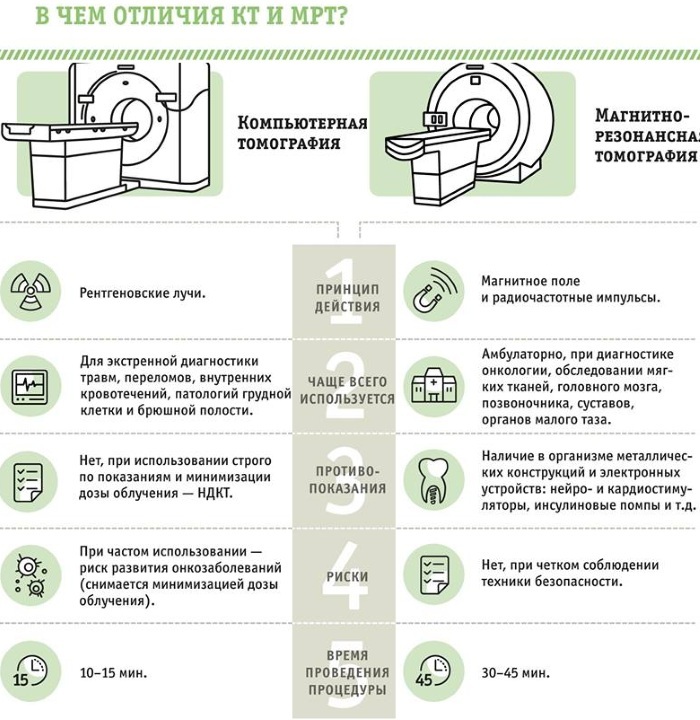
- Ultrasound examination (ultrasound) is an affordable, safe and fast method, but it has its own difficulties in the diagnosis of joints. Some structural elements of the joint can interfere with the passage of the ultrasound wave, making it difficult to obtain high-quality images. In MRI, bone tissue, due to its low water content, does not affect the scan results.
- X-rays and computed tomography (CT) examinations are based on the action of X-rays. These methods are effective mainly for finding pathologies of bone structures. The disadvantages of these studies in the diagnosis of joints are the harmful effects on the body (radiation), poor visualization of soft tissues, the inability to use during pregnancy. By sending a radio signal of a very low frequency, MRI does not provide X-ray irradiation to the human body, while maintaining information content.
- The advantage of MRI is the ability to identify a pathological process in the early stages, which facilitates and speeds up treatment for a person.
- MRI is a safe and painless alternative to arthroscopy, in which surgery is performed using special instruments to examine the joint cavity from the inside.
Indications for the procedure
An MRI of the shoulder joint should be done when:
- soreness in the joint area when moving or at rest;
- swelling or redness of the joint;
- injury of the shoulder girdle;
- inflammatory changes (arthrosis, arthritis);
- tumor processes of bone, muscle or cartilage tissue, including for the search for metastases;
- autoimmune diseases accompanied by joint damage (rheumatoid arthritis);
- entrapment of nerves;
- anomalies in the development of the shoulder joint;
- for control after surgical interventions on the joint.
Basic limitations
Before referral for research, the doctor must carefully collect information about the patient and exclude possible contraindications.
They include 2 groups:
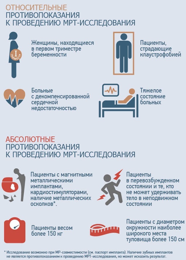
- Absolute contraindications, which prohibit the passage of magnetic research methods. This is the presence in the patient's body of devices or objects containing metal elements:
- heart valves;
- dental prostheses;
- Hearing Aids;
- metal clips applied to the vessels;
- implants;
- foreign bodies of the gastrointestinal tract and other hollow organs;
- foreign bodies of soft tissues (bullets, fragments);
- serious condition of the patient;
- contrast study during pregnancy, breastfeeding, allergies to the components of the contrast medium, renal failure.
In the area of the magnetic field, medical devices can malfunction, and metal objects are exposed to heat and movement, which can cause trouble for the patient. MRI for people in serious condition is prohibited due to the unstable work of the life-supporting systems of the body.
- The second group of contraindications - relative, in which magnetic resonance imaging is possible, but with certain conditions. This includes patients with:
- claustrophobia (fear of enclosed spaces);
- neurological diseases if they prevent a person from controlling their movements;
- pregnancy up to 12 weeks;
- people weighing more than 130 kg.
An alternative for a person with a fear of confined spaces is the study in an open-type tomograph. Such tomographs are used much less often, therefore it is necessary to clarify their availability in diagnostic centers. If it is impossible to control movements, the study can be carried out under general anesthesia or with the help of sedatives.
Despite the proven safety of the magnetic resonance imaging method, its use in the first trimester of pregnancy is a controversial issue among many diagnosticians, therefore it is justified only in complex or urgent clinical cases.
As for the weight of a person, it depends on the power of the table on which the patient is placed to be immersed in the tomograph cabin. The permissible weight varies from 130 to 160 kg, therefore, this item also needs to be clarified in the center before signing up for the procedure.
Preparation for research
Magnetic resonance imaging of the knee joint does not require any special training, so you can go through this procedure at any time. days, observing only some of the rules:
- for the patient's convenience, make an appointment for a tomography in advance;
- take with you a doctor's referral, pictures and results of previous examinations;
- before scanning, you must put any electronic or metal objects out of your pockets: a mobile phone, a player, a watch, keys, bank cards;
- remove all jewelry: earrings, chains, bracelets, piercings, rings;
- remove permanent makeup (may contain metal particles).
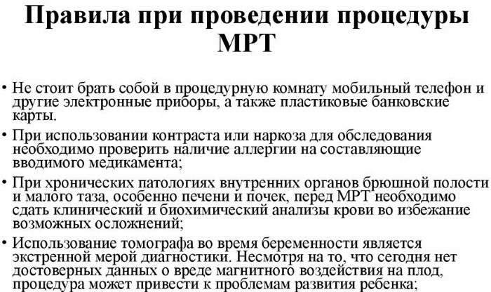
The presence of electronic devices during the research can disrupt their operation, and metal components can interfere with the research.
Diagnostic steps
During the procedure, the patient will need to go through the following steps:
- Fill out the form and familiarize yourself with the rules of the procedure at the reception.
- Remove all jewelry, metal and electronic devices, clothing with metal fittings, if necessary, change into a disposable gown.
- If a contrast study is prescribed, enter it immediately before scanning.
- Take the correct position in the tomograph booth.
- Lie motionless throughout the entire procedure.
- Waiting for the doctor to describe the images.
- Getting your hands on pictures and conclusions.
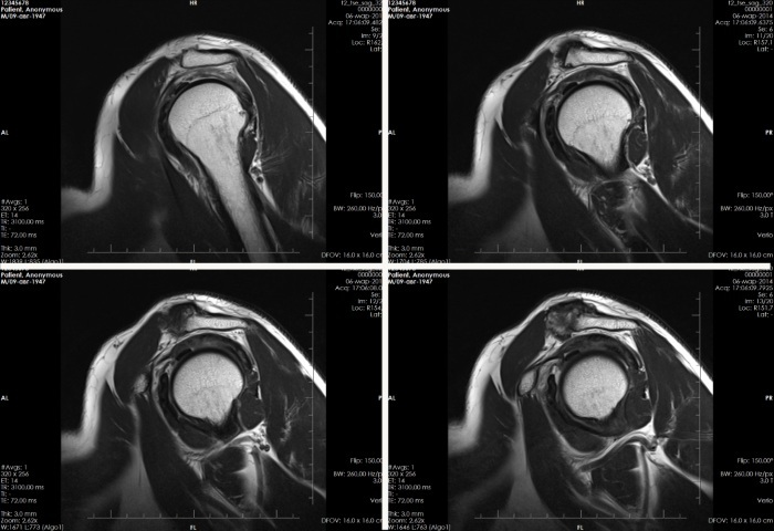
Routinely, the study is performed in three planes: sagittal, axial and oblique, this allows the most detailed examination of the area of interest from all sides. The duration of the procedure depends on the area under study and the complexity of the pathological process, on average it lasts 20-40 minutes.
The study will take place in the presence of a laboratory assistant who will help you to take the correct position depending on the area being examined, fix the limbs with special belts and give instructions. During the entire procedure, it is necessary to lie strictly still, this is what will ensure obtaining clear pictures.
In some pathologies, images are also taken in the position of abducting the arm and turning the shoulder outward. CT scans for young children who cannot lie still are performed under short-term general anesthesia.
The tomography booth is equipped with video communication and a camera, so, if necessary, the patient can contact a health worker and tell about his anxiety and well-being. The magnetic head of the tomograph creates quite loud knocking sounds when rotating around the patient, you should not be afraid of them.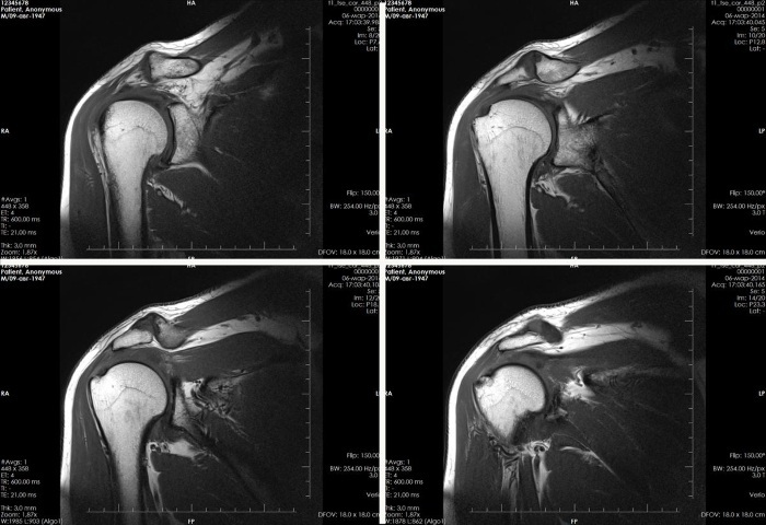
After scanning, the diagnostician reports the waiting time for the result and proceeds to describe the images obtained. This can take up to 40 minutes. up to several hours, depending on the complexity of the clinical case.
If waiting for the results causes difficulties for the patient, it is possible to receive them by e-mail. mail or download in your personal account on the website of the diagnostic center in which study.
Purpose of examination with contrast media
For a more accurate detailing of the pathological process, the patient can be assigned a tomography of the joint with contrast. As a contrast, so-called ferromagnets are used, which are substances with their own magnetic field.
These include iron oxide, manganese compounds, gadolinium salts. The contrast agent is injected intravenously before the study, it selectively accumulates in different tissues of the joint, gives a brighter and clearer image and helps in the diagnosis of function joint. This substance does not cause allergic reactions, is easily tolerated and quickly excreted from the body.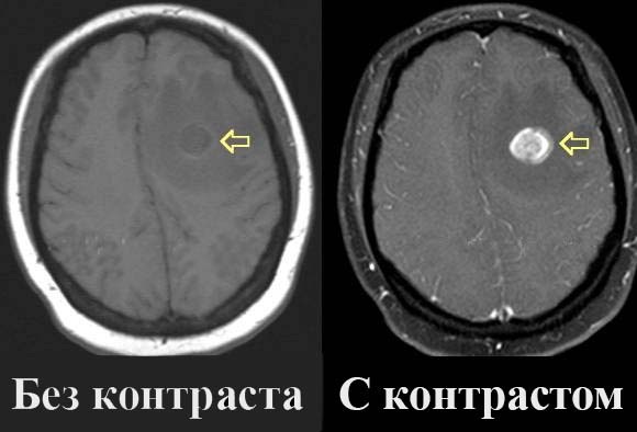
A study with a contrast agent is of great diagnostic value in the search for oncological diseases of the tissues of the joint in the early stages or metastases from other organs.
Tumor formations have an abundant blood supply, which contributes to the accumulation of contrast agent in them and gives a clear visualization of the process. In rare cases, it is possible to inject a contrast agent directly into the joint cavity, this technique allows for a better assessment of changes in the ligamentous apparatus.
Possible complications
Numerous studies in this field of diagnostics have not revealed the negative impact of MRI on the human body.
Complications are possible only if the diagnostic rules are not followed:
- malfunction of medical devices in the human body (valves, pacemakers, hearing aids), electronic devices or unreliable research results;
- aggravation of mental disorders, for example, with claustrophobia;
- very rarely, allergic reactions with the introduction of contrast in the form of a rash, itching, nausea or headache with individual intolerance to any component of the contrast mixture.
After the examination, the patient receives a film with pictures in his hands and the conclusion of a functional diagnostics doctor. If necessary, you can get the results in electronic form on disk or flash media.
Survey result
MRI in the absence of pathological changes in the images determines the normal anatomical structure of the shoulder joint. It is made up of the head of the humerus and the glenoid cavity of the scapula. A cartilaginous lip is located along the edge of the cavity, which softens the movement of the head.
Normally, the integrity and ratio of bones in the joint are not compromised. The head of the humerus must be of the correct shape, in the correct position and of a uniform structure. The cartilaginous surfaces and the cartilaginous lip are smooth and distinct.
The joint capsule, consisting of the ligaments and muscles surrounding the joint, is clearly visible, not deformed. Normally, a small amount of fluid (effusion) is also detected in the joint.
MRI of the shoulder joint due to the use of various planes and modes makes it possible to accurately assess such pathologies:
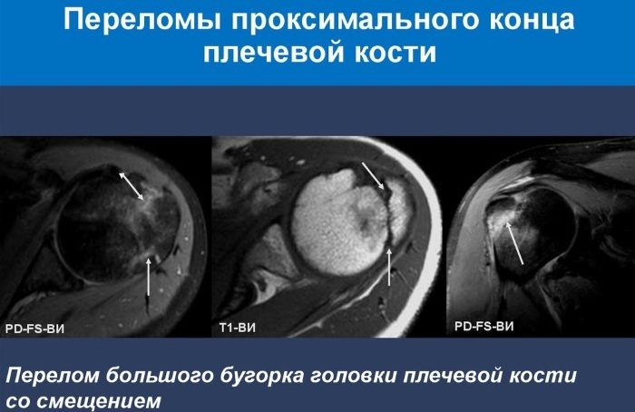
- Bone fractures. If there are defects in the bone components of the joint, the doctor diagnoses fractures or cracks in the humerus, glenoid cavity of the scapula or clavicle. It is also possible to establish the prescription of the fracture (fresh or old) and the presence of displacement of bone elements, which helps to determine the treatment tactics very accurately.
- Dislocations and joint instability. In this case, the head moves or slides off the cavity. Due to the structural features, the shoulder joint is more often than others susceptible to this pathology. This is predominantly found in early childhood and in athletes.
- Sprains, tears of ligaments or tendons due to domestic or sports injuries. MRI makes it possible to very clearly examine each fiber of the ligaments and diagnose the extent of the lesion.
- Bankart damage - is a lesion of the cartilaginous lip of the joint, as a result of which the head of the humerus is displaced. The volume of the defect can be different: from partial to complete separation of the fibrous part of the lip.
- Rotator cuff injury or rupture (impingement syndrome). The shoulder cuff is a group of muscles that surround the joint. If it is damaged, circular movements in the joint are disrupted.
- Violation of the integrity of the cartilaginous surfaces makes it possible to diagnose arthritis and arthrosis of the shoulder joint.
- Tumors and metastases of the soft tissues of the joint.
- Nerve entrapment.
- Pathological processes in the surrounding tissues, for example, neoplasms, enlarged lymph nodes.
- Possible complications after shoulder surgery: hematoma, damage to muscle bundles, axillary nerve, infection.
- Vascular abnormalities and circulatory disorders in the joint.
- Also, tomography makes it possible to recognize bursitis (inflammation of the articular bag), the accumulation of inflammatory fluid in the joint cavity, fibrotic changes and adhesions as a result of the postponed inflammatory process.
MRI of the shoulder joint
With the received conclusion and images, it is necessary to go to a doctor (traumatologist, surgeon, rheumatologist or oncologist) for further diagnosis and treatment, based on the identified pathology.
Where to make and what does the cost depend on?
MRI of the shoulder joint can be performed in private diagnostic centers or in diagnostic buildings at regional and city clinical hospitals. It is possible to clarify whether this type of medical services is provided by a phone call or on the official websites of diagnostic centers.
By region, the pricing policy looks like this:
| Region | Without contrast (rub.) | With contrast (rub.) |
| Moscow and region | 5100-7700 | 10600-22000 |
| Krasnodar | 1760-4000 | 4360-14100 |
| Krasnoyarsk | 3250-4200 | 6950-13400 |
| Vladivostok | 4400-5200 | 8300-14700 |
Given the safety of the method, this study can be completed a large number of times with a minimum time interval, depending on the nature of the disease and the characteristics of its course. The frequency of the examination is determined by the attending physician.
The cost of diagnosing the shoulder joint using MRI varies and depends on several factors: the equipment used for the tomography (class, power of the device, closed or open type of tomograph), consumables, the examined part of the body, the need to use contrast.
Shoulder MRI video
Detailed analysis of MRI of the shoulder joint: