Head MRI examination is used in modern medicine in the diagnosis of pathologies at various stages. This is a modern method based on magnetic resonance imaging, which has a very high resolution, that is, it shows even minor deviations from the norm.
They may indicate a particular pathology. The doctor gets the opportunity to study in detail the structure of the brain and other parts of the head. Thanks to modern tomographs, it is possible to obtain a layer-by-layer image (slices up to 1 mm).
During the examination, the patient is not exposed to X-rays or ultrasound beams, which can cause serious harm. Thanks to this, the MRI examination can be called a safe event.
This is confirmed by numerous studies that have proven that during the entire time of MRI there was not a single case of complication caused directly by magnetic resonance imaging tomography.
But the equipment for conducting such a study is very expensive, so not every medical institution has the opportunity to undergo diagnostic measures of this type. Usually you have to register or undergo a survey on a paid basis.
Record content:
-
1 Indications for research
- 1.1 Indications for anesthesia during MRI
-
2 How is it determined
- 2.1 Types of MRI
-
3 Preparation and analysis
- 3.1 MRI
- 4 Decoding the results
- 5 When to see a doctor
- 6 Possible contraindications
- 7 Head MRI video
Indications for research
Any doctor who diagnoses a patient can refer to an MRI. However, most often neurosurgeons, endocrinologists, vascular surgeons and other highly specialized doctors are referred for this examination. To them, in turn, the patient gets in the direction of the therapist.
An MRI of the head shows a variety of abnormalities, so the doctor can refer the patient to this examination if you suspect:
- neoplasm of a malignant or benign type;
- stroke;
- the injury received;
- single or multiple cyst;
- pathologies that affect the pituitary gland;
- diseases of the vascular system of the brain;
- the presence in the patient's body of parasites that could penetrate the brain, moving along the vascular bed;
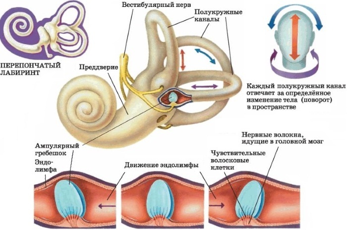
- the development of pathology, which entails impairment of hearing or vision;
- multiple sclerosis;
- dementia.
Also, a similar study is carried out if the patient complains of frequent headaches, but any other diagnostic measures taken did not help him to make an accurate diagnosis. Therefore, MRI is prescribed for migraines, as well as in the case of thrombosis, adenoma.
Also, MRI of the brain is performed for:
- The onset of metastases. As a rule, in this case, the patient complains of simultaneous problems with hearing, vision, smell, touch. He may have hallucinations, reading disorder, and other symptoms of brain damage.
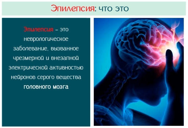
- Epilepsy and other pathologies that manifest themselves in the form of confusion of consciousness, speech. Also, MRI is mandatory for those who suffer from convulsions and other seizures.
- Meningitis, myelitis, encephalitis and other inflammations that affect the brain and are extremely life-threatening. Also, MRI is prescribed for lesions that could be provoked by measles, tuberculosis, toxoplasmosis.
- Rehabilitation after suffering a traumatic brain injury or stroke. Thanks to the examination, the doctor can assess how quickly the recovery is going and whether he has chosen the right drug therapy.
- Degenerative processes, which may indicate the development of pathologies such as multiple sclerosis, Alzheimer's disease and other diseases.
- Congenital abnormalities or signs of hydrocephalus in children.
Indications for anesthesia during MRI
As a rule, during the examination, the person is conscious. He is asked to lie down quietly while he is in a special tomograph chamber. However, in some situations, the patient is physically or psychologically unable to lie quietly in a confined space and keep his body motionless.
General anesthesia is indicated when:
- Claustrophobia. This is a pathological condition characterized by a fear of enclosed spaces. In this case, the patient will be in a state of extreme stress and panic if asked to take a horizontal position in the tomograph. In this case, it is impossible to conduct diagnostic studies. In addition, the patient himself can harm himself, therefore it is recommended to carry out the examination under general anesthesia.
- The presence of mental disorders. If it is impossible to predict how the patient may behave inside the tomograph chamber, then it is better to use anesthesia. In addition, if a person is constantly in uncontrolled movement, this will smear the resulting image, and it will still not be possible to make a diagnosis.
- Nervous tics and other uncontrolled physiological features. In this case, the patient also cannot take a calm and immobile state.
- Epilepsy and other pathologies that are accompanied by seizures. In this case, only intravenous anesthesia is indicated.
- Early childhood. A small child also cannot follow the recommendations of doctors and is not able to take an immobile state. Therefore, for young patients, it is sometimes recommended to use light anesthesia, which is influenced by a short use of the mask.
- The presence of severe pain syndrome. In the event that the patient literally writhes from pain, he will also not be able to take an even position.
How is it determined
An MRI of the head is a study that shows those changes in the human body that a doctor may not notice with other research methods. The very principle of operation of magnetic resonance equipment is based on the characteristics of the human body.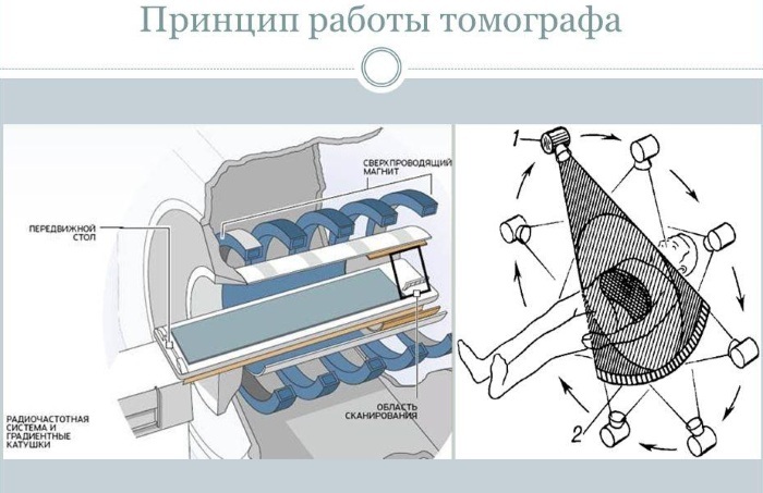
Each cell of the human body contains water molecules, which, in turn, consist of hydrogen (2 atoms) and oxygen (1 atom) atoms. If we consider them from a physical point of view, then these atoms are magnets. This means that when these atoms find themselves in a strong magnetic field, their electrons begin to rotate along the axis of the external field.
At this moment, there is a so-called deviation from the orbit along which they were moving before. When such a system is exposed to radio waves, the tilt angle of the moving particles changes.
When the external influence ceases, the electrons inside the atoms return to their previous orbit and begin to emit electromagnetic waves. At this moment, they are fixed by a tomograph. As a result, the device generates a clear three-dimensional image based on the received data.
An MRI of the head shows the water content in the cells and other parameters. Based on them, you can make a more accurate diagnosis, evaluate the quality of treatment and choose the course of the most optimal therapeutic measures for the patient.
Types of MRI
The most accurate image is obtained when the machine can make very thin sections. This parameter depends on several things - the model of the tomograph (how modern it is) and the type of MRI.
There are several types of magnetic resonance therapy:
- Standard. The specialist receives the required amount of information. No contrast medium is injected. In this case, the patient spends about 20-30 minutes in the tomograph.
-
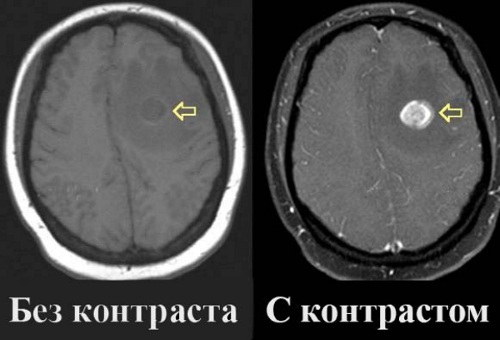
With contrast. This type of examination produces a sharper image. To do this, a special drug with gadolinium salts, Omniscan or other means that enhance the contrast of the picture is injected into the patient's vein. Solutions of this type enter the bloodstream and begin to light up when the beams of the tomograph hit them. This allows better decoding of data and timely pay attention to various anomalies in the structure of the brain. As a rule, this method is used for vascular diseases, multiple sclerosis, and cancers.
 The dose of the contrast agent is determined by the doctor on an individual basis, depending on the data of a particular patient. In the case of using a contrast agent, the duration of the procedure can be increased to 1 hour. In addition, this type of procedure differs from the standard form of MRI in that after the contrast is injected, a person can feel a metal taste in the mouth. This is a perfectly normal reaction. There are usually no other unpleasant sensations. Therefore, people quite easily tolerate scanning using contrast.
 - MR angiography. A type of procedure that is performed if necessary to assess the state of the vascular system in if you suspect that the patient is suffering from atherosclerosis, aneurysm, or there is concern thrombus formation.
- MRI of the pituitary gland. In this case, special attention is paid to the epididymis, which is the endocrine gland. Thanks to the pituitary gland, hormones are synthesized that determine the quality of the reproductive system of the body. MRI of the pituitary gland is recommended for suspected benign tumors, hormonal system disruptions, gigantism, obesity. Also, this type of procedure allows you to confirm or exclude the so-called pituitary formations.
Preparation and analysis
It is important to consult with a specialist before performing a brain scan with a CT scanner. Recommendations for preparing for an examination may differ depending on the specific situation.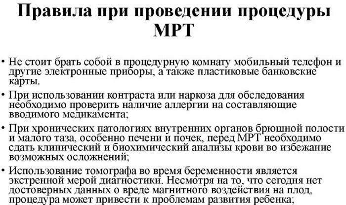
If we talk about standard recommendations for preparation, then it is worth considering the following tips:
- It is important to choose the right clothing before the examination. Since you should not keep metal objects next to a powerful magnet, you need to carefully approach the choice of clothing for examination. It should not have metal zippers, buttons, rivets or other decorations. In this case, it is worth giving preference to comfortable, loose clothing, since you may have to stay in the tomograph for up to 1 hour. Some modern clinics have disposable MRI clothing kits, but this issue should be clarified in a specific medical institution.
- It is recommended not to wear jewelry, watches or other accessories. Or you can remove them immediately before the study, but it is better to think about this in advance, since there is the risk that, being in a medical center, a person may forget to remove one or another metal element. It is also worth checking for coins, keys, and other items in your pockets.
- Before undergoing an MRI, you should exclude the intake of alcoholic beverages. If during the examination it is supposed to use a contrast medium, then nothing should be eaten 3 hours before the MRI scan. If the patient has a hearty lunch before the MRI, then during the tomography he may begin to experience severe discomfort.
In some cases, additional fluids may need to be injected prior to MRI to further enhance the contrast of the image. Also, the doctor can pre-administer a very small dose of contrast agent to the patient in order to understand if the person is allergic to the selected drug.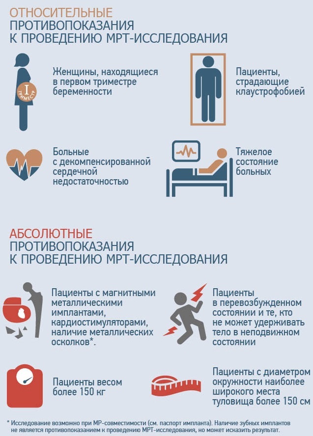
If an allergic reaction begins directly during the scanning procedure, this will distort the result and will have to be re-examined. However, in most cases, prior administration of contrast is not required.
MRI
MRI of the head shows the presence of brain lesions and developing pathologies. Since the procedure is a magnetic resonance imaging type, this means that all metal objects must be removed before the examination. In addition, the patient must remove even dentures, if any.
After that, the person is offered to take a horizontal position on a movable table, which will later go to the tomograph itself. Additionally, the patient can be secured with straps so that he does not move during the examination. After that, a special device with wires is put on the person's head. It is necessary in order to transmit and receive signals.
Also, the patient is recommended to use earplugs, since during the operation of the tomograph, not very much are constantly published pleasant clicking sounds that can get tired after a rather long stay inside the research apparatus.
When the patient is fixed on the table and is ready for examinations, the table enters the tomograph, and the medical staff moves to the next room. The doctor monitors the progress of the study using a computer. Already during the procedure itself, the specialist can evaluate the data and draw preliminary conclusions about the patient's condition.
During the operation of the unit, layer-by-layer photographs are taken. Their quality and information content depend on what kind of MRI method is used.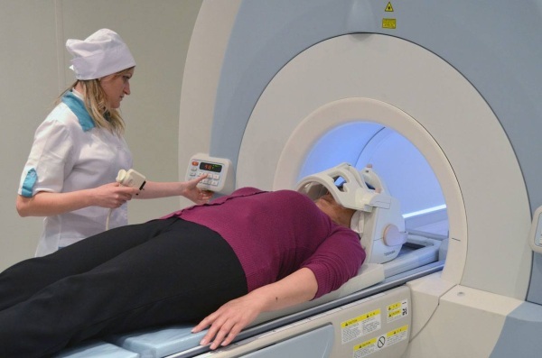
After the MRI scan, the patient can immediately go home and lead a normal life. Research of this type does not have any side effects, does not cause pain. Some discomfort can only be from a long stay in one position.
Research results can also be obtained immediately. In some medical centers, the images obtained are copied to a disk, flash card, or can be sent by e-mail. As a rule, the doctor who conducts the examination can indicate certain deviations from the norm, however, it is the attending physician who makes the final diagnosis. It is he who should be shown the results of the MRI.
Decoding the results
An MRI of the head shows a lot of information, but it is very difficult for someone who is far from medicine to understand it. Of course, in no case should you diagnose yourself on the basis of your own assumptions or when comparing your picture with those images that are on the Internet.
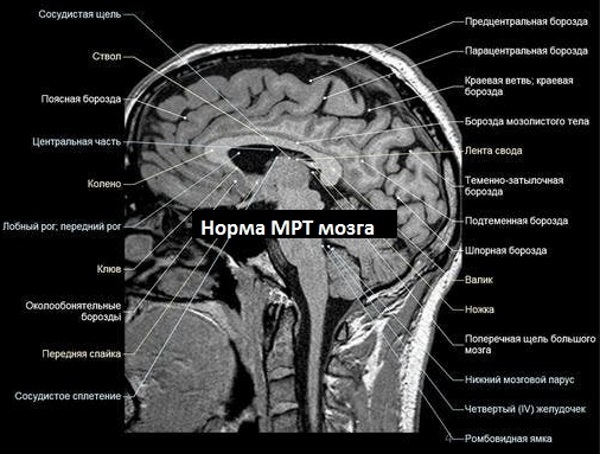
Nevertheless, several parameters can be roughly estimated that can indicate possible deviations from the norm.
| MRI indicators | Decryption features |
| Fabrics | Normally, fabrics have a characteristic gray tint, which should be lighter or darker depending on the specific area. |
| Cerebral fluid | It is a kind of light gray rivulets. |
| Intracerebral sinuses | Should look like cavities of the same black color. In this case, the intensity of the signal for the sinuses should be the same. |
| Tumors | In the image, they are light spots with uneven outlines. |
| Atrophy | Due to the too latent development of pathology, it is impossible to determine its presence. However, if you have some knowledge in the field of medicine, it is worth paying attention to dying cells. |
| Vessels | Should be evenly lit. |
| Atherosclerosis | The picture clearly shows that the vessels are narrowed. You may also notice the presence of plaques. |
| Aneurysm | The vessels are dilated, and the walls of the vessels, on the contrary, look thinner. |
| Hypertensive angiopathy | Small round cavities can be seen near the vessels. |
| Hydrocephalus | The ventricular cavities are greatly expanded.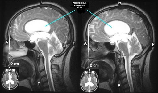
|
| Stroke | In the event of such a lesion, the brain suffers greatly from oxygen starvation. In the picture, you can determine the area that is more affected by hypoxia by the lighter shade. Vascular ruptures will also be visible in the image. They represent shaded areas with peripheral stripes. Gradually, these rings may begin to fade, so it is important to get an MRI scan right after a stroke. |
| Multiple sclerosis | With pathology on the nerve fibers, the myelin layer disappears. In the picture, you can see such lesions, which can be of various shades. It all depends on the volume of chemical components in the tissues. These lesions can be found in a wide variety of white matter zones. At the initial stage of multiple sclerosis, there may be several foci, but their number is rapidly increasing as the pathology progresses. |
It is better to entrust the interpretation of analyzes to a specialist, since only he can pay attention to the nuances. It should also be borne in mind that the same pathologies and deviations can manifest themselves in different ways, depending on a huge number of factors, including gender, age and other patient data. Other pathologies from which the patient suffers are also taken into account.
When to see a doctor
Regardless of whether the patient was able to decipher the results himself, he should make an appointment with the MRI diagnostician. Only this doctor will be able to fully assess the state of human health.
In order to understand why only a specialist can deal with decoding, it is enough to know that before starting to diagnose MRI, a person should not only get an education in a medical institute, but also get a specialty radiologist.
After that, the doctor additionally studies in courses (last from 3 to 6 months, depending on the qualifications of the candidate). Then the specialist spends another 2 years and deciphers the MRI under the supervision of more experienced colleagues, and only after that he can independently engage in diagnostics.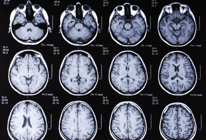
The MRI diagnostician will diagnose, but he does not prescribe treatment. Therefore, after visiting a specialist, you need to go to the attending physician and present him with the results of the study and the conclusion of the diagnostician. Which particular doctor to contact depends on the specific pathology identified in the picture.
You may need to talk to a neurosurgeon or cardiologist. To whom it is better to turn can be suggested by the diagnostician.
Possible contraindications
In this case, there are absolute and relative contraindications.
The first group includes patients:
- who suffer from heart failure in the decompensated stage;
- in the body of which there are pacemakers or the Ilizarov apparatus for fixing bones;
- suffering from increased weight, which is more than 120 kg.
Also, MRI is by no means performed if there are bullets, fragments and other metal elements in the human body that can begin to move under the influence of the equipment. In some cases, removing such elements is more dangerous than leaving them in the body. In this case, other research activities are carried out.
Relative limitations include situations when a person's body also contains elements that, nevertheless, do not always interfere with the examination.
For example, if a person has a stimulant that improves the functioning of the nervous system. Also, the doctor may allow or prohibit the MRI scan if the patient has dentures, tattoos with dyes containing metals.
Thus, MRI of the head is sometimes contraindicated. However, when you consider how much information this study shows and a small list contraindications, it becomes obvious that this procedure is really safe for person.
Head MRI video
What does an MRI of the brain show and why: