Vertebral hemangioma - This is a dangerous pathology of the spinal column, which is characterized by slow development and a latent form of the course. In most cases, this ailment does not manifest itself with any symptoms until a critical state of health occurs or the first complications associated with the presence of a tumor appear.
By its nature, the origin of the spinal hemangioma is an extraneous neoplasm of benign etiology, which rarely degenerates into oncology.
Despite this, in medical practice, there have been isolated cases when, on the basis of hemangioma tissues in vertebra, mutated cells began to form, which indicated the onset of a malignant process. The detection of this pathology is carried out using MRI diagnostics.
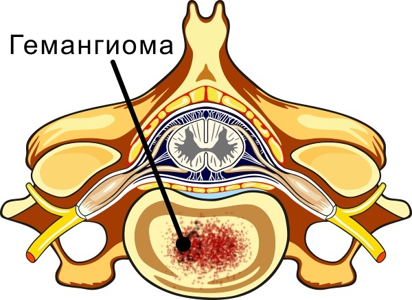
Vertebral hemangioma consists of many capillary vessels that are connected to the general circulatory system of the human body. Violation of the integrity of a benign neoplasm leads to the discovery of bleeding and injury to the spinal column.
In this regard, patients in whom the presence of gamangioma is detected on MRI are shown complete rest and minimal physical exertion on all elements of the musculoskeletal system. Otherwise, the risk of a compression fracture of the spinal column remains.
Even 30 years ago, benign neoplasms of this type could not be diagnosed using hardware equipment of that time, which led to an extensive proliferation of vascular tumors and the emergence of a large number complications.
Modern examination methods using MRI and CT equipment make it possible to identify tumor lesions of the vertebrae at the early stages of hemangioma formation. Timely removal of this neoplasm does not lead to disability of the patient, and also does not entail a long-term health disorder.
Record content:
- 1 Views
-
2 Stages and degrees
- 2.1 Proliferation
- 2.2 Stabilization
- 2.3 Spontaneous regression
- 3 Symptoms
- 4 Causes
- 5 Diagnostics
- 6 When to see a doctor
-
7 Treatment methods
-
7.1 Medications
- 7.1.1 Ibuklin
- 7.1.2 Pentalgin
- 7.1.3 Nurofen express
- 7.2 Traditional methods
- 7.3 Other methods
-
7.1 Medications
- 8 Possible complications
- 9 Video about spinal hemangioma
Views
Hemangioma of the vertebra on MRI is displayed as a dark spot of irregular shape, which can have different sizes depending on the stage and severity of the development of the disease. The table below describes the main types of this disease that are periodically encountered in medical practice.
| Types of vertebral hemangioma | Characteristics of pathology |
| Benign | Hemangioma of a benign type is the most common type of this ailment, which does not cause acute symptoms, pain attacks, or other discomfort. This type of tumor occurs in 97% of patients who, according to the results of the examination, showed signs of proliferation of vascular tissue in the region of the spinal column. |
| Malignant | Malignant hemangioma is a dangerous disease of the musculoskeletal system, which is extremely rare in medical practice. The degeneration of a tumor to a state of oncology is possible in the presence of concomitant diseases of the body, or the same prolonged exposure to negative factors directly on the vascular tissue in the circumference vertebra. |
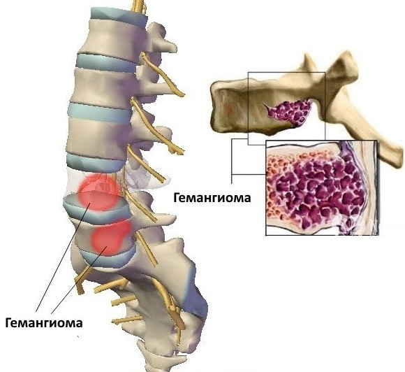
In most cases, the formation of a hemangioma, regardless of its nature of origin, occurs in the thoracic and lumbar spine. These parts of the body are the most vulnerable to tumor neoplasms of this type.
Stages and degrees
Vertebral hemangioma on MRI is detected within the first minutes from the beginning of the diagnostic examination process. As this formation progresses, there are 3 main stages of its development.
Proliferation
The proliferation stage is the period of the most active growth of vascular tissue, when the tumor gains mass and covers most of the affected vertebra. At this stage, a slow growth of the hemangioma with the addition of its volume up to 1 mm per year is possible, or a more active growth of this neoplasm.
 In the latter case, the risk of severe complications associated with impaired blood supply to the tissues of the spinal column, as well as a compression fracture of the spine, increases. A hemangioma found at the stage of proliferation must be removed as soon as possible.
Stabilization
The stage of stabilization of the growth of hemangioma is characterized by the complete cessation of the pathological process for the further growth of the tumor body. The patient may still not feel any symptoms of the disease, or he has the first signs of discomfort in the vertebra, which is surrounded by an excessive amount of vascular tissue.
At the stage of stabilization, benign hemangiomas respond well to surgical treatment with minimal risk of complications.
Spontaneous regression
The stage of spontaneous regression is characterized by the onset of the process of degenerative changes in the structure of the tumor. At this stage, the vascular tissue of the vertebral hemangioma begins to dissolve with a gradual decrease in its volume.
The cause of regression of an extraneous neoplasm of this type may be associated with the activation of the defense mechanism or increased activity of the body's immune system. Despite a partial reduction in tumor volumes, its complete disappearance without surgery is unlikely.
Symptoms
Vertebral hemangioma on MRI can be identified by a surgeon, traumatologist or neurologist who performs a diagnostic examination of the patient. In most cases, these tumors, which are of a benign origin, develop completely asymptomatic. This is the main danger of this neoplasm.
In rare cases, when the tumor reaches a sufficiently large size, patients begin to feel the following symptoms of the disease:
- a feeling of discomfort in the area of the vertebra, which is surrounded by an excessive amount of foreign vascular tissue;
- decreased mobility of the musculoskeletal system;
- numbness of epithelial tissues around the tumor;
- aching pain that appears after prolonged physical exertion;
- the presence of seals in the tissues that surround the vertebra affected by the hemangioma.
The most severe symptomatology of hemangioma is loss of sensitivity of the lower extremities or paralysis of other parts of the body, caused by a complication of the disease in the form of a spinal fracture. Similar signs of the disease appear at the later stages of tumor growth, the presence of which the patient did not suspect, or did not take proper response measures.
Causes
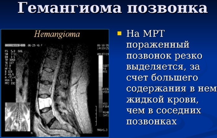
Hemangioma of the vertebra on MRI is clearly visible regardless of the part of the spinal column in which the main part of its tissues is concentrated. To date, there is no single scientific version of what exactly causes the growth of a tumor body of this type.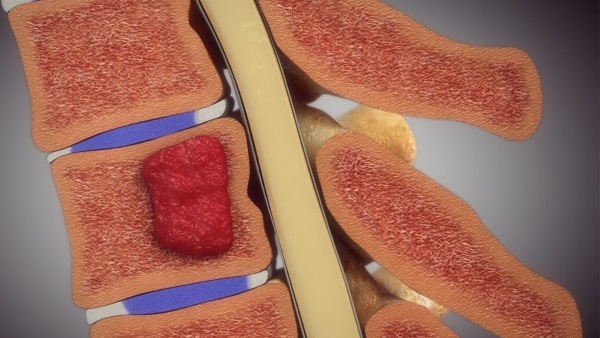
There are theories that the occurrence of hemangioma is associated with the negative influence of the following factors:
- hereditary predisposition to the appearance of neoplasms, consisting of an excessive amount of vascular tissue;
- previously suffered spinal injuries, the healing of which was allowed to take its course, and no methods of therapy were undertaken in relation to the patient;
- complications and negative consequences of surgical intervention, which was performed on the vertebrae of the thoracic or lumbar spine;
- hard physical labor, which is associated with daily weight lifting, creating loads on the spinal column;
- violation of local blood circulation, which leads to a nutritional deficiency of tissues located in the circumference of the spine;
- imbalance of sex hormones (according to medical statistics, in women with high estrogen levels, the appearance of spinal hemangiomas is recorded 5 times more often than in patients of other categories);
- concomitant diseases of the blood and hematopoietic system, which may be caused by other causative factors;
- the consequences of prolonged exposure to radioactive or other ionizing radiation that causes cell mutations and the manifestation of various kinds of genetic abnormalities;
- genetic failure, when at a certain stage of life a program of uncontrolled proliferation of tissues consisting of many blood vessels (in this case, hemangiomas can appear not only in the circumference of the vertebrae, but also cover other parts body).
Much less often, this disease is provoked by concomitant diseases of the spine in the form of chronic osteochondrosis, osteoporosis, scoliosis, kyphosis. In order for the therapy of tumor neoplasm to be as effective as possible, as well as minimized the risk of recurrence of the disease, it is recommended to establish the causal factors causing the appearance of a hemangioma.
Diagnostics
Detection of vertebral hemangioma requires a comprehensive examination of the entire spinal column. The table below shows all the methods of hardware and instrumental diagnostics that are indicated for use in relation to patients with signs of this disease.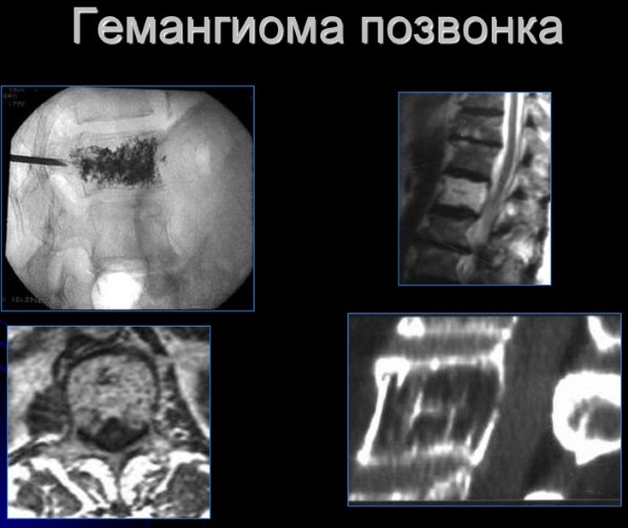
| Examination type | Diagnostic efficiency and average cost |
| MRI | With the help of MRI diagnostics, the primary definition and study of the tumor takes place. The localization of the gamangioma, as well as its location in relation to the affected vertebra, is established. The advantage of using this research method is the qualitative identification of the direct presence of an extraneous neoplasm. The disadvantage of MRI diagnostics is that the doctor receives information about the presence of a tumor, but does not have data about the degree of damage to the spinal column, and also knows nothing about the nature of an extraneous neoplasm. The average cost of an examination using MRI is 7,000 rubles. |
| CT scan | Computed tomography is the most informative diagnostic method, which is prescribed to be carried out immediately after examining the spinal column using MRI. The use of CT is advisable due to the fact that with the help of this method it is possible to maximally accurately measure the size of the tumor and assess the potential damage that was caused to the supporting ability of the vertebral pillar. The periodic use of CT diagnostics allows for effective control of the further growth of an extraneous neoplasm. For example, if the hemangioma is small and at this stage of its development, the use of surgical intervention is impractical. The average cost of CT for diagnosing a tumor of this type using a contrast agent is from 4000 to 7000 rubles. |
| X-ray | X-ray is one of the oldest, but at the same time proven methods of diagnosing the spinal column. This method of examination of the musculoskeletal system is necessary to confirm the presence of tumor and duplication of previous results obtained during the study with CT and MRI. The X-ray image shows the position of the tumor body and its approximate dimensions. In public hospitals, this diagnostic method is free. In a private clinic, its average cost varies between 600-800 rubles. |
| Spine scintigraphy | Spine scintigraphy is an innovative method for diagnosing tumor neoplasms, which allows you to determine not only the localization of the hemangioma and its size, but also the density of the vascular fabrics. The principle of this type of examination is that a liquid solution containing particles of radioactive substances is injected into the patient's vein. After the diagnostic medication has spread throughout the body, special sensors are used that pick up gamma radiation. Based on the results of the examination, an appropriate medical report is drawn up on the state of the tumor. The average cost of spinal column scintigraphy is 550 rubles. |
| Tissue biopsy | A biopsy of the hemangioma tissue is mandatory in order to confirm the fact of the benign or oncological nature of the origin of this neoplasm. The principle of this type of diagnostics is the performance of a small surgical operation. The doctor, being in the sterile conditions of the manipulation room, using special medical equipment, penetrates the structure of the hemangioma, and then selects a particle of its tissues. The collected biological material is transferred to a biochemical laboratory, where it is analyzed to determine the nature of the origin of cells. The average cost of a biopsy is 2,900 rubles. |
In the process of examining a patient, the attending physician independently decides on the use of one or another diagnostic method. To obtain the largest possible amount of information data on the state of an extraneous neoplasm in the form hemangiomas, which can disrupt the function of the vertebra, are used at once by all types of hardware and laboratory research.
When to see a doctor
An appeal to a doctor should take place immediately, as soon as a person has discovered the first signs of an extraneous neoplasm in the thoracic, lumbar or any other part of the spine.
Delay in a visit to a doctor can lead to the development of negative complications, the most severe of which is the loss of the ability to move independently and the onset of lifelong disability.
Treatment methods
The main method of treating vertebral hemangioma is performing a surgical operation to eliminate the excess amount of vascular tissue. Drug therapy can be used as an auxiliary element to achieve the effect of pain relief in the postoperative period.
Patients who have a vertebral hemangioma are categorically contraindicated to perform physiotherapy exercises, as well as to carry out various kinds of physiotherapeutic procedures.
Massage is also absolutely prohibited. These limitations are justified by the significant likelihood of an even greater decrease in the functions of the spinal column with the onset of its pathological fracture.
Medications
During the treatment of hemangioma, drugs can be used, but only for the purpose symptomatic therapy to eliminate pain syndrome, which manifests itself after surgical operation. In this case, analgesic medications are used, which also have a mild anti-inflammatory effect.
Ibuklin
Ibuklin is a pain reliever that comes in the form of tablets coated with a protective layer. This medication contains 400 mg of ibuprofen and 325 mg of paracetamol. Ibuklin has a pronounced analgesic and antispasmodic effect. The drug is prescribed for oral administration, 1 tablet up to 3 times a day after meals.

The maximum duration of a therapeutic course should not exceed 5 days. Ibuklin is contraindicated in women who are pregnant and breastfeeding. The active substances of the medication are not compatible with ethyl alcohol. The average cost of the drug Ibuklin is 142 rubles. for 10 tab.
Pentalgin
Pentalgin is a drug that has a powerful analgesic formula, which includes several active components at once, namely:
- Paracetamol - 325 mg;
- Pheniramine maleate - 10 mg;
- Caffeine anhydrous - 50 mg;
- Naproxen 100 mg
- Drotaverine hydrochloride at a dosage of 40 mg.
This drug has not only an analgesic effect, but also acts as a safe antispasmodic, eliminating hypertonicity of smooth muscles. Pentalgin is recommended to be taken in the first 3 days after the operation to remove the hemangioma.
Longer therapy with this medication is fraught with the development of side effects. The drug is taken 1-3 tablets a day immediately after a meal with a sufficient amount of liquid. The average price of the drug Pentalgin is 177 rubles. for 24 tab.
Nurofen express
Nurofen express is a gelatin capsule with a dosage of 200 mg of the active substance, which is the anti-inflammatory and analgesic ibuprofen. This drug is characterized by rapid absorption, which occurs in the intestinal cavity.
Nurofen express is contraindicated in people who have an individual intolerance to its main component in the form of ibuprofen. Taking this medication is shown in a dosage of 1 capsule 3-4 times a day.
At the same time, it is forbidden to chew the medication, or break its protective shell in any other way. The course of treatment with the drug is from 2 to 3 days, and its average cost is 225 rubles. for 16 capsules.
Traditional methods
Alternative methods of treating vertebral hemangioma are ineffective, since this disease requires surgical intervention.
Other methods
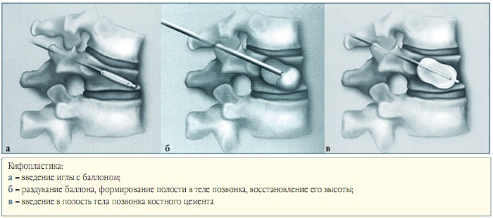
The main method of hemangioma treatment is surgery, which is performed in the spinal unit of the neurosurgical department. Modern technologies make it possible to remove tumors of this type using minimally invasive surgery.
Surgical intervention takes place in a sterile operating room with the use of local anesthesia. The patient is fully conscious all the time.
A neurosurgeon, using special medical instruments and equipment, performs vertebroplasty the affected vertebra with the removal of an excess amount of vascular tissue that affected a separate segment of the vertebral pillar.
To carry out these actions, it is enough for the doctor to perform several skin punctures and a small incision in the area of surgery. The average duration of vertebroplasty is 40 minutes. up to 2 hours, depending on the complexity of the clinical case and the degree of growth of an extraneous neoplasm.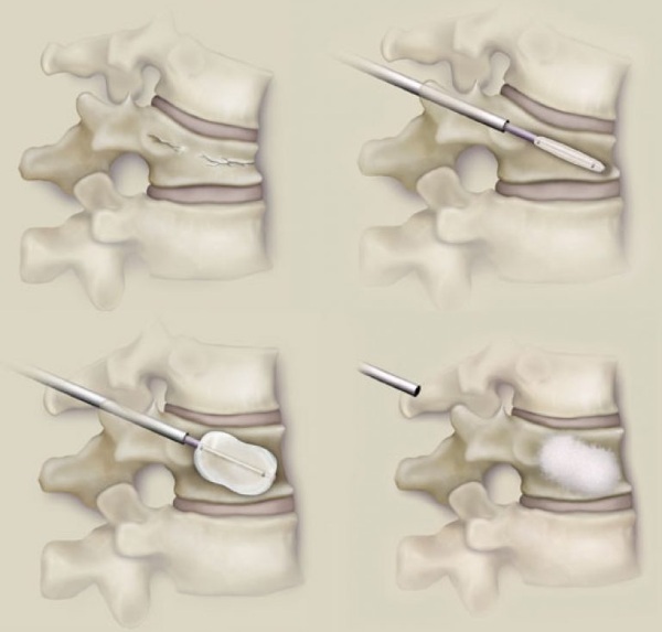
A particle of the removed hemangioma is immediately sent for histological examination to check its cells for degeneration and latent oncological process. The patient's recovery period lasts no more than 2 days. After a specified period of time, the patient can return to his usual way of life without the risk of negative consequences.
Possible complications
Lack of timely access to a doctor, or delay in performing a surgical operation to remove a hemangioma can lead to the development of the following complications and negative consequences for the body:
- loss of sensitivity of the lower extremities;
- compression fracture of the vertebra;
- chronic pain in the area of the affected part of the spinal column;
- violation of the process of urination and defecation;
- erectile dysfunction in men;
- paralysis of various parts of the body.
Vertebral hemangioma is a tumor lesion of the spinal column, which is characterized by a latent course with minimal manifestation of pathological symptoms. The main danger of this ailment is that the first signs of a functional disorder musculoskeletal system occur at the moment when an extraneous neoplasm has already reached critical dimensions.
To be examined for lesions of the spine with hemangioma, it is necessary to undergo MRI diagnostics, which displays the location of the tumor and also gives the doctor basic information about the health status the patient. The only effective treatment for this disease is the surgical removal of the extraneous neoplasm.
Video about spinal hemangioma
Doctor about spinal hemangioma: