
Diseases of the gastrointestinal tract in humans it is difficult to diagnose, since the anatomy of these organs is rather complicated.
Record content:
- 1 Human gastrointestinal tract anatomy
-
2 The structure and functions of the gastrointestinal tract
- 2.1 Oral cavity
- 2.2 Pharynx
- 2.3 Esophagus
- 2.4 Stomach
- 2.5 Intestines
- 2.6 Anal hole
-
3 Subsidiary bodies
- 3.1 Liver
- 3.2 Gall bladder
- 3.3 Glands
- 3.4 Ducts
- 3.5 The walls of the gastrointestinal tract
- 4 How does digestion and excretion take place?
- 5 Development of the digestive system
-
6 Diseases of the gastrointestinal tract and their symptoms
- 6.1 Stomatitis
- 6.2 Esophagitis
- 6.3 Gastritis
- 6.4 Duodenitis
- 6.5 Cholecystitis
- 6.6 Biliary dyskinesia
- 6.7 Pancreatitis
- 6.8 Enteritis
- 6.9 Colitis
- 6.10 Proctitis
- 6.11 Other diseases
- 7 Which doctors specialize in tract work?
- 8 Diagnostic methods
- 9 Treatment
- 10 Video about the gastrointestinal tract
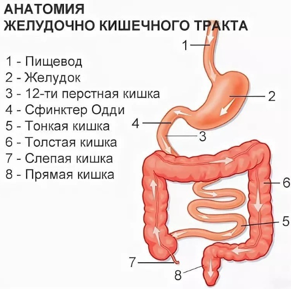
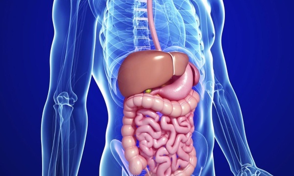
Human gastrointestinal tract anatomy
The human gastrointestinal tract (its anatomy is a collection of organs designed to carry out the digestion process) has an average length of 8 to 10 m.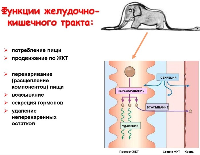
The gastrointestinal tract performs the following functions:
- Motor. Consists in the movement of food through the organs of the tract with the help of muscles.
- Secretory. It is the production of juices by glandular cells.
- Suction function. It is the process of penetration of nutrients and fluids into the bloodstream.
- Excretory (excretory).
- Endocrine (the formation of various hormones - gastrin, secretin).
The structure and functions of the gastrointestinal tract
Different parts of the gastrointestinal tract have different structures and perform strictly defined functions.
Oral cavity
In front, the mouth is covered with lips. On the sides, it is bordered by the cheeks, from above it is separated by a hard palate. Behind is the pharynx, which leads to the pharynx.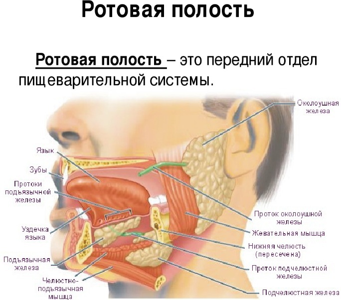
The functions of the oral cavity are food chopping as well as enzymatic function.
Pharynx
It is an unpaired organ that looks like a funnel. The pharynx is located between the mouth and the esophagus. In front of the pharynx is the larynx, nasal cavity and oral cavity. It is attached from above to the occipital bone. The upper part of the pharynx is called the vault. The pharynx is divided into the nasopharynx, the oropharynx, and the hypopharynx.
Esophagus
It is a thin muscular tube. In the cervical region, it is narrowed, then gradually begins to expand, passing into the abdominal part. The wall of the esophagus is lined with mucous membrane, submucosa and muscles.
Stomach
In its structure, it has 2 walls (back and front) and 2 holes. A large and small curvature is formed along the back wall of the stomach. The stomach has a cardiac opening through which it communicates with the esophagus. The second exit is the gatekeeper's area.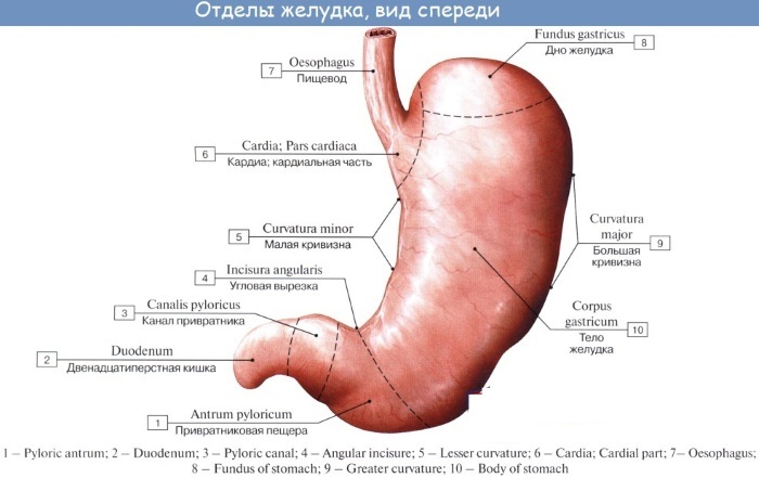
The convex opening of the stomach is represented by the fundus of the stomach. The body of the organ is located between the bottom and the gatekeeper.
The wall of the stomach is formed by a mucous membrane that forms folds. On the mucous membrane there are pits in which the glands pass. The stomach is covered with mucous, submucous, muscle layers and serous membrane.
Intestines
The human gastrointestinal tract (the anatomy includes the entire intestine) is complex. The small intestine is represented by the blind, colon, sigmoid and rectum. The small intestine consists of the duodenum, small intestine and jejunum.
Anal hole
The anus is the final anatomical formation of the digestive tract. It is surrounded by sphincters, which are formed from muscle fibers: the inner one, which is formed by smooth muscles, and the outer one, which consists of striated muscles.
Subsidiary bodies
In addition to the main ones that are directly part of the digestive system, auxiliary organs are endowed with an important role.
Liver
The liver is the largest gland in the human body. It is located in the right hypochondrium, in the abdominal cavity. It is divided into 2 lobes, which are separated from each other by a falciform ligament.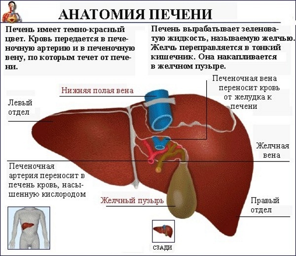
The liver is covered with a peritoneum and fibrous membrane. It is formed by hepatocytes (cells), which are involved in the formation of bile. Large nerves and blood vessels pass through the liver.
Gall bladder
The gallbladder is located at the back of the liver and is connected by a duct. In its shape, this organ resembles a pear. The gallbladder performs the function of producing bile.
Glands
The whole group is formed by the glands of the intestine, stomach, salivary glands, liver and pancreas. The largest are the pancreas and liver. Small ones are located in the mucous layer of the intestine and stomach. They are involved in the digestion and absorption of food elements.
Ducts
The gastrointestinal tract ducts are intrahepatic and extrahepatic bile ducts that merge into one hepatic. Subsequently, it connects to the cystic duct and they form a common bile duct.
With the help of the ducts, bile is able to circulate throughout the body. The sphincter of Oddi is responsible for the regulation of its intake.
The walls of the gastrointestinal tract
The walls of the gastrointestinal tract are represented by 3 layers. From the inside, they are covered with a mucous membrane, the middle layer is muscle fibers, and the outside of the tract is lined with a serous layer.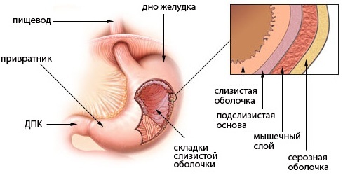
- The mucous membrane is formed by the epithelium.
- The muscular layer is represented by striated muscles and smooth muscle fibers.
- The serous layer is composed of connective tissue. It has a protective function.
How does digestion and excretion take place?
When food enters the mouth, the digestion process begins. The teeth are crushed to the smallest particles, moistened with saliva, which contains enzymes. Further, food is moved by swallowing into the esophagus, and then into the stomach.
In the stomach, food begins to break down under the influence of hydrochloric acid and enzymes. Having broken down to proteins, it moves further into the intestine already in a liquid state. In the small intestine, the process of splitting with the help of enzymes continues.
Everything else goes to the large intestine. In it, the process of fluid absorption and the formation of feces takes place. Subsequently, they are excreted from the body during bowel movements.
Development of the digestive system
The development of the digestive system begins in the embryonic period, at 4 weeks of intrauterine development. The future digestive system develops from the intestinal tube. It is attached with one side to the yolk sac.
Subsequently, it develops and is divided into the following departments:
- Primary intestinal tube.
- Anterior section.
- Middle department.
- Back section.
Diseases of the gastrointestinal tract and their symptoms
The human gastrointestinal tract is highly susceptible to the development of pathological changes. Both adults and children are exposed to them. The latter suffer more often from inflammatory diseases, since their anatomy contributes to this.
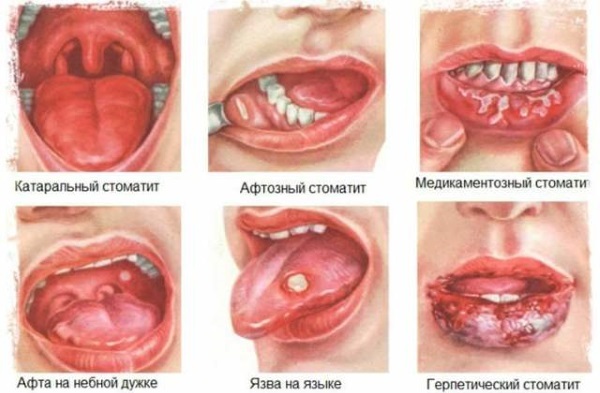
Stomatitis
One of the types of gastrointestinal diseases is stomatitis. It is an inflammatory disease of the oral mucosa.
There are several types of stomatitis:
- Viral. The main cause is various viral diseases. For example, "childhood infections" (measles, rubella, scarlet fever), herpes viruses, including varicella-zoster virus, herpes virus, and cytomegalovirus. If the stomatitis is caused by the herpes virus, it will be called herpetic.
- Bacterial. This type of stomatitis is caused by staphylococci and streptococci. Most often it occurs in people with chronic foci of infection in the oral cavity and pharynx (caries, chronic tonsillitis), as well as in children, mainly due to lack of hygiene.
- Aphthous. The etiological factors of this type of stomatitis are not fully understood.
- Allergic. Is a manifestation of any allergic reaction.
-
Candidal stomatitis. The cause of its development is a fungus. Most often, it occurs against the background of immunodeficiency (with chronic diseases or after past diseases). There is a frequent occurrence of stomatitis in people undergoing chemotherapy.
Clinical symptoms of stomatitis include the following manifestations:
- Pain in the mouth. The pain syndrome can be so severe that the person cannot speak, drink or eat. In infants, there is a rejection of the breast and any fluid. And also in children, fever is possible with a rise in temperature up to 38 ° C and an increase in the submandibular lymph nodes.
- Rash. With the development of herpetic stomatitis, a papular rash filled with serous contents is observed, after a while it opens and ulcers form on the mucous membrane. With aphthous stomatitis, ulcers (aphthae) are found on the mucous membrane, which after a while scar and disappear. With other types of stomatitis (bacterial and allergic), rashes in the form of red spots are possible.
- Hyperemia of the oral mucosa. On examination, the mucous membrane is hyperemic, edematous.
- White plaque on the mucous membrane. With candidal stomatitis, white plaque is observed on the inner surface of the cheeks, gums, and hard palate.
- Fever (mostly in children).
- Profuse salivation.
Esophagitis
Esophagitis is an inflammation of the lining of the esophagus. It can be acute or chronic. More often, esophagitis is a manifestation of other diseases of the gastrointestinal tract.
Symptoms of pathology include:
- Chest pain. The patient complains of a burning sensation in the esophagus. The pain syndrome increases when food is swallowed and goes away at rest.
- Heartburn. It occurs when hydrochloric acid is thrown into the esophagus, due to reflux.
- Nausea and vomiting. With trauma to the esophagus, vomiting occurs with an admixture of fresh blood.
- Increased body temperature.
- Dysphagia. The patient has difficulty swallowing food or liquids.
Gastritis
Gastritis is an inflammation of the stomach lining. It arises against the background of a variety of reasons. The development of the disease may be associated with nutritional disorders, Helicobacter pylori bacteria, and medication. Gastritis is common in people undergoing chemotherapy or radiation therapy. In this case, the disease is called "radiation gastritis".
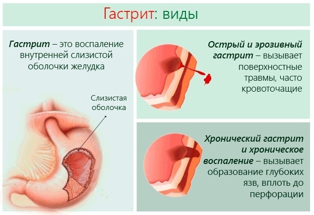
Acute and chronic forms of the disease are distinguished along the course. Erosive and non-erosive gastritis is also distinguished, with increased or decreased acidity. Depending on the type of pathology, clinical manifestations will differ.
In an acute course, the symptoms run brightly, causing the patient serious discomfort. With a chronic illness, the patient will experience only minor disturbances in well-being.
Erosive gastritis occurs with the occurrence of erosions on the wall of the stomach. This condition very often turns into a peptic ulcer, which is fraught with the development of complications, including gastric bleeding.
Symptoms of gastritis:
- Pain in the epigastric region. Usually has a mild character. With a chronic course, periodic dull pains are possible.
- Nausea.
- Heartburn, sour belching.
- Dyspeptic disorders. Diarrhea is observed. The stool may be watery and foul-smelling, but there is no increase in body temperature.
- Loss of appetite and a feeling of fullness in the stomach.
Duodenitis
Duodenitis manifests itself in inflammatory processes that involve the mucous membrane of the duodenum. It rarely occurs as a separate disease. Most often, the pathology manifests itself in conjunction with peptic ulcer, as well as with the development of intestinal infections.
Sources of duodenitis are many factors:
- Intestinal toxic infections.
- Acute and chronic diseases of the gastrointestinal tract.
- Alcohol abuse.
- Eating fatty foods.
Distinguish between catarrhal, phlegmonous and ulcerative duodenitis. The most dangerous is phlegmonous, it can lead to the development of serious complications, including peritonitis (inflammation of the peritoneum).
Duodenitis is manifested by vomiting, abdominal pain, pain on palpation is possible. There is weakness, lethargy, fever, pale skin.
Cholecystitis
Cholecystitis is an inflammation of the gallbladder due to blockage of its ducts with a stone. This pathology is a consequence of cholelithiasis, but it can also be an independent disease in various infections, immunodeficiency states, and tumors. Such cholecystitis is called non-calculous.
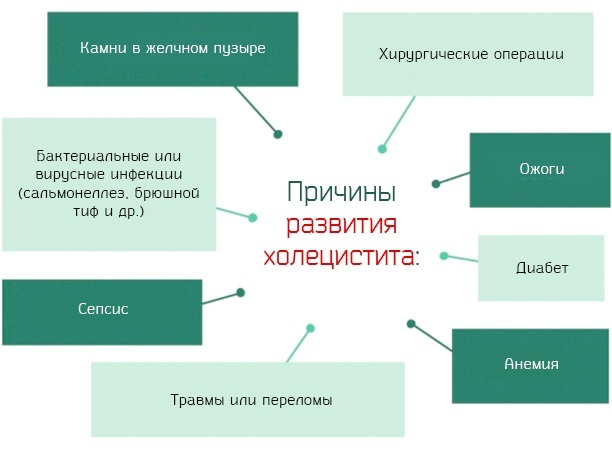
Clinical manifestations are:
- Pain syndrome in the right hypochondrium. The pain is similar to that of biliary colic but lasts much longer. Plus, after a few hours, Murphy's syndrome develops (during inhalation, palpation in the right hypochondrium, spontaneous cessation of breathing is noted).
- Attacks of nausea and vomiting.
- Bitter taste in the mouth.
- Increase in body temperature up to 38 ° C.
- Loss of appetite.
- Diarrhea.
How strongly the clinical manifestations of the disease will be expressed depends on the course of cholecystitis and its type. So, with phlegmonous or gangrenous cholecystitis, a pronounced fever (up to 40 ° C), irritation of the peritoneum, and severe intoxication of the body are possible. With this development of the disease, complications may arise.
In the chronic course of cholecystitis, short-term pain in the right hypochondrium, decreased appetite, weight loss, yellowness of the skin and mucous membranes are possible.
Biliary dyskinesia
Dyskinesia of the biliary tract is a violation of motility in the work of the gallbladder, which manifests itself in a violation of the outflow of bile. The causes of occurrence are disorders of the autonomic nervous system, pathological changes in the gallbladder.
Distinguish between hypokinetic and hyperkinetic forms of the disease. In the first type, the motor function is reduced, and in the second, on the contrary, it is increased.
Clinical manifestations are:
- Pain syndrome in the epigastric region with irradiation to the right hypochondrium. An attack of pain occurs after eating or increasing physical activity. Has the character of dull periodic pain.
- Nausea, sometimes vomiting, bitterness in the mouth.
- Icteric staining of the skin. This symptom is not observed in all cases of the development of pathology.
Pancreatitis
Pancreatitis is inflammation of the pancreas. Often, recurrence of acute pancreatitis occurs from foods with high fat content. And also the development of inflammation of the pancreas can provoke injury, cancer and poisoning.
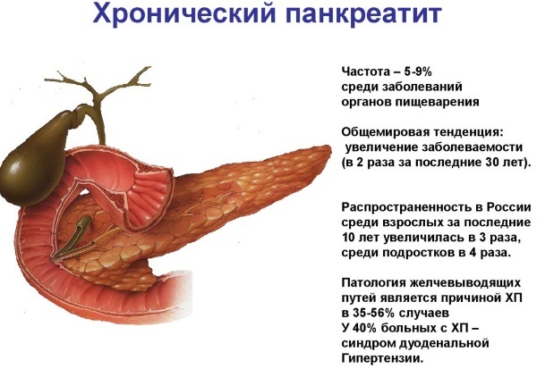
Clinical manifestations are:
- Pain syndrome. The pain is localized at the beginning of the attack in the epigastric region, then takes on the character of a girdle.
- Increased body temperature.
- Vomiting of bile, not relieving.
- Yellowness of the skin (may not be).
Pancreatitis is dangerous for its complications, namely peritonitis. With peritonitis, there is a sharp deterioration in the patient's well-being, an increase in the symptoms of intoxication of the body. If within 24 hours. do not provide such a patient with medical care, death is possible.
Enteritis
Enteritis is an inflammation of the mucous membrane of the small intestine. Distinguish between acute and chronic course of the disease. Acute occurs in infectious diseases (salmonellosis, typhoid, cholera), food poisoning.
Chronic enteritis occurs with certain intestinal diseases (Crohn's disease, irritable bowel syndrome), with tumors, improper diet, taking certain medications, as well as infection with lamblia and others parasites.
Clinical picture:
- Abdominal pain.
- Diarrhea.
- Vomiting and nausea.
- Temperature increase.
- Weakness.
In a chronic course, as a rule, the symptoms are latent. The patient may be disturbed by mild pain in the abdomen, loose stools.
Colitis
Colitis is considered an inflammation of the lining of the large intestine. There are acute and chronic colitis. Acute colitis occurs in infectious diseases (dysentery), poisoning, diseases of the digestive system, and in some autoimmune diseases.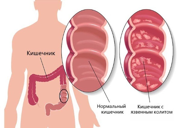
Colitis symptoms:
- Stomach ache.
- Flatulence. And also the patient has a constant "rumbling" in the abdominal cavity.
- Diarrhea. In an acute course, frequent bowel movements, tenesmus (false urge to defecate) are observed. In a chronic course, stool can be observed up to 5 times a day. Most often, bowel movements occur after eating.
- Pallor of the skin. And also there is dryness of the skin, the appearance of cracks. This is due to impaired absorption of vitamins into the body.
- Increased body temperature in acute course.
Proctitis
Proctitis refers to inflammatory diseases of the rectum. It occurs against the background of inflammatory bowel diseases, infectious, radiation exposure to the rectum.
With the development of the disease, the patient complains of a constant urge to defecate. However, they are false and defecation does not occur. Blood or mucus can be seen in the stool. In some cases, the patient may feel severe pain in the rectum and anus.
Other diseases
Other diseases of the digestive system include:
-
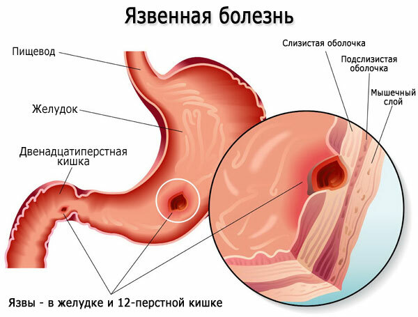
Stomach ulcer (this is a chronic disease characterized by the formation of ulcers on the stomach lining). Symptoms of the disease are pain in the epigastric region, heartburn, "hunger pains". The ulcer is dangerous for its complications. Namely, bleeding, peritonitis and degeneration into cancer of the stomach.
- Cirrhosis Is a chronic liver disease characterized by its gradual destruction. This disease often occurs against the background of alcohol abuse, viral hepatitis, cancer, and some autoimmune diseases.
- Haemorrhoids. It is a varicose veins of the hemorrhoidal plexus. It occurs after childbirth or during pregnancy, due to heavy lifting and excessive straining during bowel movements. Symptoms are the presence of blood in the stool, pain in the anus.
Which doctors specialize in tract work?
Doctors - gastroenterologists are engaged in diagnostics and treatment of gastrointestinal diseases. And also when infectious diseases of the gastrointestinal tract are detected, infectious disease doctors are involved in the treatment of such patients. And in case of liver pathology, doctors - hepatologists will deal with diagnostics and treatment.
Diagnostic methods
The human gastrointestinal tract (its anatomy has a complex structure) for any pathology requires specific diagnostics:
- EFGDS (esophagogastroduodenoscopy) - This study is an examination of the esophagus, stomach and duodenum using an endoscope. This method is used for suspected esophagitis, gastritis, ulcers, tumors. With its help, you can assess the state of the mucous membrane and identify the slightest deviation from the norm.
-
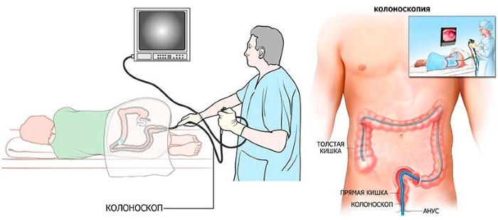
Colonoscopy - This is an endoscopic examination that allows you to examine the mucous membrane of the colon. With the help of it, you can assess the condition of the intestines, see if there is inflammation, neoplasms and other defects. And also with the help of a colonoscopy, you can conduct a diagnostic biopsy.
- Sigmoidoscopy Is a method in which a doctor examines the rectum.
- Irrigoscopy - This is an x-ray examination of the large intestine. It is carried out using a contrast agent. This method is used when the results of colonoscopy are questionable or when endoscopic examination is impossible.
- Fluoroscopy of the stomach. Allows you to determine the presence of tumors, hiatal hernias, stenosis.
- Ultrasound.
Treatment
The human gastrointestinal tract, the anatomy of which is represented by complex structures, requires complex treatment:
| Principles | Methods |
| Medicines |
|
| Diet |
|
| Surgical intervention | For diseases that do not respond to conservative treatment or carry a risk of complications. |
The effectiveness of diagnosing diseases of the human gastrointestinal tract directly depends on the correct understanding of its anatomy.
Video about the gastrointestinal tract
Digestive system:


