
MRI allows you to diagnose congenital and acquired defects in the sacral regioniliac joint at an early stage. But for it to be correct, you need to know how to prepare for the examination.
Record content:
- 1 What is diagnostics?
- 2 What does the scan show?
- 3 Diagnostic benefits
- 4 Indications for the procedure
- 5 Basic limitations
- 6 Preparation for research
- 7 Diagnostic steps
- 8 Suppression of the signal from fat
- 9 MRI for children
- 10 Where to make and what does the cost depend on?
- 11 MRI video
What is diagnostics?
The sacroiliac joint is a paired joint that connects the ilium and the lateral sacrum. It is considered a tight joint.
Diseases that affect this part of the musculoskeletal system are rarely diagnosed. Therefore, the procedure for examining this section is carried out during the study of the bones of the pelvis and the lower part of the spinal column. The doctor can refer the patient to examinations of this particular joint if he has signs indicating pathology of this part of the musculoskeletal system.
MRI is a safe and painless method for diagnosing pathologies of the musculoskeletal system, which was developed by Peter Mansfield and Paul Lauterbur in 2004. For this they received the Nobel Prize.
The essence of the method is that bones and adjacent soft tissues are studied using a magnetic field.
The person is placed on the tomograph table, which can be open or closed. During the examination, the patient must be motionless. This allows you to get a high-contrast, high-quality image.
To conduct a deeper diagnosis, MRI can be performed using a contrast agent. For this purpose, medications based on iodine or gadolinium are used.
What does the scan show?
An MRI of the sacroiliac joints may show:
- inflammatory process in the spinal cord, joints and intervertebral discs;
- expansion of the joint space;
- neoplasms;
- bone growths;
- injuries and diseases of the locomotor system;
- deposition of calcium in the musculoskeletal system;
- osteochondrosis;
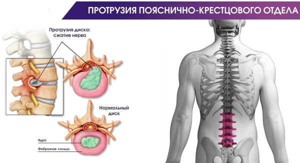
- protrusion and intervertebral hernia;
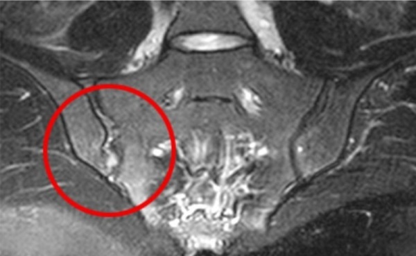
MRI. Edema of the sacroiliac joint - pinching of the nerve endings of the spinal cord;
- multiple sclerosis;
- vascular pathology;
- lumbarization of the vertebrae.
Diagnostic benefits
The advantages of MRI include the following:
- non-invasiveness, during the procedure, the integrity of the skin is not violated;
- the possibility of early detection of pathological changes;
- harmlessness, so it can be carried out as often as necessary.
When comparing MRI with other diagnostic methods, such as CT or ultrasound, then each method has its pros and cons.
With the help of MRI, it is better to examine soft tissues and assess the condition of the muscles of the tendon-ligamentous apparatus, while CT allows you to determine whether the bones are healthy.
When a doctor suspects a fracture, joint inflammation, or bone growth, computed tomography is the preferred choice. But it should be borne in mind that CT is a more harmful diagnostic method, since X-rays are used during the procedure, so the procedure can be carried out every six months. In addition, computed tomography is contraindicated for children and women in position.
Another safe diagnostic method is ultrasound, it can also be done often, it is allowed for children and patients who are expecting a baby. But in comparison with MRI, the technique is less informative and does not always allow detecting the disease at an early stage.
But ultrasound can be done at any stage of pregnancy, while MRI is contraindicated in the first trimester (but if there are diseases that threaten life, then the gynecologist may decide to apply the technique in the first 3 months pregnancy).
Often, to identify pathologies of the sacroiliac joint, an x-ray is prescribed, but on an x-ray the picture does not always show the affected joint in detail and it can be problematic for the doctor to detect defects in this area. This is due to the structural features of the pelvic bones, due to which the shadows of different bone structures are superimposed.
And if with the help of an X-ray it is possible to see well injuries of the pelvic region and sacroiliac joints, then it is problematic to identify arthrosis or inflammation.
It is impossible to say unequivocally which of these methods is better. Therefore, if necessary, the doctor can simultaneously prescribe several diagnostic methods.
Indications for the procedure
Indications for MRI may be as follows:
- deterioration of flexibility and mobility of the spinal column;
- injuries of the lower spine and pelvic bones;
- back pain, which causes an increase in the load on the joints and a decrease in their mobility;
- arthritis of the legs, especially inflammation of the ankle;
- osteochondrosis, due to which pain appears, there are lumbago in the neck and lower back, the load on the joints increases;
- infections of the pelvic organs;
- pain in the lower abdomen and in the region of the sacrum, radiating to the lower extremities;
- unexpected attacks of lameness that occur after prolonged sitting or physical exertion;
- malignant neoplasms giving metastases;
- inflammation of the musculoskeletal system in this area;
- ankylosing spondylitis;
- sacroiliitis;
- multiple sclerosis;
- hereditary predisposition to the appearance of the HLA-B27 gene;
- rheumatoid arthritis;
- pain in the lower back of unclear etiology;
- arthrosis;
- Crohn's disease, in which there is pain in the pelvis, ulcerative colitis;
- a foreign object in the area of the sacroiliac joint, which is found on an X-ray;
- osteophytes and exostoses;
- protrusion and intervertebral hernia;
- vascular pathologies in the region of the spine.
The study is carried out in order to track the disease in dynamics, to monitor the effectiveness of conservative therapy. It is prescribed before and after surgery.
MRI of the sacroiliac joints of this area is also performed if a person is experiencing excessive physical exertion on this area.
Basic limitations
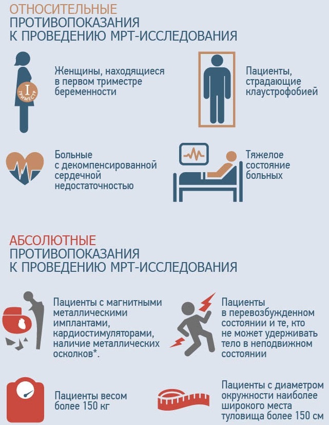
MRI for some groups of patients with defects of the sacroiliac joints is contraindicated:
- The presence of pacemakers, hemostatic clips or insulin pump. The fact is that the magnetic field can cause malfunction of these devices.
- The presence of any metal objects in the patient's body that cannot be removed, for example, fragments, metal pins, as they can become hot and cause burns.
- Tattoos made with inks containing ferromagnetic particles.
- 1st trimester of pregnancy, when the internal organs of the fetus are laid.
In addition, there are relative contraindications to the appointment of MRI, when it is possible under certain conditions:
- Claustrophobia. If a person has a fear of confined spaces, then the procedure is recommended using open-type tomographs.
- A person cannot lie still during an MRI scan, which is why the image on the images will be indistinct and it will be impossible to determine the presence or absence of pathology from them. In this case, before the procedure, the patient is given medications that have a sedative effect or the examination is carried out under general anesthesia.
- The body weight of a person is greater than the weight for which the tomograph table is designed. If there is such a problem, it is necessary to select equipment that is designed for a greater weight. For obese people, open scanners are usually recommended, as they tend to hold more weight. There are tomographs that are not designed for weighing more than 130-150 kg.
Also, on an individual basis, the doctor must decide whether an MRI can be performed if the patient is diagnosed with the following health problems:
- heart failure in the stage of decompensation;
- serious condition of a person;
- inappropriate behavior, for example, the patient is in a state of alcoholic or drug intoxication.
A relative contraindication to the procedure is the presence of an intrauterine device in a woman's body.
MRI with contrast should not be performed on women in a position and supporting breastfeeding, children who are not who have reached the age of 12, as well as patients suffering from renal failure or allergy to the injected medication. MRI with iodine-containing contrast agents should not be performed with iodine and seafood intolerance.
If the patient has diabetes mellitus, and he takes anti-hypoglycemic medications in tablets, then before magnetic resonance imaging with a contrast agent should be consulted with endocrinologist.
Women who are breastfeeding, after examination with contrast medication, need to express milk twice, and breastfeeding can be resumed only a day after the MRI.
Preparation for research
MRI of the sacroiliac joints does not require special training. At the same time, a number of rules should be observed that must be observed when preparing for the examination.
You do not need to follow any diet before the study, limit fluid intake, refuse to take medications and physical activity, that is, a person can lead a normal Lifestyle.
If an MRI is performed using a contrast agent, then before the procedure, it is necessary to check if there is any allergy to it.
Before you go to the examination, you need to:
- To get rid of all metal elements on clothes, in some hospitals they give special disposable pajamas for an MRI scan. If you have your own clothes, then it can be pajamas, a tunic, a robe with ties.
- Remove all metal jewelry, glasses and watches.
- Leave outside the office, where the diagnostics will take place, plastic cards, cell phones, other electronic devices and devices, as they may be damaged by a magnetic field.
- Women are advised to remove all makeup, as some products may contain metal particles.
- The doctor should be informed about the tattoos on the body, as they can be made with metal-containing paints, after the procedure, skin irritation may appear at the site of the tattoo.
In some medical centers, the patient may be checked with a metal detector before the procedure to detect metal particles.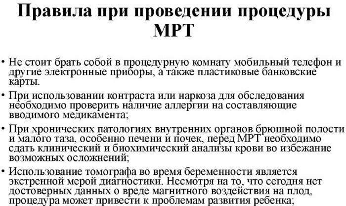
It is necessary to take documents with you to the MRI, such as:
- a referral from a doctor for an MRI (if you wish, you can sign up for examinations without a referral from a doctor);
- medical card;
- the results of previous examinations, if any.
If the patient is to undergo an MRI scan with a contracted substance, then a creatine test is needed, which was passed no later than 14 days ago.
Diagnostic steps
MRI of the sacroiliac joints is performed as follows:
- Before the procedure, a person needs to remove all metal objects from himself.
- Then the patient needs to lie down on the tomograph table, which, together with the person, is rolled into the rotating element of the device. In this case, the area from which the magnetic radiation emanates must be inside the apparatus.
- Throughout the entire procedure, the person must lie motionless in order to obtain clearer and higher-quality images, allowing an accurate diagnosis to be made.
- After receiving the images, the doctor decides whether to inject a contrast agent in order to obtain a clearer image, allowing to assess the condition of the musculoskeletal system.
- When carrying out diagnostics, a person should not have unpleasant sensations, but the tomograph makes sounds. To keep them out of the way, the patient can insert earplugs into the ear canal. To make the person comfortable during the procedure, you can take someone close to you. If an MRI is performed on a child, then the presence of the parents is mandatory.
- As a rule, the procedure lasts from half an hour to 1 hour in time. The time depends on the size of the study area and on the need to inject a contrast agent.
- After the procedure, a person can go home immediately.
- The patient receives the finished pictures with the conclusion in his hands. Usually they are ready within an hour, but if contrast enhancement was used, this time can be lengthened. In this case, the results will be ready the next day. Most paid medical centers send diagnostic results by email.
When a person decides to undergo an MRI for himself without a referral from a doctor, then either a rheumatologist or a traumatologist can make a diagnosis.
Contrast enhancement
The contrast consists of isotonic sodium solution and a special drug that is administered intravenously. With the blood flow, it is delivered to the research site.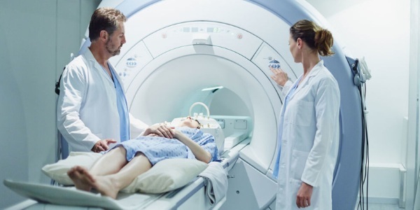
Under the influence of a magnetic field, the contrast agent in the pathological area is "highlighted", as a result a clear image is obtained that allows one to examine the shape and size of the defect. Such a study helps well to consider neoplasms, blood clots, hematomas, foci in which the inflammatory process is taking place.
Suppression of the signal from fat
When conducting an MRI, you cannot do without methods that allow you to suppress the signal from fat. If this is not done, then it may cause a diagnostic error.
There are several methods of fat suppression, of which the doctor chooses the most optimal, depending on the capabilities of the tomograph.
MRI for children
Magnetic resonance imaging has no age restrictions (although some experts do not recommend such an examination for patients under 5-7 years of age).
During the MRI process, you need to lie still for 15-30 minutes, which is not always within the power of the child, therefore, the procedure can be carried out for children under anesthesia.
Also, many young patients are afraid of confined spaces, so they are advised to carry out diagnostics using open-type tomographs. During the procedure, a parent can hold the child's hand.
Survey result
The entire examination process is recorded on a laser disk. The results of an MRI are a series of images, according to which the doctor writes a description, in which he lists the structures where the pathology was found. In the conclusion, the doctor writes what disease the identified changes correspond to.
According to the results of the examination, symptoms of pathologies such as:
- Sacroiliitis (inflammation of the sacroiliac joint). It can be either an independent disease or a symptom of ankylosing spondylitis.
- Degenerative-dystrophic processes in the spinal column and intervertebral discs.
- Musculoskeletal system injuries.
- Neoplasms and metastases in this area.
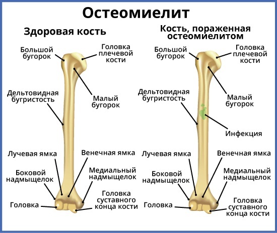
- Purulent-inflammatory processes in the region of the sacrum and pelvic bones, such as osteomyelitis, tuberculosis.
- Anomalies of the musculoskeletal system, such as not overgrowing of the arches of the vertebrae, underdevelopment of the segments of the sacrum, an extra vertebra.
- Herniated discs, exostosis and other formations.
The specialist who performs the MRI makes a descriptive conclusion, but he does not make a diagnosis, since the magnetic resonance imaging only shows structures that do not comply with the norms.
To make an accurate diagnosis, the results of the study should be evaluated by the attending physician in combination with others. data: clinical manifestations of the disease, the results of other analyzes and examinations, including CT, ultrasound, x-ray.
Where to make and what does the cost depend on?
If there is a referral from a doctor, then an MRI can be done free of charge at the hospital at the place of residence, but since there is a long queue for this procedure, it can be passed for a fee both in the state and in frequent clinic.
The cost of the examination starts from 2 thousand. rub. The price depends on the power of the tomograph, the qualifications of the doctor, the prestige of the clinic, the need to use a contrast agent (in this case, the cost will be 2 times higher).
Approximate prices:
| City | Clinic name and service cost |
| Moscow | Diomag R, price 3900 rubles; Elena Malyshey Medical Center, price 6000 rubles. |
| St. Petersburg | Simed, price 2690 rubles; MARCH, 3200 rubles. |
| Kazan | Clinic Am Medica, price 2610 rubles; LDS MIBTs, price 3600 rubles. |
MRI is considered one of the most accurate, safe and reliable methods for diagnosing sacroiliac joints, but it helps to make the correct diagnosis only in combination with other methods survey.
MRI video
Analysis of MRI of the lumbar spine:


