
The vascular mesh located in the human body resembles a tree. The trunk is the main artery that receives oxygen-rich blood from the heart. Thick main branches branch off from it, passing into small blood vessels. The abdominal aorta is partthat runs through the abdominal cavity.
Record content:
- 1 What is the abdominal aorta and where is it located?
-
2 Abdominal aorta anatomy
- 2.1 Ascending department
- 2.2 Aortic arch
- 2.3 Descending department
- 3 Microscopic structure
- 4 What is the function of the aorta?
- 5 Size norm for ultrasound
- 6 Branches of a large unpaired arterial vessel
-
7 Possible pathologies and complications of the abdominal aorta, their symptoms
- 7.1 Aneurysm
- 7.2 Atherosclerosis
- 7.3 Abdominal aortic aortitis
- 7.4 Leriche syndrome
- 7.5 Mesenteric artery infarction
- 7.6 Past injuries
- 7.7 Transferred operations
- 7.8 Abdominal tumors
- 8 Methods for diagnosing diseases
- 9 Abdominal aorta video
What is the abdominal aorta and where is it located?
As the main artery, the aorta develops early in the embryonic stage and is responsible for the transfer of blood from the heart. The aorta is the central artery of the body. It exits the left ventricle of the heart and distributes oxygenated blood. It begins between the 2 collarbones in the upper chest, curves around the heart, and after crossing the diaphragm, the abdominal cavity enters.
The abdominal aorta is located on the posterior abdominal wall in the retroperitoneal space, where it bifurcates at the end into paired iliac arteries in the lower abdomen.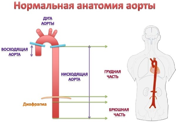
This large vessel regulates blood flow: blood from the heart is ejected in leaps and bounds, and later, in the aorta, it flows in a continuous stream. This becomes possible due to the inner elastic walls of the vessel.
Abdominal aorta anatomy
The aorta is divided into several areas. The ascending part is called the ascending aorta. It runs vertically upward from the heart and is interrupted by the aortic valve. The coronary arteries that feed the heart leave the bulbous portion of this area. The descending part extends down to the pelvic region. From here 2 large pelvic arteries branch off.
The descending aorta, in turn, is divided into:
- thoracic aorta;
- renal arteries;
- the abdominal aorta below the renal arteries.
Other vessels supplying the surrounding organs and nerves branch off from these 3 divisions. Two pelvic arteries deliver fresh blood to the legs.
The aorta is not only highly branched to reliably supply blood to the entire body, but its vessels are also extremely elastic. This elasticity is necessary to maintain constant pressure in the bloodstream. Sensitive pressure sensors on the inner walls respond to blood flow and signal to muscle fibers that blood flow is stopped.
Regional division of the aorta occurs in the direction of blood flow through the vessel.
Ascending department
The ascending part of the vessel is the first of 4 regions. Here the blood is under high pressure as it leaves the left ventricle due to its strong contraction.
The beginning of this area is marked by the aortic valve of the heart, and just below it there are 3 small openings known as sinuses that supply blood to the coronary heart arteries. They give rise to the right and left coronary arteries that supply the myocardium.
Aortic arch
The ascending section merges with the arch, which changes the direction of the main artery by about 180˚. Vessels that supply blood to the head and arms extend from the upper part of the arch.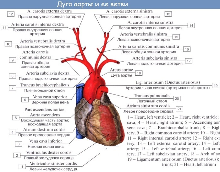
The function of this part of the vessel is to regulate the blood pressure leaving the left ventricle of the heart. The aortic arch has 3 branches. They supply blood to the brain and upper body.
Descending department
The third region is known as the descending thoracic aorta. This segment is located in the chest and supplies blood to the organs and other structures of this cavity.
The descending section makes up most of the main artery. It runs vertically down through the entire chest and abdomen. In the pelvic area, it is divided into 2 large pelvic arteries.
Descending is divided into sections:
- Thoracic aorta. This part lies entirely in the chest and supplies blood to the pericardium, esophagus, lung tissue and intercostal space. The thoracic reaches the diaphragm at the level of the 12th thoracic vertebra.
- Abdominal aorta located initially at the diaphragm. Thus, when the descending aorta passes through the diaphragm, its name changes to abdominal. Here she creates branches that supply blood to the organs and structures of the abdominal cavity.
At the level of the 4th lumbar vertebra, the abdominal part is divided into 2 large pelvic arteries.
Microscopic structure
The aorta is a complex structure formed by a combination of various tissues and components such as nerves, extracellular matrix, endothelial cells, intimal cells, and smooth muscle.
The walls of this "pipeline" consist of 3 layers:
- Inner lining or endothelium, consisting of 1 layer of epithelial cells and in contact with blood. It provides a soft lining surface for blood flow.
- Middle shell. Consists of 5-7 layers of smooth muscles surrounding the lumen of the aorta and elastic tissue. This special composition provides the flexibility of the vessel, which is necessary for it to contract or expand with each heartbeat. The tissue layer also contributes to the correct distribution of blood.
- Outer sheath or adventitia - This is the outer layer of the vessel, formed mainly by collagen. The substance is necessary for the artery to remain attached to the surrounding tissues and organs.
What is the function of the aorta?
The main role of the vessel in the human body is to supply blood to the entire structure of the body, with the exception of the lungs. The main artery provides blood supply, continuous blood flow, and maintains AD.
The beating heart creates large pressure drops in the circulatory system through contraction (systole) and relaxation (diastole). Due to its elasticity, the aorta can compensate for this and thus provide a continuous blood flow. With the help of the "Windkessel" function, it maintains blood pressure in the normal range.
Size norm for ultrasound
The abdominal aorta is located below the diaphragm, which can be seen on an ultrasound scan. When scanning, anomalies of its structure are clearly distinguishable: narrowing of the lumen, aneurysm. And you can also determine the presence of blood clots and assess the blood flow inside the vessel.
The normal aorta of an adult is measured in cross section by the maximum inner diameter, which is about 3 cm at the level of the xiphoid process and up to 1 cm at the level of the bifurcation (its separation). The diameter of the transverse and vertical cut must be the same.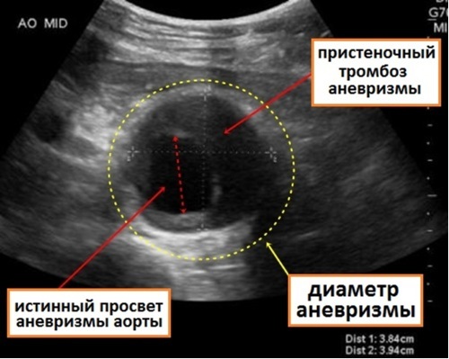
Measurements should be taken at different levels along the entire length of the main artery. Any significant increase in diameter is pathological.
The aorta can be displaced with scoliosis, tumors of the retroperitoneal space, or damage to the lymph nodes located in the front of the lumbar vertebrae. If the aorta has a cross-sectional diameter of more than 5 cm, an urgent need to consult a clinician, because there is a high risk of rupture.
If the diameter is 3 to 4 cm, it should be checked after 12 months, and if there are larger aneurysms, after 6 months. Indications for surgery, taking into account the individual situation: for men - a diameter of 5 to 5.5 cm, for women - from 4.5 cm.
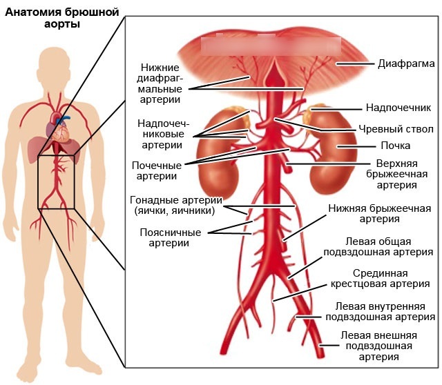
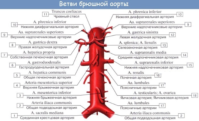
Branches of a large unpaired arterial vessel
The abdominal part of the aorta, all the main arterial branches are very elastic. During systole, the walls of the aorta and arteries dilate to accommodate the increased blood flow. During diastole, nearby vessels contract, and elastin fibers help the blood flow through the arterial vessels.
Overview of the branches of the aorta:
| Parts of the aorta | Branches | What organs supply blood |
| Upstream department: coronary arteries |
Left | Left atrium, left ventricle |
| Right | Right atrium, right ventricle | |
| Aortic arch | Brachiocephalic trunk | The right half of the head, neck and brain, right arm |
| Carotid artery | Brain, organ of vision, neck | |
| Left subclavian artery | Parts of the neck and thoracic region | |
| Descending | Intercostal | Intercostal space |
| Subcostal arteries | Hypochondrium | |
| Superior artery (diaphragm) | Diaphragm | |
| Bronchial | Connective tissue and lymph nodes of the lungs, bronchial mucosa | |
| Esophageal | Esophagus | |
| Pericardial | The back of the pericardium | |
| Mediastinal | Lymph nodes of the posterior mediastinum, connective tissue | |
| Abdominal | Lower diaphragmatic (pair) | Diaphragm |
| Celiac trunk | Pancreas, duodenum, spleen and adjacent mesentery, stomach, liver | |
| Superior mesenteric | Small intestine below the duodenum, as well as in part of the cecum and large intestine | |
| Adrenal | Adrenal glands | |
| Renal | Adrenal glands | |
| Ovarian (female), testicular (male) |
Ovary and epididymis | |
| Inferior mesenteric | Distal third of the transverse colon, descending colon, sigmoid colon, and upper rectum | |
| Lumbar | Lumbar spine, spinal canal, parts of the lumbar muscles of the back and side walls of the trunk | |
| Common iliac | Pelvic area and lower limb | |
| Sacral median (tail) |
Sacral spine |
The receipt of blood by each organ or tissue is precisely regulated by the body. The brain receives about 13%, and sometimes more, sometimes less blood is delivered to other organs, depending on how "active" they are. After eating, for example, quite a lot of blood enters the digestive tract, about 24%. When performing physical work, more blood flows into the skeletal muscles than at rest.
Possible pathologies and complications of the abdominal aorta, their symptoms
Aortic disease primarily affects the elderly, and women are less affected than men. The development of pathology is a rather lengthy process. Sometimes diseases develop so slowly that they do not bother a person for years. All abdominal aortic lesions are always associated with impaired blood flow.
Aneurysm
An aneurysm is an enlargement of an artery that can become very large and then rupture over time and cause severe internal bleeding, shock, or death.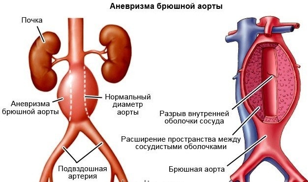
Blood clots (thrombi) very often form inside an unruptured aneurysm sac, which can lead to vasoconstriction in:
- legs;
- hands;
- organs of the abdominal cavity.
About 80% of all main artery aneurysms are located in the abdominal region below the renal arteries.
In the vast majority of cases, aortic aneurysm is asymptomatic.
But sometimes there are complaints such as:
- hoarseness of the voice;
- dyspnea;
- shoulder pain;
- back problems or hemoptysis.
Most often, the lesion is discovered by chance during X-ray examination.
The main cause of aneurysm formation is arteriosclerosis, slow "calcification" of blood vessels.
An aneurysm can present with abdominal pain, but diffuse back pain that looks more like "lumbago" or renal colic is equally likely. A very large bulge may appear as a throbbing lump on the abdomen. On rupture, life-threatening shock quickly ensues. Because of the large blood loss after a rupture, people die from internal bleeding in a very short time.
Atherosclerosis
Fat, calcium, connective tissue, blood clots are deposited on the walls and the aorta narrows. This causes disruption of the flow, damage to the walls of the vessels and their rupture. The deposits interfere with the elastic movement of the aorta and prevent it from contracting normally.
Many people with atherosclerosis have no symptoms. They can only be diagnosed after a heart attack or stroke. Early atherosclerosis is usually asymptomatic. In advanced stages, a person may develop symptoms, depending on the location and severity of the blockage.
Abdominal aortic aortitis
Inflammation of the aorta is extremely rare, but it can cause vascular stenosis, leading to rupture.
Some of the common causes of inflammation are:
- Infection. A number of organisms cause infection, which leads to vascular inflammation. In most cases, a bacterial infection in adjacent tissues can travel towards the aorta through the bloodstream. It is known that syphilis, salmonella, staphylococcal and streptococcal infections spread bacteria, attack the immune system and damage the aorta, which causes its inflammation.
- Connective tissue diseases. Autoimmune diseases, in which the immune system attacks its own tissues, lead to an inflammatory condition. Rheumatoid arthritis, sarcoidosis, systemic lupus erythematosus are largely responsible for the formation of the inflammatory process.
At the initial stages, symptoms do not appear, often inflammation is found at the stage of surgery for an aneurysm.
Leriche syndrome
Peripheral artery disease is caused by a buildup of plaque in the iliac arteries. The constriction of blood vessels caused by deposits in them leads to a decrease in blood flow to the legs.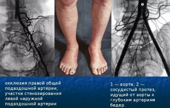
Over time, other symptoms appear:
- the lower limbs grow cold and blue;
- pain and cramps appear in the legs, buttocks;
- erectile dysfunction occurs.
Mesenteric artery infarction
Mesenteric artery ischemia occurs when one or more of the 3 main arteries that supply blood to the small and large intestines are narrowed or blocked. They are called the mesenteric arteries.
Arterial strengthening occurs when fat, cholesterol, and other substances build up in the walls of the arteries. It is more common in smokers and people with high blood pressure or high blood cholesterol.
Symptoms occur when oxygen supply slows down due to the gradual hardening of the mesenteric arteries:
- stomach pain after eating;
- diarrhea;
- vomit.
Past injuries
Blunt injury to the abdominal aorta is rare and accounts for only 5% of all vascular injuries. This is partly due to its protected position. Patients who have suffered such an injury are rarely admitted to the hospital alive. Such injuries are often seen in accidents and are associated with severe blunt intra-abdominal injuries and fractures of the lumbar-thoracic spine.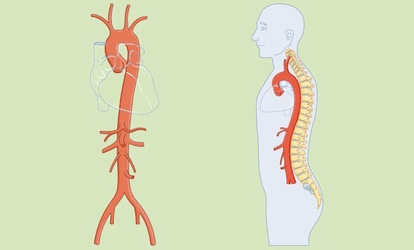
The mechanism of damage is associated with biomechanical direct and indirect forces acting on the vessel. They destroy the intima and, depending on the magnitude of the blow, can cause the aorta to be cut. Most often, areas are injured at the level of the inferior mesenteric artery, renal arteries and at the site of its division into the iliac arteries.
Transferred operations
As with any invasive procedure, surgery to correct an abdominal aortic aneurysm is risky and fraught with possible complications. Some of them are associated with clamping the aorta and, accordingly, a violation of the blood supply to the organs located below the clamping zone (kidneys, spinal cord, intestines, lower extremities).
Abdominal tumors
Malignant neoplasms and aneurysms are common diseases, especially among the aging population.
With pancreatic cancer, it is possible for the tumor to grow into the surrounding arteries. This is especially dangerous because cancer cells spread through the blood and the lymphatic system throughout the body (metastasis). In liver cancer, the tumor can go beyond and enter the hepatic vessels. There is often a blockage of the blood vessels supplying the tumor to the liver.
Retroperitoneal fibrosis is a rare condition also known as Ormond's disease. It occurs when an excess of fibrous tissue forms behind the stomach and intestines. Fibrosis is the proliferation of excess connective tissue, the mass of which causes compression and blockage of the ureters. The condition usually begins with inflammation and fibrosis of the abdominal aorta.
The abdominal aorta is located in the abdominal cavity and has branches. On one of them, blood enters the area under the kidneys. As the disease progresses, the arteries that carry blood to the legs and kidneys are affected. Pain, swelling of the legs, and decreased kidney function may occur.
Methods for diagnosing diseases
The doctor has different options for recognizing abnormalities in the main artery. Some information can already be obtained by palpation, but an imaging procedure is usually required.
Various options are possible:
- chest x-ray with angiography;
- ultrasound scanning;
- magnetic resonance imaging (MRI)
- computed tomography (CT)
- echocardiography through the esophagus;
- cardiac catheterization.
The altered diameter of the abdominal aorta, located around vital organs, causes dangerous pathologies that cannot be ignored.
Any occurrence of ailments that do not have a logical explanation in the form of poisoning or digestive disorders is a reason to consult a doctor and undergo an examination. The sooner changes are recognized, the less negative consequences they will have.
Abdominal aorta video
Abdominal aorta anatomy:


